Tau Antibody (1D5) - Pre-formed Fibrils - BSA Free
Novus Biologicals | Catalog # NBP2-79825
![Western Blot: Tau Antibody (1D5)Pre-formed Fibrils [NBP2-79825]](https://resources.rndsystems.com/images/products/Tau-Antibody-1D5-Pre-formed-Fibrils-Western-Blot-NBP2-79825-img0004.jpg "Western Blot: Tau Antibody (1D5)Pre-formed Fibrils [NBP2-79825]")
Loading...
Key Product Details
Species Reactivity
Human, Mouse, Rat
Applications
Western Blot, ELISA, Immunocytochemistry/ Immunofluorescence, Dot Blot
Label
Unconjugated
Antibody Source
Monoclonal Mouse IgG1 Clone # 1D5
Format
BSA Free
Loading...
Product Specifications
Immunogen
Human Recombinant Tau441 (2N4R), P301S mutant Protein Pre-formed Fibrils
Specificity
Detects Multiple Bands. Antibody detects monomer under denaturing conditions but preferentially detects fibril under native conditions (dot blot).
Clonality
Monoclonal
Host
Mouse
Isotype
IgG1
Scientific Data Images for Tau Antibody (1D5) - Pre-formed Fibrils - BSA Free
Western Blot: Tau Antibody (1D5)Pre-formed Fibrils [NBP2-79825]
Western Blot: Tau Antibody (1D5) - Pre-formed Fibrils [NBP2-79825] - Western Blot analysis of Human Breast Cancer Cell line and Mouse Brain showing detection of Tau protein using Mouse Anti-Tau Monoclonal Antibody, Clone 1D5 (NBP2-79825). Lane 1: MW Marker. Lane 2: Human T-47d (10ug). Lane 3: Mouse Brain (20ug). Block: 5% Skim Milk powder in TBST. Primary Antibody: Mouse Anti-Tau Monoclonal Antibody (NBP2-79825) at 1:1000 for 2 hours at RT with shaking. Secondary Antibody: Goat anti-mouse IgG:HRP at 1:5000 for 1 hour at RT with shaking. Color Development: Chemiluminescent for HRP (Moss) for 5 min in RT.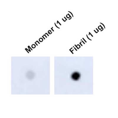
Dot Blot: Tau Antibody (1D5) - Pre-formed Fibrils [NBP2-79825] - Dot Blot analysis using Mouse Anti-Tau Monoclonal Antibody, Clone 1D5 (NBP2-79825). Tissue: Recombinant Protein. Species: Human. Primary Antibody: Mouse Anti-Tau Monoclonal Antibody (NBP2-79825) at 1:1000 for 2 hours at RT with shaking. Secondary Antibody: Goat anti-mouse IgG:HRP at 1:5000 for 1 hour at RT with shaking.
Applications for Tau Antibody (1D5) - Pre-formed Fibrils - BSA Free
Application
Recommended Usage
Western Blot
1:1000
Formulation, Preparation, and Storage
Purification
Protein G purified
Formulation
PBS (pH 7.4), 50% Glycerol
Format
BSA Free
Preservative
0.09% Sodium Azide
Concentration
Please see the vial label for concentration. If unlisted please contact technical services.
Shipping
The product is shipped with polar packs. Upon receipt, store it immediately at the temperature recommended below.
Stability & Storage
Store at -20C.
Background: Tau
Long Name
Microtubule-Associated Protein Tau
Alternate Names
MAPT, MSTD, MTBT1, Neurofibrillary tangle protein, PPND
Gene Symbol
MAPT
Additional Tau Products
Product Documents for Tau Antibody (1D5) - Pre-formed Fibrils - BSA Free
Certificate of Analysis
To download a Certificate of Analysis, please enter a lot or batch number in the search box below.
Product Specific Notices for Tau Antibody (1D5) - Pre-formed Fibrils - BSA Free
This product is for research use only and is not approved for use in humans or in clinical diagnosis. Primary Antibodies are guaranteed for 1 year from date of receipt.
Related Research Areas
Customer Reviews for Tau Antibody (1D5) - Pre-formed Fibrils - BSA Free
There are currently no reviews for this product. Be the first to review Tau Antibody (1D5) - Pre-formed Fibrils - BSA Free and earn rewards!
Have you used Tau Antibody (1D5) - Pre-formed Fibrils - BSA Free?
Submit a review and receive an Amazon gift card!
$25/€18/£15/$25CAN/¥2500 Yen for a review with an image
$10/€7/£6/$10CAN/¥1110 Yen for a review without an image
Submit a review
Protocols
Find general support by application which include: protocols, troubleshooting, illustrated assays, videos and webinars.
- Appropriate Fixation of IHC/ICC Samples
- Cellular Response to Hypoxia Protocols
- ClariTSA™ Fluorophore Kits
- Detection & Visualization of Antibody Binding
- ELISA Sample Preparation & Collection Guide
- ELISA Troubleshooting Guide
- How to Run an R&D Systems DuoSet ELISA
- How to Run an R&D Systems Quantikine ELISA
- How to Run an R&D Systems Quantikine™ QuicKit™ ELISA
- ICC Cell Smear Protocol for Suspension Cells
- ICC Immunocytochemistry Protocol Videos
- ICC for Adherent Cells
- Immunocytochemistry (ICC) Protocol
- Immunocytochemistry Troubleshooting
- Immunofluorescence of Organoids Embedded in Cultrex Basement Membrane Extract
- Immunohistochemistry (IHC) and Immunocytochemistry (ICC) Protocols
- Preparing Samples for IHC/ICC Experiments
- Preventing Non-Specific Staining (Non-Specific Binding)
- Primary Antibody Selection & Optimization
- Protocol for VisUCyte™ HRP Polymer Detection Reagent
- Protocol for the Fluorescent ICC Staining of Cell Smears - Graphic
- Protocol for the Fluorescent ICC Staining of Cultured Cells on Coverslips - Graphic
- Protocol for the Preparation and Fluorescent ICC Staining of Cells on Coverslips
- Protocol for the Preparation and Fluorescent ICC Staining of Non-adherent Cells
- Protocol for the Preparation and Fluorescent ICC Staining of Stem Cells on Coverslips
- Protocol for the Preparation of a Cell Smear for Non-adherent Cell ICC - Graphic
- Quantikine HS ELISA Kit Assay Principle, Alkaline Phosphatase
- Quantikine HS ELISA Kit Principle, Streptavidin-HRP Polymer
- R&D Systems Quality Control Western Blot Protocol
- Sandwich ELISA (Colorimetric) – Biotin/Streptavidin Detection Protocol
- Sandwich ELISA (Colorimetric) – Direct Detection Protocol
- TUNEL and Active Caspase-3 Detection by IHC/ICC Protocol
- The Importance of IHC/ICC Controls
- Troubleshooting Guide: ELISA
- Troubleshooting Guide: Western Blot Figures
- Western Blot Conditions
- Western Blot Protocol
- Western Blot Protocol for Cell Lysates
- Western Blot Troubleshooting
- Western Blot Troubleshooting Guide
- View all Protocols, Troubleshooting, Illustrated assays and Webinars
Loading...