TAZ/WWTR1 Antibody - BSA Free
Novus Biologicals | Catalog # NB110-58359
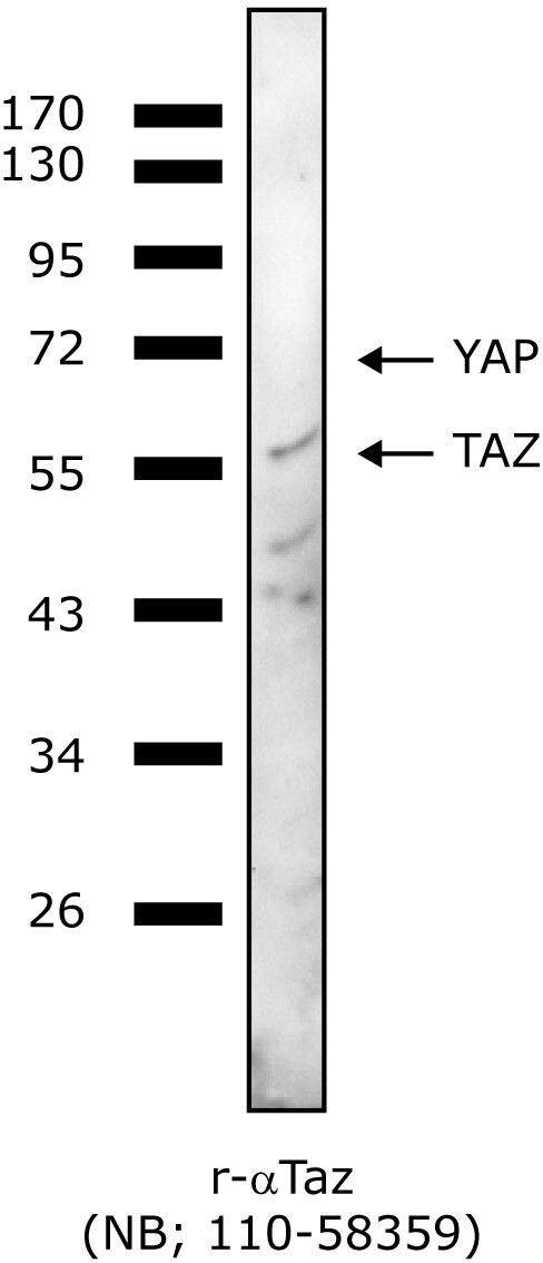
![Western Blot: TAZ/WWTR1 AntibodyBSA Free [NB110-58359]](https://resources.rndsystems.com/images/products/TAZ-WWTR1-Antibody-Western-Blot-NB110-58359-img0006.jpg "Western Blot: TAZ/WWTR1 AntibodyBSA Free [NB110-58359]")
Key Product Details
Species Reactivity
Validated:
Cited:
Applications
Validated:
Cited:
Label
Antibody Source
Format
Product Specifications
Immunogen
Localization
Clonality
Host
Isotype
Scientific Data Images for TAZ/WWTR1 Antibody - BSA Free
Western Blot: TAZ/WWTR1 AntibodyBSA Free [NB110-58359]
Western Blot: TAZ/WWTR1 Antibody [NB110-58359] - Detection of TAZ on HEK 293 cell lysate using NB110-58359.![Immunocytochemistry/ Immunofluorescence: TAZ/WWTR1 Antibody - BSA Free [NB110-58359]](https://resources.rndsystems.com/images/products/TAZ-WWTR1-Antibody-Immunocytochemistry-Immunofluorescence-NB110-58359-img0005.jpg "Immunocytochemistry/ Immunofluorescence: TAZ/WWTR1 Antibody - BSA Free [NB110-58359]")
Immunocytochemistry/ Immunofluorescence: TAZ/WWTR1 Antibody - BSA Free [NB110-58359]
Immunocytochemistry/Immunofluorescence: TAZ/WWTR1 Antibody [NB110-58359] - Detection of TAZ (Green) in HepG2 cells using NB110-58359. Nuclei (Blue) are counterstained with Hoecsht 33258.![Immunohistochemistry-Paraffin: TAZ/WWTR1 Antibody - BSA Free [NB110-58359]](https://resources.rndsystems.com/images/products/TAZ-WWTR1-Antibody-Immunohistochemistry-Paraffin-NB110-58359-img0010.jpg "Immunohistochemistry-Paraffin: TAZ/WWTR1 Antibody - BSA Free [NB110-58359]")
Immunohistochemistry-Paraffin: TAZ/WWTR1 Antibody - BSA Free [NB110-58359]
Immunohistochemistry-Paraffin: TAZ/WWTR1 Antibody [NB110-58359] - IHC analysis of formalin fixed paraffin-embedded (FFPE) human kidney using TAZ/WWTR1 antibody at 1:500 on a Bond Rx autostainer (Leica Biosystems). The assay involved 20 minutes of heat induced antigen retrieval (HIER) using 10mM sodium citrate buffer (pH 6.0) and endogenous peroxidase quenching with peroxide block. The sections were incubated with primary antibody for 30 minutes and Bond Polymer Refine Detection (Leica Biosystems) with DAB was used for signal development followed by counterstaining with hematoxylin. Whole slide scanning and capturing of representative images was performed using Aperio AT2 (Leica Biosystems). Nuclear and cytoplasmic staining was observed. Staining was performed by Histowiz.![Simple Western: TAZ/WWTR1 AntibodyBSA Free [NB110-58359]](https://resources.rndsystems.com/images/products/TAZ-WWTR1-Antibody-Simple-Western-NB110-58359-img0008.jpg "Simple Western: TAZ/WWTR1 AntibodyBSA Free [NB110-58359]")
Simple Western: TAZ/WWTR1 AntibodyBSA Free [NB110-58359]
Simple Western: TAZ/WWTR1 Antibody [NB110-58359] - Simple Western lane view shows a specific band for TAZ in 0.5 mg/ml of HeLa lysate. This experiment was performed under reducing conditions using the 12-230 kDa separation system.
Immunoprecipitation: TAZ/WWTR1 Antibody - BSA Free [NB110-58359] -
Immunoprecipitation: TAZ/WWTR1 Antibody - BSA Free [NB110-58359] - TAZ knockout in muscle decreases mitochondrial mass, respiration, & exercise ability. b Proteins of gastrocnemius muscle of WT & mKO mice analysed via immunoblotting for detection of mitochondrial marker proteins. Alpha-tubulin used as loading control. Representative data shown & experiment performed twice w/ similar results. Image collected & cropped by CiteAb from following publication (https://pubmed.ncbi.nlm.nih.gov/35115527), licensed under a CC-BY license. Not internally tested by Novus Biologicals.
Western Blot: TAZ/WWTR1 Antibody - BSA Free [NB110-58359] -
RhoV and WWTR1 enhance ZIKV infection in A549 cells. Reconstituted (A) RhoV or (B) WWTR1 A549 KO clones were treated with different amounts of Dox (0, 0.001, 0.01, 0.1, 1, and 10 ug/mL) for 24 h to induce the expression of N-terminally V5-tagged RhoV or WWTR1 through the ePiggyBac transposon system. RhoV, WWTR1, and beta -actin (loading control) protein expression was determined by immunoblotting with V5, WWTR1, and beta -actin antibodies. The data are representative of two independent experiments. Reconstituted (C) RhoV or (D) WWTR1 A549 KO clones were treated with or without 1 ug/mL of Dox for 24 h prior to 1 h adsorption with ZIKV (MOI = 1 PFU/cell) and harvested at 12, 18, 24, and 48 h.p.i. to quantify infection levels. Dox was added back to the media during the course of infection. ZIKV infected cells were then fixed and permeabilized to stain with the pan-flavivirus envelope antibody prior to flow cytometry analysis. Infection levels of cells treated with Dox were normalized to that of the respective untreated condition (no Dox) at each timepoint and reported as fold change in ZIKV infection. The data are combined from three independent experiments. (E,F) Supernatant of infected cells from (C,D) was collected at 12, 18, 24, and 48 h.p.i. and titered on Vero cells by plaque assay. The data are representative of three independent experiments. Asterisks indicate statistically significant differences (two-way ANOVA and Sidak’s multiple comparisons test: *, p < 0.05; **, p < 0.01; ****, p < 0.0001). Image collected and cropped by CiteAb from the following open publication (https://pubmed.ncbi.nlm.nih.gov/34834920), licensed under a CC-BY license. Not internally tested by Novus Biologicals.
Western Blot: TAZ/WWTR1 Antibody - BSA Free [NB110-58359] -
A genome-wide CRISPR activation ZIKV screen in cells defective in IFN signaling. (A) Schematic flow of the CRISPR activation screen setup and conditions. STAT1−/− fibroblasts were transduced with lentiviruses carrying the SAM complex followed by the sgRNA library upregulating all known human gene isoforms. After antibiotic selection, these cells were challenged with ZIKV (strain: PRVABC59) at MOI of 0.5 PFU/cell and incubated for 14 days. Genomic DNA of mock-infected and ZIKV-infected cells was extracted, amplified, sequenced, and bioinformatically analyzed to determine potential antiviral and proviral candidate genes from the sgRNAs enriched or depleted in surviving cells, respectively. (B,C) Scatter plots showing negative selection of sgRNAs targeting the top candidate genes identified by MAGeCK VISPR, a quality control and analysis workflow for CRISPR screens, compared with other sgRNAs in the library after ZIKV infection (p-value < 0.001 and false discovery rate (FDR) < 0.05). (D) RhoV mRNA levels were measured by RT-qPCR in STAT1−/− fibroblasts transduced with lentiviruses carrying RhoV promoter targeting sgRNAs 1, 2, and 3. mRNA fold changes relative to the RhoV mRNA levels in NT sgRNA transduced STAT1−/− fibroblasts are shown. The data are from one independent experiment performed in triplicate. (E) WWTR1 protein expression in STAT1−/− fibroblasts transduced with lentiviruses carrying WWTR1 promoter targeting sgRNAs 1, 2, and 3 as well as NT sgRNA was determined by immunoblotting using WWTR1 antibody. beta -actin serves as a loading control. The data are from one experiment. Image collected and cropped by CiteAb from the following open publication (https://pubmed.ncbi.nlm.nih.gov/34834920), licensed under a CC-BY license. Not internally tested by Novus Biologicals.Applications for TAZ/WWTR1 Antibody - BSA Free
Chromatin Immunoprecipitation
Immunocytochemistry/ Immunofluorescence
Immunohistochemistry
Immunohistochemistry-Paraffin
Immunoprecipitation
Proximity Ligation Assay
Simple Western
Western Blot
See Simple Western Antibody Database for Simple Western validation: Tested in HeLa lysate 0.5 mg/mL, separated by Size, antibody dilution of 1:100, apparent MW was 58 kDa. Separated by Size-Wes, Sally Sue/Peggy Sue.
Reviewed Applications
Read 1 review rated 3 using NB110-58359 in the following applications:
Formulation, Preparation, and Storage
Purification
Formulation
Format
Preservative
Concentration
Shipping
Stability & Storage
Background: TAZ/WWTR1
Long Name
Alternate Names
Entrez Gene IDs
Gene Symbol
UniProt
Additional TAZ/WWTR1 Products
Product Documents for TAZ/WWTR1 Antibody - BSA Free
Certificate of Analysis
To download a Certificate of Analysis, please enter a lot or batch number in the search box below.
Product Specific Notices for TAZ/WWTR1 Antibody - BSA Free
This product is for research use only and is not approved for use in humans or in clinical diagnosis. Primary Antibodies are guaranteed for 1 year from date of receipt.
Related Research Areas
Citations for TAZ/WWTR1 Antibody - BSA Free
 Powered by Bioz
Customer Reviews for TAZ/WWTR1 Antibody - BSA Free (1)
Have you used TAZ/WWTR1 Antibody - BSA Free?
Submit a review and receive an Amazon gift card!
$25/€18/£15/$25CAN/¥2500 Yen for a review with an image
$10/€7/£6/$10CAN/¥1110 Yen for a review without an image
Submit a review
Customer Images
-
Application: Western BlotSample Tested: HEK293T cell lysateSpecies: HumanVerified Customer | Posted 11/06/2013

There are no reviews that match your criteria.
Protocols
View specific protocols for TAZ/WWTR1 Antibody - BSA Free (NB110-58359):
Culture cells to appropriate density in 35 mm culture dishes or 6-well plates.
1. Remove culture medium and wash the cells briefly in PBS. Add 10% formalin to the dish and fix at room temperature for 10 minutes.
2. Remove the formalin and wash the cells in PBS.
3. Permeablize the cells with 0.1% Triton X100 or other suitable detergent for 10 min.
4. Remove the permeablization buffer and wash three times for 10 minutes each in PBS. Be sure to not let the specimen dry out.
5. To block nonspecific antibody binding, incubate in 10% normal goat serum from 1 hour to overnight at room temperature.
6. Add primary antibody at appropriate dilution and incubate overnight at 4C.
7. Remove primary antibody and replace with PBS. Wash three times for 10 minutes each.
8. Add secondary antibody at appropriate dilution. Incubate for 1 hour at room temperature.
9. Remove secondary antibody and replace with PBS. Wash three times for 10 minutes each.
10. Counter stain DNA with DAPi if required.
Antigen Unmasking:
Bring slides to a boil in 10 mM sodium citrate buffer (pH 6.0) then maintain at a sub-boiling temperature for 10 minutes. Cool slides on bench-top for 30 minutes (keep slides in the sodium citrate buffer at all times).
Staining:
1. Wash sections in deionized water three times for 5 minutes each.
2. Wash sections in PBS for 5 minutes.
3. Block each section with 100-400 ul blocking solution (1% BSA in PBS) for 1 hour at room temperature.
4. Remove blocking solution and add 100-400 ul diluted primary antibody. Incubate overnight at 4 C.
5. Remove antibody solution and wash sections in wash buffer three times for 5 minutes each.
6. Add 100-400 ul HRP polymer conjugated secondary antibody. Incubate 30 minutes at room temperature.
7. Wash sections three times in wash buffer for 5 minutes each.
8. Add 100-400 ul DAB substrate to each section and monitor staining closely.
9. As soon as the sections develop, immerse slides in deionized water.
10. Counterstain sections in hematoxylin.
11. Wash sections in deionized water two times for 5 minutes each.
12. Dehydrate sections.
13. Mount coverslips.
1. Perform SDS-PAGE on samples to be analyzed, loading 10-25 ug of total protein per lane.
2. Transfer proteins to PVDF membrane according to the instructions provided by the manufacturer of the membrane and transfer apparatus.
3. Stain the membrane with Ponceau S (or similar product) to assess transfer success, and mark molecular weight standards where appropriate.
4. Rinse the blot TBS -0.05% Tween 20 (TBST).
5. Block the membrane in 5% Non-fat milk in TBST (blocking buffer) for at least 1 hour.
6. Wash the membrane in TBST three times for 10 minutes each.
7. Dilute primary antibody in blocking buffer and incubate overnight at 4C with gentle rocking.
8. Wash the membrane in TBST three times for 10 minutes each.
9. Incubate the membrane in diluted HRP conjugated secondary antibody in blocking buffer (as per manufacturer's instructions) for 1 hour at room temperature.
10. Wash the blot in TBST three times for 10 minutes each (this step can be repeated as required to reduce background).
11. Apply the detection reagent of choice in accordance with the manufacturer's instructions.
Find general support by application which include: protocols, troubleshooting, illustrated assays, videos and webinars.
- Antigen Retrieval Protocol (PIER)
- Antigen Retrieval for Frozen Sections Protocol
- Appropriate Fixation of IHC/ICC Samples
- Cellular Response to Hypoxia Protocols
- ChIP Protocol Video
- Chromatin Immunoprecipitation (ChIP) Protocol
- Chromatin Immunoprecipitation Protocol
- Chromogenic IHC Staining of Formalin-Fixed Paraffin-Embedded (FFPE) Tissue Protocol
- Chromogenic Immunohistochemistry Staining of Frozen Tissue
- ClariTSA™ Fluorophore Kits
- Detection & Visualization of Antibody Binding
- Fluorescent IHC Staining of Frozen Tissue Protocol
- Graphic Protocol for Heat-induced Epitope Retrieval
- Graphic Protocol for the Preparation and Fluorescent IHC Staining of Frozen Tissue Sections
- Graphic Protocol for the Preparation and Fluorescent IHC Staining of Paraffin-embedded Tissue Sections
- Graphic Protocol for the Preparation of Gelatin-coated Slides for Histological Tissue Sections
- ICC Cell Smear Protocol for Suspension Cells
- ICC Immunocytochemistry Protocol Videos
- ICC for Adherent Cells
- IHC Sample Preparation (Frozen sections vs Paraffin)
- Immunocytochemistry (ICC) Protocol
- Immunocytochemistry Troubleshooting
- Immunofluorescence of Organoids Embedded in Cultrex Basement Membrane Extract
- Immunofluorescent IHC Staining of Formalin-Fixed Paraffin-Embedded (FFPE) Tissue Protocol
- Immunohistochemistry (IHC) and Immunocytochemistry (ICC) Protocols
- Immunohistochemistry Frozen Troubleshooting
- Immunohistochemistry Paraffin Troubleshooting
- Immunoprecipitation Protocol
- Preparing Samples for IHC/ICC Experiments
- Preventing Non-Specific Staining (Non-Specific Binding)
- Primary Antibody Selection & Optimization
- Protocol for Heat-Induced Epitope Retrieval (HIER)
- Protocol for Making a 4% Formaldehyde Solution in PBS
- Protocol for VisUCyte™ HRP Polymer Detection Reagent
- Protocol for the Fluorescent ICC Staining of Cell Smears - Graphic
- Protocol for the Fluorescent ICC Staining of Cultured Cells on Coverslips - Graphic
- Protocol for the Preparation & Fixation of Cells on Coverslips
- Protocol for the Preparation and Chromogenic IHC Staining of Frozen Tissue Sections
- Protocol for the Preparation and Chromogenic IHC Staining of Frozen Tissue Sections - Graphic
- Protocol for the Preparation and Chromogenic IHC Staining of Paraffin-embedded Tissue Sections
- Protocol for the Preparation and Chromogenic IHC Staining of Paraffin-embedded Tissue Sections - Graphic
- Protocol for the Preparation and Fluorescent ICC Staining of Cells on Coverslips
- Protocol for the Preparation and Fluorescent ICC Staining of Non-adherent Cells
- Protocol for the Preparation and Fluorescent ICC Staining of Stem Cells on Coverslips
- Protocol for the Preparation and Fluorescent IHC Staining of Frozen Tissue Sections
- Protocol for the Preparation and Fluorescent IHC Staining of Paraffin-embedded Tissue Sections
- Protocol for the Preparation of Gelatin-coated Slides for Histological Tissue Sections
- Protocol for the Preparation of a Cell Smear for Non-adherent Cell ICC - Graphic
- R&D Systems Quality Control Western Blot Protocol
- TUNEL and Active Caspase-3 Detection by IHC/ICC Protocol
- The Importance of IHC/ICC Controls
- Troubleshooting Guide: Immunohistochemistry
- Troubleshooting Guide: Western Blot Figures
- Western Blot Conditions
- Western Blot Protocol
- Western Blot Protocol for Cell Lysates
- Western Blot Troubleshooting
- Western Blot Troubleshooting Guide
- View all Protocols, Troubleshooting, Illustrated assays and Webinars
FAQs for TAZ/WWTR1 Antibody - BSA Free
-
Q: I am looking for a WWTR1 antibody and NB110-58359 looks like the best one. I wonder if you guys can provide me the target sequence information of the antibody?
A: WWTR1 antibody (NB110-58359) is generated against residues 178-197 of WWTR1 (immunogen accession number Q9GZV5). The exact immunogen sequence for this antibody is: CPQRSMAVSQPNLVMNHQHQQ
-
Q: I want to know which TAZ primary antibody is best produced for WB (human and mouse)!
A:
I would recommend this TAZ antibody: NB110-58359. This is also comes in a sample size.
-
Q: Our customer was interested in the TAZ Antibody (Cat.NB110-58359). Could you please help to confirm if it has been tested with rat cells?
A: The TAZ antibody, NB110-58359, has been tested and validated for use in rat and is therefore guaranteed. Our 100% guarantee covers all of our products for the listed species and applications.
-
Q: We have an inquiry for your product #NB110-58359, TAZ Antibody. Our customer would like to try this product with rat cells lysate. Could you please let me know if you have data image of rat cells lysate can provide?
A: This antibody has been validated and is therefore guaranteed for use in human, rat and mouse in the following applications: Western blot, Immunoprecipitation and ICC/IF. We do not currently have a WB image that we can share however rat is one of the guaranteed species.
-
Q: I am looking for a WWTR1 antibody and NB110-58359 looks like the best one. I wonder if you guys can provide me the target sequence information of the antibody?
A: WWTR1 antibody (NB110-58359) is generated against residues 178-197 of WWTR1 (immunogen accession number Q9GZV5). The exact immunogen sequence for this antibody is: CPQRSMAVSQPNLVMNHQHQQ
-
Q: I want to know which TAZ primary antibody is best produced for WB (human and mouse)!
A:
I would recommend this TAZ antibody: NB110-58359. This is also comes in a sample size.
-
Q: Our customer was interested in the TAZ Antibody (Cat.NB110-58359). Could you please help to confirm if it has been tested with rat cells?
A: The TAZ antibody, NB110-58359, has been tested and validated for use in rat and is therefore guaranteed. Our 100% guarantee covers all of our products for the listed species and applications.
-
Q: We have an inquiry for your product #NB110-58359, TAZ Antibody. Our customer would like to try this product with rat cells lysate. Could you please let me know if you have data image of rat cells lysate can provide?
A: This antibody has been validated and is therefore guaranteed for use in human, rat and mouse in the following applications: Western blot, Immunoprecipitation and ICC/IF. We do not currently have a WB image that we can share however rat is one of the guaranteed species.