TBK1 Antibody (108A429) - BSA Free
Novus Biologicals | Catalog # NB100-56705
Key Product Details
Validated by
Species Reactivity
Validated:
Cited:
Predicted:
Applications
Validated:
Cited:
Label
Antibody Source
Format
Product Specifications
Immunogen
Clonality
Host
Isotype
Theoretical MW
Disclaimer note: The observed molecular weight of the protein may vary from the listed predicted molecular weight due to post translational modifications, post translation cleavages, relative charges, and other experimental factors.
Scientific Data Images for TBK1 Antibody (108A429) - BSA Free
![Knockout Validated: TBK1 Antibody (108A429) - BSA Free [NB100-56705]](https://resources.rndsystems.com/images/products/TBK1-Antibody-108A429-Knockout-Validated-NB100-56705-img0023.jpg "Western Blot: TBK1 Antibody (108A429) - BSA Free [NB100-56705]")
Western Blot: TBK1 Antibody (108A429) - BSA Free [NB100-56705]
Western Blot: TBK1 Antibody (108A429) [NB100-56705] - Western blot using lysates from U20S parental cell line and TBK1 knockout U20S cell line (KO), collected in RIPA buffer. Nitrocellulose membrane was probed with Mouse Anti-Human/Mouse/Rat TBK1 Monoclonal Antibody (Catalog # NB100-56705) at 1 ug/ul O/N at 4C, followed by HRP-conjugated Anti-Mouse IgG Secondary Antibody and ECL detection. A specific band at 83 kDa was detected for TBK1 in the parental U20S cell line, but is not detectable in knockout U20S cell line. The Ponceau stained transfers of each blot are shown to confirm equal protein loading. Image, protocol and testing courtesy of YCharOS Inc. (ycharos.com).![Simple Western: TBK1 Antibody (108A429)BSA Free [NB100-56705]](https://resources.rndsystems.com/images/products/TBK1-Antibody-108A429-Simple-Western-NB100-56705-img0014.jpg "Simple Western: TBK1 Antibody (108A429)BSA Free [NB100-56705]")
Simple Western: TBK1 Antibody (108A429)BSA Free [NB100-56705]
Simple Western: TBK1 Antibody (108A429) [NB100-56705] - Lane view shows a specific band for NAK/TBK1 in 0.5 mg/ml of Daudi (left) and Raw 264.7 (right) lysate. This experiment was performed under reducing conditions using the 12-230 kDa separation system. * Non-specific interaction with the 230 kDa Simple Western standard may be seen with this antibody![Immunohistochemistry-Paraffin: TBK1 Antibody (108A429) - BSA Free [NB100-56705]](https://resources.rndsystems.com/images/products/TBK1-Antibody-108A429-Immunohistochemistry-Paraffin-NB100-56705-img0021.jpg "Immunohistochemistry-Paraffin: TBK1 Antibody (108A429) - BSA Free [NB100-56705]")
Immunohistochemistry-Paraffin: TBK1 Antibody (108A429) - BSA Free [NB100-56705]
Immunohistochemistry-Paraffin: TBK1 Antibody (108A429) [NB100-56705] - Analysis of FFPE human testis tissue section using Peripheral Node Addressin antibody (clone MECA-79R) at 1:200. The staining was developed with HRP-DAB detection method and the counterstaining was performed using hematoxylin.![Western Blot: TBK1 Antibody (108A429)BSA Free [NB100-56705]](https://resources.rndsystems.com/images/products/TBK1-Antibody-108A429-Western-Blot-NB100-56705-img0016.jpg "Western Blot: TBK1 Antibody (108A429)BSA Free [NB100-56705]")
Western Blot: TBK1 Antibody (108A429)BSA Free [NB100-56705]
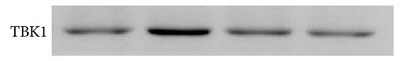
Western Blot: TBK1 Antibody (108A429) [NB100-56705] - Analysis using Azide and BSA Free version of NB100-56705. Lysate from 293 cells untransfected (lane 1) and transfected with human TBK1 cDNA (lane 2) probed with TBK1 antibody at 2 ug/ml.![Immunocytochemistry/ Immunofluorescence: TBK1 Antibody (108A429) - BSA Free [NB100-56705]](https://resources.rndsystems.com/images/products/TBK1-Antibody-108A429-Immunocytochemistry-Immunofluorescence-NB100-56705-img0013.jpg "Immunocytochemistry/ Immunofluorescence: TBK1 Antibody (108A429) - BSA Free [NB100-56705]")
Immunocytochemistry/ Immunofluorescence: TBK1 Antibody (108A429) - BSA Free [NB100-56705]
Immunocytochemistry/Immunofluorescence: TBK1 Antibody (108A429) [NB100-56705] - Antibody was tested in HeLa cells with DyLight 488 (green). Nuclei and alpha-tubulin were counterstained with DAPI (blue) and DyLight 550 (red). A dilution of 1:10 was used. Image objective 40x.![Immunohistochemistry-Paraffin: TBK1 Antibody (108A429) - BSA Free [NB100-56705]](https://resources.rndsystems.com/images/products/TBK1-Antibody-108A429-Immunohistochemistry-Paraffin-NB100-56705-img0022.jpg "Immunohistochemistry-Paraffin: TBK1 Antibody (108A429) - BSA Free [NB100-56705]")
Immunohistochemistry-Paraffin: TBK1 Antibody (108A429) - BSA Free [NB100-56705]
Immunohistochemistry-Paraffin: TBK1 Antibody (108A429) [NB100-56705] - Immunohistochemistry: TBK1 Antibody (108A429) - Azide and BSA Free [NBP2-33243] - Staining on human testis (Formalin-Fixed Paraffin-Embedded (FFPE)) Image from the Azide and BSA Free version of this antibody.![Western Blot: TBK1 Antibody (108A429)BSA Free [NB100-56705]](https://resources.rndsystems.com/images/products/TBK1-Antibody-108A429-Western-Blot-NB100-56705-img0012.jpg "Western Blot: TBK1 Antibody (108A429)BSA Free [NB100-56705]")
Western Blot: TBK1 Antibody (108A429)BSA Free [NB100-56705]
Western Blot: TBK1 Antibody (108A429) [NB100-56705] - Analysis of Human Daudi (A) and mouse RAW (B) cell lysate probed with TBK1 antibody at 2 ug/ml.![Immunocytochemistry/ Immunofluorescence: TBK1 Antibody (108A429) - BSA Free [NB100-56705]](https://resources.rndsystems.com/images/products/TBK1-Antibody-108A429-Immunocytochemistry-Immunofluorescence-NB100-56705-img0019.jpg "Immunocytochemistry/ Immunofluorescence: TBK1 Antibody (108A429) - BSA Free [NB100-56705]")
Immunocytochemistry/ Immunofluorescence: TBK1 Antibody (108A429) - BSA Free [NB100-56705]
Immunocytochemistry/Immunofluorescence: TBK1 Antibody (108A429) [NB100-56705] - HeLa cells were fixed for 10 minutes using 10% formalin and then permeabilized for 5 minutes using 1X PBS + 0.05% Triton X-100. The cells were incubated with anti-TBK1 at 20 ug/ml overnight at 4C and detected with an anti-mouse DyLight 488 (Green) at a 1:500 dilution. Actin was detected with Phalloidin 568 (Red) at a 1:200 dilution. Nuclei were counterstained with DAPI (Blue). Cells were imaged using a 40X objective. - BSA Free [NB100-56705] -")
Western Blot: TBK1 Antibody (108A429) - BSA Free [NB100-56705] -
STING pathway analysis. (A) STING mRNA relative expression in uninfected (CTR), HHV-6A, HHV-6B or HHV-7 infected NK92 cells 3d.p.i. ∗p values Student t test; (B) Western Blot analysis for house-keeping actin (upper blot) and STING (lower blot) expression in uninfected (CTR), HHV-6A, HHV-6B or HHV-7 infected NK92 cells 3d.p.i. For stimulation of the cytoplasmic DNA sensing pathways, we used 2′,3′-cGAMP. The molecular weights were determined by protein ladder (14.4-97.4kDa) (BioRad). Actin was evidenced at 44kDa, STING 35kDa. The images were acquired by Geliance 600 (Perkin Elmer, MA, United States). The complete Western Blots are reported in Supplementary Figure S3. ∗p value Student t test. The histogram represents the STING band intensity in relation with the corresponding actin band. (C) Western Blot analysis for house-keeping actin (upper blot) and cGas, IFI16, TBK1, IRF3, pIRF3Ser396, STAT6, pSTAT6Tyr641 (lower blots) expression in uninfected (CTR), HHV-6A, HHV-6B or HHV-7 infected NK92 cells 3 d.p.i. The molecular weights were determined by protein ladder (25-250kDa; 14.4-97.4 kDa). Actin was evidenced at 44kDa, cGas at 68kDa, IFI16 at 100kDa, TBK1 at 80kDa, IRF3 at 55kDa, STAT6 at 120kDa. The histogram represents the STING band intensity in relation with the corresponding actin band. ∗p value Student t test. The images were acquired by Geliance 600 (Perkin Elmer, MA, United States). The complete Western Blots are reported in Supplementary Figure S3. (D) HHV-6A infected NK92 cells were characterized by immunofluorescence for STING [anti-STING PE Ab (Clone T3-680)], STAT6 [anti-STAT6 FITC (Clone D-1)] and gp116 (Clone 6A5) expression. (Nikon Eclipse TE2000S) equipped with a digital camera. Original magnification 100×. Uninfected NK92 cells were used as control. Image collected and cropped by CiteAb from the following open publication (https://pubmed.ncbi.nlm.nih.gov/32140147), licensed under a CC-BY license. Not internally tested by Novus Biologicals.Applications for TBK1 Antibody (108A429) - BSA Free
Immunocytochemistry/ Immunofluorescence
Immunohistochemistry
Immunohistochemistry-Paraffin
Simple Western
Western Blot
In Simple Western only 10 - 15 uL of the recommended dilution is used per data point.
See Simple Western Antibody Database for Simple Western validation: Tested in Daudi and Raw 2647 lysate 0.5 mg/mL, separated by Size, antibody dilution of 10 ug/mL. Separated by Size-Wes, Sally Sue/Peggy Sue.
Reviewed Applications
Read 2 reviews rated 5 using NB100-56705 in the following applications:
Formulation, Preparation, and Storage
Purification
Formulation
Format
Preservative
Concentration
Shipping
Stability & Storage
Background: TBK1
Long Name
Alternate Names
Gene Symbol
UniProt
Additional TBK1 Products
Product Documents for TBK1 Antibody (108A429) - BSA Free
Certificate of Analysis
To download a Certificate of Analysis, please enter a lot or batch number in the search box below.
Product Specific Notices for TBK1 Antibody (108A429) - BSA Free
This product is for research use only and is not approved for use in humans or in clinical diagnosis. Primary Antibodies are guaranteed for 1 year from date of receipt.
Related Research Areas
Citations for TBK1 Antibody (108A429) - BSA Free
 Powered by Bioz
Customer Reviews for TBK1 Antibody (108A429) - BSA Free (2)
Have you used TBK1 Antibody (108A429) - BSA Free?
Submit a review and receive an Amazon gift card!
$25/€18/£15/$25CAN/¥2500 Yen for a review with an image
$10/€7/£6/$10CAN/¥1110 Yen for a review without an image
Submit a review
Customer Images
-
Application: Western BlotSample Tested: Human Corneal Fibroblast CellsSpecies: HumanVerified Customer | Posted 08/10/2021TBK1 Antibody. Human Corneal Fibroblast Cells. Western blot.

-
Application: Western BlotSample Tested: 293 whole cell lysateSpecies: HumanVerified Customer | Posted 12/21/2017293 T cells were transfected with TBK1 vector for 24h, whole cell lysate was prepared using RIPA and run on western blot. 1:2000 dilution of antibody used.
There are no reviews that match your criteria.
Protocols
Find general support by application which include: protocols, troubleshooting, illustrated assays, videos and webinars.
- Antigen Retrieval Protocol (PIER)
- Antigen Retrieval for Frozen Sections Protocol
- Appropriate Fixation of IHC/ICC Samples
- Cellular Response to Hypoxia Protocols
- Chromogenic IHC Staining of Formalin-Fixed Paraffin-Embedded (FFPE) Tissue Protocol
- Chromogenic Immunohistochemistry Staining of Frozen Tissue
- ClariTSA™ Fluorophore Kits
- Detection & Visualization of Antibody Binding
- Fluorescent IHC Staining of Frozen Tissue Protocol
- Graphic Protocol for Heat-induced Epitope Retrieval
- Graphic Protocol for the Preparation and Fluorescent IHC Staining of Frozen Tissue Sections
- Graphic Protocol for the Preparation and Fluorescent IHC Staining of Paraffin-embedded Tissue Sections
- Graphic Protocol for the Preparation of Gelatin-coated Slides for Histological Tissue Sections
- ICC Cell Smear Protocol for Suspension Cells
- ICC Immunocytochemistry Protocol Videos
- ICC for Adherent Cells
- IHC Sample Preparation (Frozen sections vs Paraffin)
- Immunocytochemistry (ICC) Protocol
- Immunocytochemistry Troubleshooting
- Immunofluorescence of Organoids Embedded in Cultrex Basement Membrane Extract
- Immunofluorescent IHC Staining of Formalin-Fixed Paraffin-Embedded (FFPE) Tissue Protocol
- Immunohistochemistry (IHC) and Immunocytochemistry (ICC) Protocols
- Immunohistochemistry Frozen Troubleshooting
- Immunohistochemistry Paraffin Troubleshooting
- Immunoprecipitation Protocol
- Preparing Samples for IHC/ICC Experiments
- Preventing Non-Specific Staining (Non-Specific Binding)
- Primary Antibody Selection & Optimization
- Protocol for Heat-Induced Epitope Retrieval (HIER)
- Protocol for Making a 4% Formaldehyde Solution in PBS
- Protocol for VisUCyte™ HRP Polymer Detection Reagent
- Protocol for the Fluorescent ICC Staining of Cell Smears - Graphic
- Protocol for the Fluorescent ICC Staining of Cultured Cells on Coverslips - Graphic
- Protocol for the Preparation & Fixation of Cells on Coverslips
- Protocol for the Preparation and Chromogenic IHC Staining of Frozen Tissue Sections
- Protocol for the Preparation and Chromogenic IHC Staining of Frozen Tissue Sections - Graphic
- Protocol for the Preparation and Chromogenic IHC Staining of Paraffin-embedded Tissue Sections
- Protocol for the Preparation and Chromogenic IHC Staining of Paraffin-embedded Tissue Sections - Graphic
- Protocol for the Preparation and Fluorescent ICC Staining of Cells on Coverslips
- Protocol for the Preparation and Fluorescent ICC Staining of Non-adherent Cells
- Protocol for the Preparation and Fluorescent ICC Staining of Stem Cells on Coverslips
- Protocol for the Preparation and Fluorescent IHC Staining of Frozen Tissue Sections
- Protocol for the Preparation and Fluorescent IHC Staining of Paraffin-embedded Tissue Sections
- Protocol for the Preparation of Gelatin-coated Slides for Histological Tissue Sections
- Protocol for the Preparation of a Cell Smear for Non-adherent Cell ICC - Graphic
- R&D Systems Quality Control Western Blot Protocol
- TUNEL and Active Caspase-3 Detection by IHC/ICC Protocol
- The Importance of IHC/ICC Controls
- Troubleshooting Guide: Immunohistochemistry
- Troubleshooting Guide: Western Blot Figures
- Western Blot Conditions
- Western Blot Protocol
- Western Blot Protocol for Cell Lysates
- Western Blot Troubleshooting
- Western Blot Troubleshooting Guide
- View all Protocols, Troubleshooting, Illustrated assays and Webinars
FAQs for TBK1 Antibody (108A429) - BSA Free
-
Q: Are the following monoclonals purified from ascites or from tissue culture supernatant? NB100-56541, NB100-56705, NB600-1298, NB100-56534, NB100-56505, NBP2-24873, NB600-1107, NBP2-24917, NB100-56524, NB100-56712
A: These antibodies are all purified from tissue culture supernatant.
-
Q: Are there any publications where this TBK1 antibody was used in western blot for mouse?
A: This TBK1 antibody was cited in a study looking at the relation between loss of TBK1 activity and increased susceptibility to LPS induced lethality. PMID 20651301
-
Q: Cant this TBK1 antibody be conjugated to PE for FLOW?
A: Although it has not been validated for flow we do offer this TBK1 antibody as a PE conjugate. If you would like to test in FLOW you may want to take advantage of our innovator's reward program.
-
Q: I'm using NAX (TBK1 antibody) why is my band different from the predicted molecular weight?
A: While this TBK1 antibody is typically observed around 83 kDA variations including PTMs, relative charges and other experimental factors can affect where the band is observed.
-
Q: Is there a smaller size of your TBK1 antibody?
A: This TBK1 antibody is offered in a smaller 0.025 mg size.
-
Q: We are looking to use your TBK1 antibody in simple western but need a good loading control also validated in simple western, which would you recommend?
A: Our alpha tubulin loading control antibody (NB100-690) would be a good choice to use with the TBK1 antibody; it detects around 55 kDa and is validated in simple western.
-
Q: What is the exact sequence this TBK1 antibody recognizes?
A: The exact epitope of this TBK1 antibody is unknown because we do not perform epitope mapping. This TBK1 antibody was made against a synthetic peptide corresponding to aa 563-577 of human TBK1.
-
Q: Are the following monoclonals purified from ascites or from tissue culture supernatant? NB100-56541, NB100-56705, NB600-1298, NB100-56534, NB100-56505, NBP2-24873, NB600-1107, NBP2-24917, NB100-56524, NB100-56712
A: These antibodies are all purified from tissue culture supernatant.
-
Q: Are there any publications where this TBK1 antibody was used in western blot for mouse?
A: This TBK1 antibody was cited in a study looking at the relation between loss of TBK1 activity and increased susceptibility to LPS induced lethality. PMID 20651301
-
Q: Cant this TBK1 antibody be conjugated to PE for FLOW?
A: Although it has not been validated for flow we do offer this TBK1 antibody as a PE conjugate. If you would like to test in FLOW you may want to take advantage of our innovator's reward program.
-
Q: I'm using NAX (TBK1 antibody) why is my band different from the predicted molecular weight?
A: While this TBK1 antibody is typically observed around 83 kDA variations including PTMs, relative charges and other experimental factors can affect where the band is observed.
-
Q: Is there a smaller size of your TBK1 antibody?
A: This TBK1 antibody is offered in a smaller 0.025 mg size.
-
Q: We are looking to use your TBK1 antibody in simple western but need a good loading control also validated in simple western, which would you recommend?
A: Our alpha tubulin loading control antibody (NB100-690) would be a good choice to use with the TBK1 antibody; it detects around 55 kDa and is validated in simple western.
-
Q: What is the exact sequence this TBK1 antibody recognizes?
A: The exact epitope of this TBK1 antibody is unknown because we do not perform epitope mapping. This TBK1 antibody was made against a synthetic peptide corresponding to aa 563-577 of human TBK1.