TEAD1 Antibody - Azide and BSA Free
Novus Biologicals | Catalog # NBP2-95204
![Immunocytochemistry/ Immunofluorescence: TEF1 Antibody - Azide and BSA Free [NBP2-95204]](https://resources.rndsystems.com/images/products/TEF1-Antibody-Immunocytochemistry-Immunofluorescence-NBP2-95204-img0002.jpg "Immunocytochemistry/ Immunofluorescence: TEF1 Antibody - Azide and BSA Free [NBP2-95204]")
Loading...
Key Product Details
Species Reactivity
Human, Mouse, Rat
Applications
Immunohistochemistry, Immunohistochemistry-Paraffin, Western Blot, ELISA, Immunocytochemistry/ Immunofluorescence, Immunoprecipitation, Chromatin Immunoprecipitation (ChIP)
Label
Unconjugated
Antibody Source
Polyclonal Rabbit IgG
Format
Azide and BSA Free
Loading...
Product Specifications
Immunogen
Recombinant fusion protein containing a sequence corresponding to amino acids 135-215 of human TEAD1 (NP_068780.2). AIHNKLGLPGIPRPTFPGAPGFWPGMIQTGQPGSSQDVKPFVQQAYPIQPAVTAPIPGFEPASAPAPSVPAWQGRSIGTTK
Clonality
Polyclonal
Host
Rabbit
Isotype
IgG
Theoretical MW
48 kDa.
Disclaimer note: The observed molecular weight of the protein may vary from the listed predicted molecular weight due to post translational modifications, post translation cleavages, relative charges, and other experimental factors.
Disclaimer note: The observed molecular weight of the protein may vary from the listed predicted molecular weight due to post translational modifications, post translation cleavages, relative charges, and other experimental factors.
Scientific Data Images for TEAD1 Antibody - Azide and BSA Free
Immunocytochemistry/ Immunofluorescence: TEF1 Antibody - Azide and BSA Free [NBP2-95204]
Immunocytochemistry/Immunofluorescence: TEF1 Antibody [NBP2-95204] - Analysis of MCF7 cells using TEF1.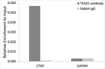
Chromatin Immunoprecipitation: TEF1 Antibody [NBP2-95204] - Analysis of extracts of HeLa cells, using TEF1 rabbit IgG. The amount of immunoprecipitated DNA was checked by quantitative PCR. Histogram was constructed by the ratios of the immunoprecipitated DNA to the input.

Immunoprecipitation: TEAD1 Antibody - Azide and BSA Free [TEAD1] -
Immunoprecipitation: TEAD1 Antibody - Azide and BSA Free [TEAD1] - Immunoprecipitation of TEAD1 in 600 ug extracts from mouse heart using 3 ug TEAD1 Rabbit pAb. Western blot analysis was performed using TEAD1 Rabbit pAb at 1:200 dilution.
Western Blot: TEAD1 Antibody - Azide and BSA Free [TEAD1] -
Western Blot: TEAD1 Antibody - Azide and BSA Free [TEAD1] - Western blot analysis of lysates from Rat lung using TEAD1 Rabbit pAb at 1:2000 dilution.Secondary antibody: HRP-conjugated Goat anti-Rabbit IgG (H+L) at 1:10000 dilution.
Lysates/proteins: 25 ug per lane.
Blocking buffer: 3% nonfat dry milk in TBST.
Detection: ECL Basic Kit.
Exposure time:90s.

Immunohistochemistry: TEAD1 Antibody - Azide and BSA Free [TEAD1] -
Immunohistochemistry: TEAD1 Antibody - Azide and BSA Free [TEAD1] - Immunohistochemistry analysis of paraffin-embedded Human liver using TEAD1 Rabbit pAb at dilution of 1:50 (40x lens). High pressure antigen retrieval performed with 0.01M Citrate Bufferr (pH 6.0) prior to IHC staining.
Immunohistochemistry: TEAD1 Antibody - Azide and BSA Free [TEAD1] -
Immunohistochemistry: TEAD1 Antibody - Azide and BSA Free [TEAD1] - Immunohistochemistry analysis of paraffin-embedded Human lung using TEAD1 Rabbit pAb at dilution of 1:50 (40x lens). High pressure antigen retrieval performed with 0.01M Citrate Bufferr (pH 6.0) prior to IHC staining.
Western Blot: TEAD1 Antibody - Azide and BSA Free [TEAD1] -
Western Blot: TEAD1 Antibody - Azide and BSA Free [TEAD1] - Western blot analysis of various lysates using TEAD1 Rabbit pAb at 1:700 dilution incubated overnight at 4C.Secondary antibody: HRP-conjugated Goat anti-Rabbit IgG (H+L) at 1:10000 dilution.
Lysates/proteins: 25 ug per lane.
Blocking buffer: 3% nonfat dry milk in TBST.
Detection: ECL Basic Kit.
Exposure time: 28s.
Applications for TEAD1 Antibody - Azide and BSA Free
Application
Recommended Usage
Chromatin Immunoprecipitation (ChIP)
5μg antibody for 10μg-15μg of Chromatin
ELISA
Recommended starting concentration is 1 μg/mL. Please optimize the concentration based on your specific assay requirements.
Immunocytochemistry/ Immunofluorescence
1:50-1:200
Immunohistochemistry
1:50 - 1:200
Immunohistochemistry-Paraffin
1:50 - 1:200
Immunoprecipitation
5μg-4μg antibody for 400μg-600μg extracts of whole cells
Western Blot
1:100 - 1:500
Formulation, Preparation, and Storage
Purification
Affinity purified
Formulation
PBS (pH 7.3), 50% glycerol
Format
Azide and BSA Free
Preservative
0.02% Sodium Azide
Concentration
Please see the vial label for concentration. If unlisted please contact technical services.
Shipping
The product is shipped with polar packs. Upon receipt, store it immediately at the temperature recommended below.
Stability & Storage
Store at -20C. Avoid freeze-thaw cycles.
Background: TEAD1
Long Name
Transcriptional enhancer factor TEF-1
Alternate Names
AA, NTEF-1, protein GT-IIC, REF1, TCF13, TCF-13, TEA domain family member 1 (SV40 transcriptional enhancer factor), TEAD1, TEAD-1, TEF-1, transcription factor 13, transcriptional enhancer factor 1, transcriptional enhancer factor TEF-1
Gene Symbol
TEAD1
Additional TEAD1 Products
Product Documents for TEAD1 Antibody - Azide and BSA Free
Certificate of Analysis
To download a Certificate of Analysis, please enter a lot or batch number in the search box below.
Product Specific Notices for TEAD1 Antibody - Azide and BSA Free
This product is for research use only and is not approved for use in humans or in clinical diagnosis. Primary Antibodies are guaranteed for 1 year from date of receipt.
Customer Reviews for TEAD1 Antibody - Azide and BSA Free
There are currently no reviews for this product. Be the first to review TEAD1 Antibody - Azide and BSA Free and earn rewards!
Have you used TEAD1 Antibody - Azide and BSA Free?
Submit a review and receive an Amazon gift card!
$25/€18/£15/$25CAN/¥2500 Yen for a review with an image
$10/€7/£6/$10CAN/¥1110 Yen for a review without an image
Submit a review
Protocols
Find general support by application which include: protocols, troubleshooting, illustrated assays, videos and webinars.
- Antigen Retrieval Protocol (PIER)
- Antigen Retrieval for Frozen Sections Protocol
- Appropriate Fixation of IHC/ICC Samples
- Cellular Response to Hypoxia Protocols
- ChIP Protocol Video
- Chromatin Immunoprecipitation (ChIP) Protocol
- Chromatin Immunoprecipitation Protocol
- Chromogenic IHC Staining of Formalin-Fixed Paraffin-Embedded (FFPE) Tissue Protocol
- Chromogenic Immunohistochemistry Staining of Frozen Tissue
- ClariTSA™ Fluorophore Kits
- Detection & Visualization of Antibody Binding
- ELISA Sample Preparation & Collection Guide
- ELISA Troubleshooting Guide
- Fluorescent IHC Staining of Frozen Tissue Protocol
- Graphic Protocol for Heat-induced Epitope Retrieval
- Graphic Protocol for the Preparation and Fluorescent IHC Staining of Frozen Tissue Sections
- Graphic Protocol for the Preparation and Fluorescent IHC Staining of Paraffin-embedded Tissue Sections
- Graphic Protocol for the Preparation of Gelatin-coated Slides for Histological Tissue Sections
- How to Run an R&D Systems DuoSet ELISA
- How to Run an R&D Systems Quantikine ELISA
- How to Run an R&D Systems Quantikine™ QuicKit™ ELISA
- ICC Cell Smear Protocol for Suspension Cells
- ICC Immunocytochemistry Protocol Videos
- ICC for Adherent Cells
- IHC Sample Preparation (Frozen sections vs Paraffin)
- Immunocytochemistry (ICC) Protocol
- Immunocytochemistry Troubleshooting
- Immunofluorescence of Organoids Embedded in Cultrex Basement Membrane Extract
- Immunofluorescent IHC Staining of Formalin-Fixed Paraffin-Embedded (FFPE) Tissue Protocol
- Immunohistochemistry (IHC) and Immunocytochemistry (ICC) Protocols
- Immunohistochemistry Frozen Troubleshooting
- Immunohistochemistry Paraffin Troubleshooting
- Immunoprecipitation Protocol
- Preparing Samples for IHC/ICC Experiments
- Preventing Non-Specific Staining (Non-Specific Binding)
- Primary Antibody Selection & Optimization
- Protocol for Heat-Induced Epitope Retrieval (HIER)
- Protocol for Making a 4% Formaldehyde Solution in PBS
- Protocol for VisUCyte™ HRP Polymer Detection Reagent
- Protocol for the Fluorescent ICC Staining of Cell Smears - Graphic
- Protocol for the Fluorescent ICC Staining of Cultured Cells on Coverslips - Graphic
- Protocol for the Preparation & Fixation of Cells on Coverslips
- Protocol for the Preparation and Chromogenic IHC Staining of Frozen Tissue Sections
- Protocol for the Preparation and Chromogenic IHC Staining of Frozen Tissue Sections - Graphic
- Protocol for the Preparation and Chromogenic IHC Staining of Paraffin-embedded Tissue Sections
- Protocol for the Preparation and Chromogenic IHC Staining of Paraffin-embedded Tissue Sections - Graphic
- Protocol for the Preparation and Fluorescent ICC Staining of Cells on Coverslips
- Protocol for the Preparation and Fluorescent ICC Staining of Non-adherent Cells
- Protocol for the Preparation and Fluorescent ICC Staining of Stem Cells on Coverslips
- Protocol for the Preparation and Fluorescent IHC Staining of Frozen Tissue Sections
- Protocol for the Preparation and Fluorescent IHC Staining of Paraffin-embedded Tissue Sections
- Protocol for the Preparation of Gelatin-coated Slides for Histological Tissue Sections
- Protocol for the Preparation of a Cell Smear for Non-adherent Cell ICC - Graphic
- Quantikine HS ELISA Kit Assay Principle, Alkaline Phosphatase
- Quantikine HS ELISA Kit Principle, Streptavidin-HRP Polymer
- R&D Systems Quality Control Western Blot Protocol
- Sandwich ELISA (Colorimetric) – Biotin/Streptavidin Detection Protocol
- Sandwich ELISA (Colorimetric) – Direct Detection Protocol
- TUNEL and Active Caspase-3 Detection by IHC/ICC Protocol
- The Importance of IHC/ICC Controls
- Troubleshooting Guide: ELISA
- Troubleshooting Guide: Immunohistochemistry
- Troubleshooting Guide: Western Blot Figures
- Western Blot Conditions
- Western Blot Protocol
- Western Blot Protocol for Cell Lysates
- Western Blot Troubleshooting
- Western Blot Troubleshooting Guide
- View all Protocols, Troubleshooting, Illustrated assays and Webinars
Loading...