Thrombospondin-1 Antibody
Novus Biologicals | Catalog # NBP1-52410
![Western Blot: Thrombospondin-1 Antibody [NBP1-52410]](https://resources.rndsystems.com/images/products/Thrombospondin-1-Antibody-Western-Blot-NBP1-52410-img0010.jpg "Western Blot: Thrombospondin-1 Antibody [NBP1-52410]")
Loading...
Key Product Details
Species Reactivity
Human, Mouse, Porcine, Bovine, Equine, Monkey
Applications
Immunohistochemistry, Immunohistochemistry-Paraffin, Western Blot, Peptide ELISA, Flow Cytometry, Immunocytochemistry/ Immunofluorescence
Label
Unconjugated
Antibody Source
Polyclonal Goat IgG
Loading...
Product Specifications
Immunogen
Peptide with sequence NRIPESGGDNSVFD-C, from the N Terminus of the Thrombospondin-1 protein sequence according to NP_003237.2.
Epitope
N-Terminus
Clonality
Polyclonal
Host
Goat
Isotype
IgG
Scientific Data Images for Thrombospondin-1 Antibody
Western Blot: Thrombospondin-1 Antibody [NBP1-52410]
Western Blot: Thrombospondin-1 Antibody [NBP1-52410] - (1ug/ml) staining of Mouse Kidney lysate (35ug protein in RIPA buffer). Detected by chemiluminescence.![Immunocytochemistry/ Immunofluorescence: Thrombospondin-1 Antibody [NBP1-52410]](https://resources.rndsystems.com/images/products/Thrombospondin-1-Antibody-Immunocytochemistry-Immunofluorescence-NBP1-52410-img0008.jpg "Immunocytochemistry/ Immunofluorescence: Thrombospondin-1 Antibody [NBP1-52410]")
Immunocytochemistry/ Immunofluorescence: Thrombospondin-1 Antibody [NBP1-52410]
Immunocytochemistry/Immunofluorescence: Thrombospondin-1 Antibody [NBP1-52410] - Analysis of paraformaldehyde fixed HepG2 cells, permeabilized with 0.15% Triton. Primary incubation 1hr (10ug/ml) followed by Alexa Fluor 488 secondary antibody (4ug/ml), showing cytoplasmic/Plasma Membrane staining.![Immunohistochemistry-Paraffin: Thrombospondin-1 Antibody [NBP1-52410]](https://resources.rndsystems.com/images/products/Thrombospondin-1-Antibody-Immunohistochemistry-Paraffin-NBP1-52410-img0006.jpg "Immunohistochemistry-Paraffin: Thrombospondin-1 Antibody [NBP1-52410]")
Immunohistochemistry-Paraffin: Thrombospondin-1 Antibody [NBP1-52410]
Immunohistochemistry-Paraffin: Thrombospondin-1 Antibody [NBP1-52410] - Analysis of anti-THBS1 / Thrombospondin antibody with human placenta at concentration 3.75 ug/ml.![Flow Cytometry: Thrombospondin-1 Antibody [NBP1-52410]](https://resources.rndsystems.com/images/products/Thrombospondin-1-Antibody-Flow-Cytometry-NBP1-52410-img0012.jpg "Flow Cytometry: Thrombospondin-1 Antibody [NBP1-52410]")
Flow Cytometry: Thrombospondin-1 Antibody [NBP1-52410]
Flow Cytometry: Thrombospondin-1 Antibody [NBP1-52410] - Analysis of paraformaldehyde fixed A431 cells (blue line), permeabilized with 0.5% Triton. Primary incubation 1hr (10ug/ml) followed by Alexa Fluor 488 secondary antibody (2ug/ml). IgG control: Unimmunized goat IgG (black line) followed by Alexa Fluor 488 secondary antibody.![Immunohistochemistry-Paraffin: Thrombospondin-1 Antibody [NBP1-52410]](https://resources.rndsystems.com/images/products/Thrombospondin-1-Antibody-Immunohistochemistry-Paraffin-NBP1-52410-img0004.jpg "Immunohistochemistry-Paraffin: Thrombospondin-1 Antibody [NBP1-52410]")
Immunohistochemistry-Paraffin: Thrombospondin-1 Antibody [NBP1-52410]
Immunohistochemistry-Paraffin: Thrombospondin-1 Antibody [NBP1-52410] - Analysis of anti-THBS1 / Thrombospondin antibody with human spleen at concentration 3.75 ug/ml.![Flow Cytometry: Thrombospondin-1 Antibody [NBP1-52410]](https://resources.rndsystems.com/images/products/Thrombospondin-1-Antibody-Flow-Cytometry-NBP1-52410-img0003.jpg "Flow Cytometry: Thrombospondin-1 Antibody [NBP1-52410]")
Flow Cytometry: Thrombospondin-1 Antibody [NBP1-52410]
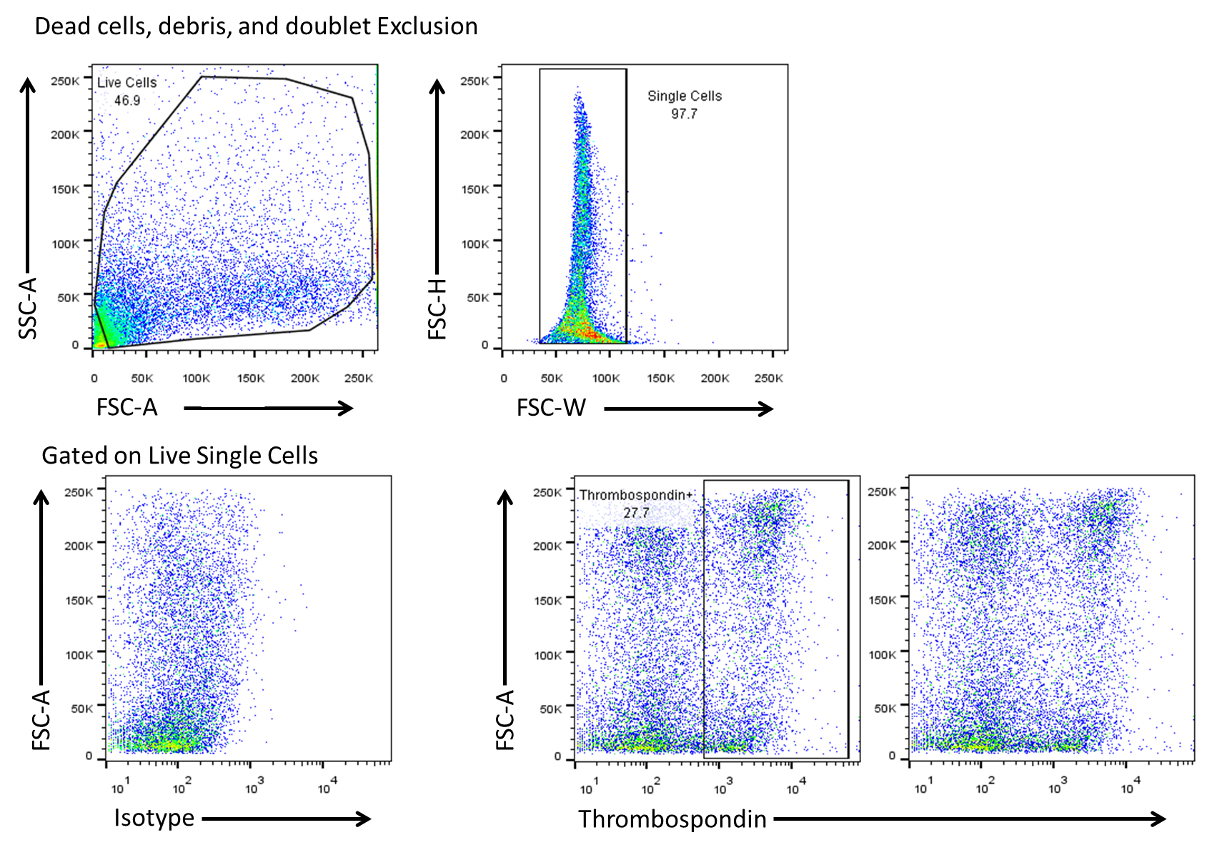
Flow Cytometry: Thrombospondin-1 Antibody [NBP1-52410] - Analysis of anti-THBS1 / Thrombospondin antibody with Spleenocytes. Image submitted by a verified customer review.Applications for Thrombospondin-1 Antibody
Application
Recommended Usage
Immunocytochemistry/ Immunofluorescence
10 ug/ml
Immunohistochemistry-Paraffin
3.75 ug/ml
Peptide ELISA
1:128000
Western Blot
0.3-1 ug/ml
Application Notes
IHC: NBP1-52410 was validated for use in immunohistochemistry on a panel of 21 formalin-fixed, paraffin-embedded (FFPE) human tissues after heat induced antigen retrieval in pH 6.0 citrate buffer. After incubation with the primary antibody, slides were incubated with biotinylated secondary antibody, followed by alkaline phosphatase-streptavidin and chromogen. The stained slides were evaluated by a pathologist to confirm staining specificity. Thrombospondin-1 antibody validated for FLOW from a verified customer review..
Reviewed Applications
Read 1 review rated 5 using NBP1-52410 in the following applications:
Flow Cytometry Panel Builder
Bio-Techne Knows Flow Cytometry
Save time and reduce costly mistakes by quickly finding compatible reagents using the Panel Builder Tool.
Advanced Features
- Spectra Viewer - Custom analysis of spectra from multiple fluorochromes
- Spillover Popups - Visualize the spectra of individual fluorochromes
- Antigen Density Selector - Match fluorochrome brightness with antigen density

Formulation, Preparation, and Storage
Purification
Antigen Affinity-purified
Formulation
TBS (pH 7.3) and 0.5% BSA
Preservative
0.02% Sodium Azide
Concentration
Concentrations vary lot to lot. See vial label for concentration. If unlisted please contact technical services.
Shipping
The product is shipped with polar packs. Upon receipt, store it immediately at the temperature recommended below.
Stability & Storage
Store at -20C. Avoid freeze-thaw cycles.
Background: Thrombospondin-1
Alternate Names
THBS1, Thrombospondin1, TSP-1
Entrez Gene IDs
7057 (Human)
Gene Symbol
THBS1
UniProt
Additional Thrombospondin-1 Products
Product Documents for Thrombospondin-1 Antibody
Certificate of Analysis
To download a Certificate of Analysis, please enter a lot or batch number in the search box below.
Product Specific Notices for Thrombospondin-1 Antibody
This product is for research use only and is not approved for use in humans or in clinical diagnosis. Primary Antibodies are guaranteed for 1 year from date of receipt.
Related Research Areas
Citations for Thrombospondin-1 Antibody
 Powered by Bioz
Customer Reviews for Thrombospondin-1 Antibody (1)
5 out of 5
1 Customer Rating
Have you used Thrombospondin-1 Antibody?
Submit a review and receive an Amazon gift card!
$25/€18/£15/$25CAN/¥2500 Yen for a review with an image
$10/€7/£6/$10CAN/¥1110 Yen for a review without an image
Submit a review
Customer Images
Showing
1
-
1 of
1 review
Showing All
Filter By:
-
Application: Flow CytometrySample Tested:Species: HumanVerified Customer | Posted 11/23/2014Flow cytometry on Spleenocytes using Thrombospondin antibody

There are no reviews that match your criteria.
Protocols
Find general support by application which include: protocols, troubleshooting, illustrated assays, videos and webinars.
- 7-Amino Actinomycin D (7-AAD) Cell Viability Flow Cytometry Protocol
- Antigen Retrieval Protocol (PIER)
- Antigen Retrieval for Frozen Sections Protocol
- Appropriate Fixation of IHC/ICC Samples
- Cellular Response to Hypoxia Protocols
- Chromogenic IHC Staining of Formalin-Fixed Paraffin-Embedded (FFPE) Tissue Protocol
- Chromogenic Immunohistochemistry Staining of Frozen Tissue
- ClariTSA™ Fluorophore Kits
- Detection & Visualization of Antibody Binding
- ELISA Sample Preparation & Collection Guide
- ELISA Troubleshooting Guide
- Extracellular Membrane Flow Cytometry Protocol
- Flow Cytometry Protocol for Cell Surface Markers
- Flow Cytometry Protocol for Staining Membrane Associated Proteins
- Flow Cytometry Staining Protocols
- Flow Cytometry Troubleshooting Guide
- Fluorescent IHC Staining of Frozen Tissue Protocol
- Graphic Protocol for Heat-induced Epitope Retrieval
- Graphic Protocol for the Preparation and Fluorescent IHC Staining of Frozen Tissue Sections
- Graphic Protocol for the Preparation and Fluorescent IHC Staining of Paraffin-embedded Tissue Sections
- Graphic Protocol for the Preparation of Gelatin-coated Slides for Histological Tissue Sections
- How to Run an R&D Systems DuoSet ELISA
- How to Run an R&D Systems Quantikine ELISA
- How to Run an R&D Systems Quantikine™ QuicKit™ ELISA
- ICC Cell Smear Protocol for Suspension Cells
- ICC Immunocytochemistry Protocol Videos
- ICC for Adherent Cells
- IHC Sample Preparation (Frozen sections vs Paraffin)
- Immunocytochemistry (ICC) Protocol
- Immunocytochemistry Troubleshooting
- Immunofluorescence of Organoids Embedded in Cultrex Basement Membrane Extract
- Immunofluorescent IHC Staining of Formalin-Fixed Paraffin-Embedded (FFPE) Tissue Protocol
- Immunohistochemistry (IHC) and Immunocytochemistry (ICC) Protocols
- Immunohistochemistry Frozen Troubleshooting
- Immunohistochemistry Paraffin Troubleshooting
- Intracellular Flow Cytometry Protocol Using Alcohol (Methanol)
- Intracellular Flow Cytometry Protocol Using Detergents
- Intracellular Nuclear Staining Flow Cytometry Protocol Using Detergents
- Intracellular Staining Flow Cytometry Protocol Using Alcohol Permeabilization
- Intracellular Staining Flow Cytometry Protocol Using Detergents to Permeabilize Cells
- Preparing Samples for IHC/ICC Experiments
- Preventing Non-Specific Staining (Non-Specific Binding)
- Primary Antibody Selection & Optimization
- Propidium Iodide Cell Viability Flow Cytometry Protocol
- Protocol for Heat-Induced Epitope Retrieval (HIER)
- Protocol for Liperfluo
- Protocol for Making a 4% Formaldehyde Solution in PBS
- Protocol for VisUCyte™ HRP Polymer Detection Reagent
- Protocol for the Characterization of Human Th22 Cells
- Protocol for the Characterization of Human Th9 Cells
- Protocol for the Fluorescent ICC Staining of Cell Smears - Graphic
- Protocol for the Fluorescent ICC Staining of Cultured Cells on Coverslips - Graphic
- Protocol for the Preparation & Fixation of Cells on Coverslips
- Protocol for the Preparation and Chromogenic IHC Staining of Frozen Tissue Sections
- Protocol for the Preparation and Chromogenic IHC Staining of Frozen Tissue Sections - Graphic
- Protocol for the Preparation and Chromogenic IHC Staining of Paraffin-embedded Tissue Sections
- Protocol for the Preparation and Chromogenic IHC Staining of Paraffin-embedded Tissue Sections - Graphic
- Protocol for the Preparation and Fluorescent ICC Staining of Cells on Coverslips
- Protocol for the Preparation and Fluorescent ICC Staining of Non-adherent Cells
- Protocol for the Preparation and Fluorescent ICC Staining of Stem Cells on Coverslips
- Protocol for the Preparation and Fluorescent IHC Staining of Frozen Tissue Sections
- Protocol for the Preparation and Fluorescent IHC Staining of Paraffin-embedded Tissue Sections
- Protocol for the Preparation of Gelatin-coated Slides for Histological Tissue Sections
- Protocol for the Preparation of a Cell Smear for Non-adherent Cell ICC - Graphic
- Protocol: Annexin V and PI Staining by Flow Cytometry
- Protocol: Annexin V and PI Staining for Apoptosis by Flow Cytometry
- Quantikine HS ELISA Kit Assay Principle, Alkaline Phosphatase
- Quantikine HS ELISA Kit Principle, Streptavidin-HRP Polymer
- R&D Systems Quality Control Western Blot Protocol
- Sandwich ELISA (Colorimetric) – Biotin/Streptavidin Detection Protocol
- Sandwich ELISA (Colorimetric) – Direct Detection Protocol
- TUNEL and Active Caspase-3 Detection by IHC/ICC Protocol
- The Importance of IHC/ICC Controls
- Troubleshooting Guide: ELISA
- Troubleshooting Guide: Fluorokine Flow Cytometry Kits
- Troubleshooting Guide: Immunohistochemistry
- Troubleshooting Guide: Western Blot Figures
- Western Blot Conditions
- Western Blot Protocol
- Western Blot Protocol for Cell Lysates
- Western Blot Troubleshooting
- Western Blot Troubleshooting Guide
- View all Protocols, Troubleshooting, Illustrated assays and Webinars
Loading...