Tight Junction Protein 1 Antibody - BSA Free
Novus Biologicals | Catalog # NBP1-85047
![Knockout Validated: Tight Junction Protein 1 Antibody [NBP1-85047]](https://resources.rndsystems.com/images/products/Tight-Junction-Protein-1-Antibody-Knockout-Validated-NBP1-85047-img0019.jpg "Western Blot: Tight Junction Protein 1 Antibody [NBP1-85047]")
Loading...
Key Product Details
Validated by
Knockout/Knockdown, Orthogonal Validation, Biological Validation
Species Reactivity
Validated:
Human
Cited:
Human, Mouse, Rat
Predicted:
Mouse (100%), Rat (100%). Backed by our 100% Guarantee.
Applications
Validated:
Immunohistochemistry, Immunohistochemistry-Paraffin, Western Blot, Immunocytochemistry/ Immunofluorescence
Cited:
Immunohistochemistry, Immunohistochemistry-Paraffin, Western Blot, Immunocytochemistry/ Immunofluorescence, Simple Western, IF/IHC
Label
Unconjugated
Antibody Source
Polyclonal Rabbit IgG
Format
BSA Free
Loading...
Product Specifications
Immunogen
This antibody was developed against Recombinant Protein corresponding to amino acids: RKLYERSHKLRKNNHHLFTTTINLNSMNDGWYGALKEAIQQQQNQLVWVSEGKADGATSDDLDLHDDRLSYLSAPGSEYSMYSTDSRHTSDYEDTDTEGGAYTDQELDETLNDEVGTPPESAITRSSEPVRED
Marker
Intercellular Junctions/Tight Junction Marker
Clonality
Polyclonal
Host
Rabbit
Isotype
IgG
Scientific Data Images for Tight Junction Protein 1 Antibody - BSA Free
Western Blot: Tight Junction Protein 1 Antibody [NBP1-85047]
Western Blot: Tight Junction Protein 1 Antibody [NBP1-85047] - Western blot shows lysates of HeLa human cervical epithelial carcinoma parental cell line and TJP1 knockout (KO) HeLa cell line. PVDF membrane was probed with 1:200 of Rabbit Anti-Human TJP1 Polyclonal Antibody (Catalog # NBP1-85047) followed by HRP-conjugated Anti-Rabbit IgG Secondary Antibody (Catalog #HAF008). Specific band was detected for TJP1 at approximately 260 kDa (as indicated) in the parental HeLa cell line, but is not detectable in the knockout HeLa cell line. This experiment was conducted under reducing conditions.![Immunohistochemistry-Paraffin: Tight Junction Protein 1 Antibody [NBP1-85047]](https://resources.rndsystems.com/images/products/Tight-Junction-Protein-1-Antibody-Immunohistochemistry-Paraffin-NBP1-85047-img0018.jpg "Immunohistochemistry-Paraffin: Tight Junction Protein 1 Antibody [NBP1-85047]")
![Simple Western: Tight Junction Protein 1 Antibody [NBP1-85047]](https://resources.rndsystems.com/images/products/Tight-Junction-Protein-1-Antibody-Simple-Western-NBP1-85047-img0006.jpg "Simple Western: Tight Junction Protein 1 Antibody [NBP1-85047]")
Simple Western: Tight Junction Protein 1 Antibody [NBP1-85047]
Simple Western: Tight Junction Protein 1 Antibody [NBP1-85047] - Simple Western lane view shows a specific band for TJP1 in 0.2 mg/ml of HeLa lysate. This experiment was performed under reducing conditions using the 66-440 kDa separation system.![Immunohistochemistry: Tight Junction Protein 1 Antibody [NBP1-85047]](https://resources.rndsystems.com/images/products/Tight-Junction-Protein-1-Antibody-Immunohistochemistry-NBP1-85047-img0025.jpg "Immunohistochemistry: Tight Junction Protein 1 Antibody [NBP1-85047]")
Immunohistochemistry: Tight Junction Protein 1 Antibody [NBP1-85047]
Tight-Junction-Protein-1-Antibody-Immunohistochemistry-NBP1-85047-img0025.jpg![Western Blot: Tight Junction Protein 1 Antibody [NBP1-85047]](https://resources.rndsystems.com/images/products/Tight-Junction-Protein-1-Antibody-Western-Blot-NBP1-85047-img0026.jpg "Western Blot: Tight Junction Protein 1 Antibody [NBP1-85047]")
![Immunocytochemistry/ Immunofluorescence: Tight Junction Protein 1 Antibody [NBP1-85047]](https://resources.rndsystems.com/images/products/Tight-Junction-Protein-1-Antibody-Immunocytochemistry-Immunofluorescence-NBP1-85047-img0024.jpg "Immunocytochemistry/ Immunofluorescence: Tight Junction Protein 1 Antibody [NBP1-85047]")
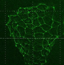
Immunocytochemistry/ Immunofluorescence: Tight Junction Protein 1 Antibody [NBP1-85047]
Immunocytochemistry/Immunofluorescence: Tight Junction Protein 1 Antibody [NBP1-85047] - Rat epithelial cells stained positively with Tight Junction Protein 1. Primary antibody at 1:100. Secondary antibody: goat anti-rabbit-Alexa 488 conjugated at 1:500. ICC/IF image submitted by a verified customer review.![Western Blot: Tight Junction Protein 1 Antibody [NBP1-85047]](https://resources.rndsystems.com/images/products/Tight-Junction-Protein-1-Antibody-Western-Blot-NBP1-85047-img0020.jpg "Western Blot: Tight Junction Protein 1 Antibody [NBP1-85047]")
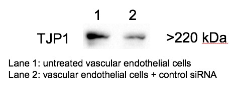
Western Blot: Tight Junction Protein 1 Antibody [NBP1-85047]
Western Blot: Tight Junction Protein 1 Antibody [NBP1-85047] - Analysis in control (vector only transfected HEK293T lysate) and TJP1 over-expression lysate (Co-expressed with a C-terminal myc-DDK tag (3.1 kDa) in mammalian HEK293T cells).![Western Blot: Tight Junction Protein 1 Antibody [NBP1-85047]](https://resources.rndsystems.com/images/products/Tight-Junction-Protein-1-Antibody-Western-Blot-NBP1-85047-img0021.jpg "Western Blot: Tight Junction Protein 1 Antibody [NBP1-85047]")
Western Blot: Tight Junction Protein 1 Antibody [NBP1-85047]
Western Blot: Tight Junction Protein 1 Antibody [NBP1-85047] - Analysis in human cell line A-431.![Immunocytochemistry/ Immunofluorescence: Tight Junction Protein 1 Antibody [NBP1-85047]](https://resources.rndsystems.com/images/products/Tight-Junction-Protein-1-Antibody-Immunocytochemistry-Immunofluorescence-NBP1-85047-img0008.jpg "Immunocytochemistry/ Immunofluorescence: Tight Junction Protein 1 Antibody [NBP1-85047]")
Immunocytochemistry/ Immunofluorescence: Tight Junction Protein 1 Antibody [NBP1-85047]
Immunocytochemistry/Immunofluorescence: Tight Junction Protein 1 Antibody [NBP1-85047] - Staining of human cell line U-2 OS shows localization to cytosol & cell junctions. Antibody staining is shown in green.![Immunocytochemistry/ Immunofluorescence: Tight Junction Protein 1 Antibody [NBP1-85047]](https://resources.rndsystems.com/images/products/Tight-Junction-Protein-1-Antibody-Immunocytochemistry-Immunofluorescence-NBP1-85047-img0022.jpg "Immunocytochemistry/ Immunofluorescence: Tight Junction Protein 1 Antibody [NBP1-85047]")
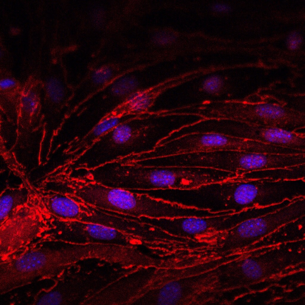
Immunocytochemistry/ Immunofluorescence: Tight Junction Protein 1 Antibody [NBP1-85047]
Immunocytochemistry/Immunofluorescence: Tight Junction Protein 1 Antibody [NBP1-85047] - The primary mouse lung endothelial cells were fixed, permeabilized and stained with 1:100 diluted anti-ZO-1 ab for overnight. Samples were washed and subsequently incubated with 1:500 diluted Alexa Fuor 546 at room temperature for 1 hour. ICC/iF image submitted by a verified customer reveiw.![Immunocytochemistry/ Immunofluorescence: Tight Junction Protein 1 Antibody [NBP1-85047]](https://resources.rndsystems.com/images/products/Tight-Junction-Protein-1-Antibody-Immunocytochemistry-Immunofluorescence-NBP1-85047-img0023.jpg "Immunocytochemistry/ Immunofluorescence: Tight Junction Protein 1 Antibody [NBP1-85047]")
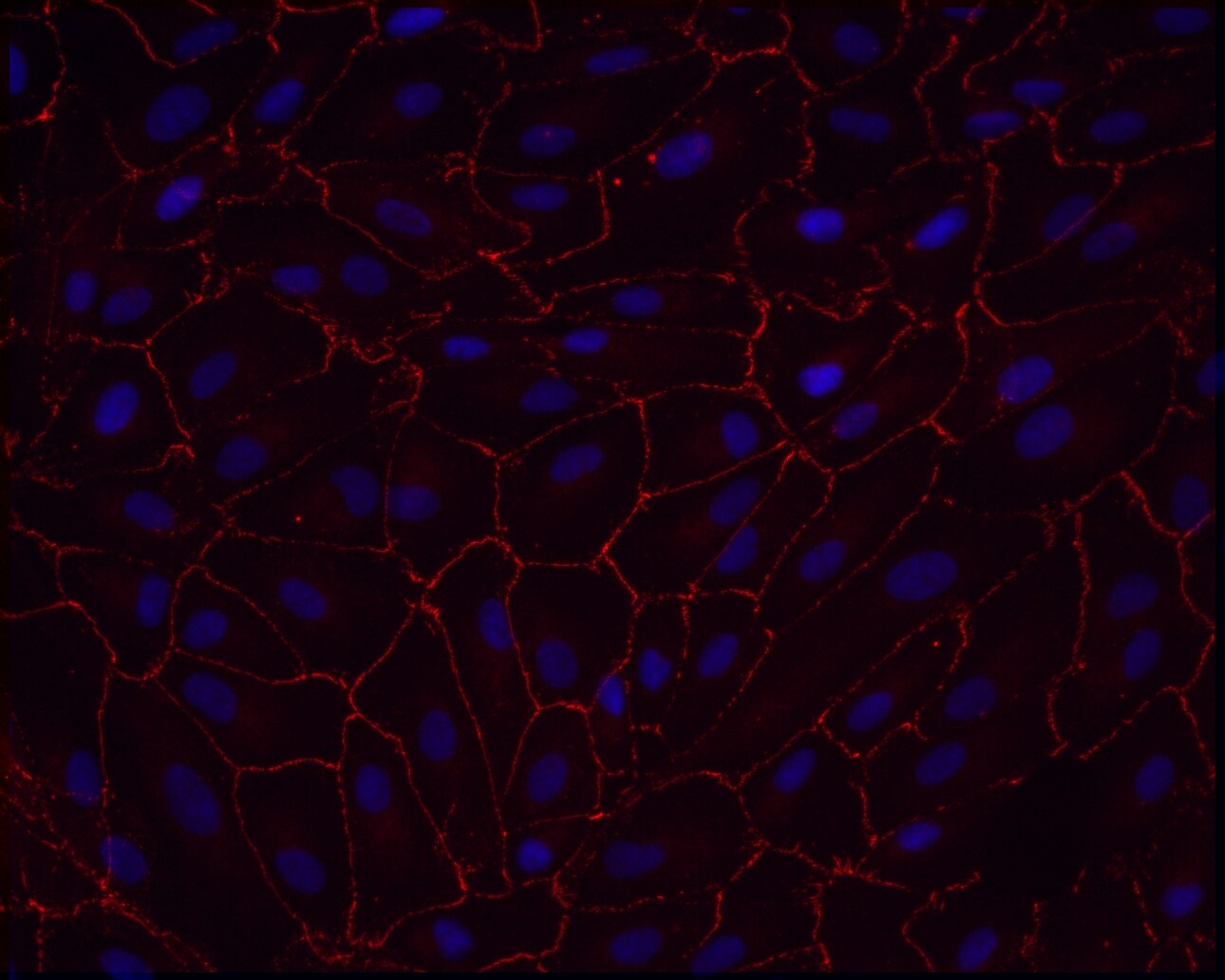
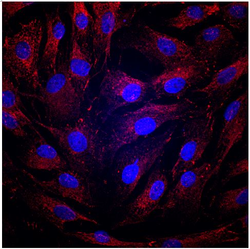
Immunocytochemistry/ Immunofluorescence: Tight Junction Protein 1 Antibody [NBP1-85047]
Immunocytochemistry/Immunofluorescence: Tight Junction Protein 1 Antibody [NBP1-85047] - Monolayer of Human ARPE-19 cells on a glass substrate. Tight Junction Protein 1 staining in red. ICC/IF image submitted by a verified customer review.![Immunohistochemistry-Paraffin: Tight Junction Protein 1 Antibody [NBP1-85047]](https://resources.rndsystems.com/images/products/Tight-Junction-Protein-1-Antibody-Immunohistochemistry-Paraffin-NBP1-85047-img0014.jpg "Immunohistochemistry-Paraffin: Tight Junction Protein 1 Antibody [NBP1-85047]")
Immunohistochemistry-Paraffin: Tight Junction Protein 1 Antibody [NBP1-85047]
Immunohistochemistry-Paraffin: Tight Junction Protein 1 Antibody [NBP1-85047] - Staining of human kidney shows strong membranous positivity in cells in glomeruli.![Immunohistochemistry-Paraffin: Tight Junction Protein 1 Antibody [NBP1-85047]](https://resources.rndsystems.com/images/products/Tight-Junction-Protein-1-Antibody-Immunohistochemistry-Paraffin-NBP1-85047-img0015.jpg "Immunohistochemistry-Paraffin: Tight Junction Protein 1 Antibody [NBP1-85047]")
Immunohistochemistry-Paraffin: Tight Junction Protein 1 Antibody [NBP1-85047]
Immunohistochemistry-Paraffin: Tight Junction Protein 1 Antibody [NBP1-85047] - Staining of human placenta shows moderate membranous positivity in trophoblastic cells.![Immunohistochemistry-Paraffin: Tight Junction Protein 1 Antibody [NBP1-85047]](https://resources.rndsystems.com/images/products/Tight-Junction-Protein-1-Antibody-Immunohistochemistry-Paraffin-NBP1-85047-img0016.jpg "Immunohistochemistry-Paraffin: Tight Junction Protein 1 Antibody [NBP1-85047]")
Immunohistochemistry-Paraffin: Tight Junction Protein 1 Antibody [NBP1-85047]
Immunohistochemistry-Paraffin: Tight Junction Protein 1 Antibody [NBP1-85047] - Staining of human skeletal muscle shows no membranous positivity in myocytes as expected.![Immunohistochemistry-Paraffin: Tight Junction Protein 1 Antibody [NBP1-85047]](https://resources.rndsystems.com/images/products/Tight-Junction-Protein-1-Antibody-Immunohistochemistry-Paraffin-NBP1-85047-img0017.jpg "Immunohistochemistry-Paraffin: Tight Junction Protein 1 Antibody [NBP1-85047]")
Immunohistochemistry-Paraffin: Tight Junction Protein 1 Antibody [NBP1-85047]
Immunohistochemistry-Paraffin: Tight Junction Protein 1 Antibody [NBP1-85047] - Staining of human testis shows moderate to strong membranous positivity in cells in seminiferous ducts.
Western Blot: Tight Junction Protein 1 Antibody [NBP1-85047] -
Western Blot: Tight Junction Protein 1 Antibody [NBP1-85047] - Western blot analysis of function protein expression on primary human renal cortical epithelial cells challenged with cytokines. Primary human renal tubular epithelial cells are challenged with cytokine cocktail (15nM IFN gamma, 6nM TNF alpha, & 3nM IL1 beta ). (A, B) Expression of Snail, E-Cad, TJP1, SMA. AhR, IDO, KMO, KY, MHCI & II. (C, D) Densitometric quantitation of protein in (A, B). All Western blots are representative of at least three independent experiments. *P < 0.05, ***P < 0.0001 versus to cells without treatment, analysis is multiple t-tests, pairwise comparison of individual time-point to control indicated same p-value. Image collected & cropped by CiteAb from the following publication (https://pubmed.ncbi.nlm.nih.gov/34305900), licensed under a CC-BY license. Not internally tested by Novus Biologicals.
Western Blot: Tight Junction Protein 1 Antibody [NBP1-85047] -
Western Blot: Tight Junction Protein 1 Antibody [NBP1-85047] - Effects of dietary Flammulina velutipes stem waste (FVS) inclusion on the relative expression of tight junction proteins in jejunum of growing pigs. a The relative expression of ZO-1 in jejunum of growing pigs fed control & 2.5% or 5% FVS diets. b The relative expression of occludin in jejunum of growing pigs fed control & 2.5% or 5% FVS diets. c The relative expression of claudin-1 in jejunum of growing pigs fed control & 2.5% or 5% FVS diets. Values are means (3 pigs per treatment) with standard errors represented by vertical bars. *P < 0.05, **P < 0.01 Image collected & cropped by CiteAb from the following publication (https://pubmed.ncbi.nlm.nih.gov/32391146), licensed under a CC-BY license. Not internally tested by Novus Biologicals.
Immunocytochemistry/ Immunofluorescence: Tight Junction Protein 1 Antibody [NBP1-85047] -
Immunocytochemistry/ Immunofluorescence: Tight Junction Protein 1 Antibody [NBP1-85047] - Differentiation of FH- & corr-FH-iPSCs into hepatocytes. a Representative pictures of cell morphology & immunostainings of the indicated markers at day 25 of differentiation. Scale bars: 50 μm. b Representative images of DCFA excretion at the biliary poles of corr-FH-iHeps. All images were taken with × 10 objective, z-stacks of xy sections of the cells were acquired with an epifluorescence microscope (Nikon Elipse) & analyzed with ImageJ software. Arrowheads indicate bile canaliculi Image collected & cropped by CiteAb from the following publication (https://pubmed.ncbi.nlm.nih.gov/31358055), licensed under a CC-BY license. Not internally tested by Novus Biologicals.
Western Blot: Tight Junction Protein 1 Antibody [NBP1-85047] -
Western Blot: Tight Junction Protein 1 Antibody [NBP1-85047] - Protective role of 3HK & 3HAA in TEC stimulated with cytokines. Primary human renal TEC pre-incubated with different doses of 3HK or 3HAA overnight; the cells are challenged with the cytokine cocktail (15 nM IFN gamma, 6 nM TNF alpha, & 3 nM IL1 beta ) for 24 h; protein expression was assayed with Western blot: (A, B) 3HK & 3HAA cannot reverse IDO, MHC I, BID expression induced by cytokines. 3HK & 3HAA effectively restore Bcl-xL & Tjp1 expression. 3HAA shows its different function in up-regulation of AhR expression in TEC in inflammatory conditions. (C) Qualification of protein expression in TEC challenged with cytokine cocktail in the presence of 3HK. ***P < 0.0001 versus cell treated with Cyto. (D) Qualification of protein expression in TEC challenged with cytokine cocktail in the presence of 3HAA. ***P < 0.0001, ****P < 0.00001 versus cell treated with Cyto. Pairwise comparison of individual dose to control indicated same p-value. All Western blots are representative of three independent experiments; analysis is multiple one-way ANOVA. Image collected & cropped by CiteAb from the following publication (https://pubmed.ncbi.nlm.nih.gov/34305900), licensed under a CC-BY license. Not internally tested by Novus Biologicals.
Western Blot: Tight Junction Protein 1 Antibody [NBP1-85047] -
Western Blot: Tight Junction Protein 1 Antibody [NBP1-85047] - Protective role of 3HK & 3HAA in TEC stimulated with cytokines. Primary human renal TEC pre-incubated with different doses of 3HK or 3HAA overnight; the cells are challenged with the cytokine cocktail (15 nM IFN gamma, 6 nM TNF alpha, & 3 nM IL1 beta ) for 24 h; protein expression was assayed with Western blot: (A, B) 3HK & 3HAA cannot reverse IDO, MHC I, BID expression induced by cytokines. 3HK & 3HAA effectively restore Bcl-xL & Tjp1 expression. 3HAA shows its different function in up-regulation of AhR expression in TEC in inflammatory conditions. (C) Qualification of protein expression in TEC challenged with cytokine cocktail in the presence of 3HK. ***P < 0.0001 versus cell treated with Cyto. (D) Qualification of protein expression in TEC challenged with cytokine cocktail in the presence of 3HAA. ***P < 0.0001, ****P < 0.00001 versus cell treated with Cyto. Pairwise comparison of individual dose to control indicated same p-value. All Western blots are representative of three independent experiments; analysis is multiple one-way ANOVA. Image collected & cropped by CiteAb from the following publication (https://pubmed.ncbi.nlm.nih.gov/34305900), licensed under a CC-BY license. Not internally tested by Novus Biologicals.Applications for Tight Junction Protein 1 Antibody - BSA Free
Application
Recommended Usage
Immunocytochemistry/ Immunofluorescence
0.25-2 ug/ml
Immunohistochemistry
1:200 - 1:500
Immunohistochemistry-Paraffin
1:200 - 1:500
Western Blot
0.04-0.4 ug/ml
Application Notes
IHC-Paraffin, HIER pH 6 retrieval is recommended. ICC/IF, Fixation Permeabilization: Use PFA/Triton X-100.
Reviewed Applications
Read 5 reviews rated 4.6 using NBP1-85047 in the following applications:
Formulation, Preparation, and Storage
Purification
Affinity purified
Formulation
PBS (pH 7.2) and 40% Glycerol
Format
BSA Free
Preservative
0.02% Sodium Azide
Concentration
Concentrations vary lot to lot. See vial label for concentration. If unlisted please contact technical services.
Shipping
The product is shipped with polar packs. Upon receipt, store it immediately at the temperature recommended below.
Stability & Storage
Store at 4C short term. Aliquot and store at -20C long term. Avoid freeze-thaw cycles.
Background: Tight Junction Protein 1
Alternate Names
DKFZp686M05161, MGC133289, Tight junction protein 1, tight junction protein 1 (zona occludens 1), tight junction protein ZO-1, TJP1, ZO1, ZO-1, zona occludens 1, Zona occludens protein 1, zonula occludens 1 protein, Zonula occludens protein 1
Gene Symbol
TJP1
Additional Tight Junction Protein 1 Products
Product Documents for Tight Junction Protein 1 Antibody - BSA Free
Certificate of Analysis
To download a Certificate of Analysis, please enter a lot or batch number in the search box below.
Product Specific Notices for Tight Junction Protein 1 Antibody - BSA Free
This product is for research use only and is not approved for use in humans or in clinical diagnosis. Primary Antibodies are guaranteed for 1 year from date of receipt.
Citations for Tight Junction Protein 1 Antibody - BSA Free
 Powered by Bioz
Customer Reviews for Tight Junction Protein 1 Antibody - BSA Free (5)
4.6 out of 5
5 Customer Ratings
Have you used Tight Junction Protein 1 Antibody - BSA Free?
Submit a review and receive an Amazon gift card!
$25/€18/£15/$25CAN/¥2500 Yen for a review with an image
$10/€7/£6/$10CAN/¥1110 Yen for a review without an image
Submit a review
Customer Images
Showing
1
-
5 of
5 reviews
Showing All
Filter By:
-
Application: ImmunocytochemistrySample Tested: Epithelial cellsSpecies: RatVerified Customer | Posted 12/10/2020Rat epithelial cells stained positively with Tight Junction Protein 1. Primary ab concentration = 1:100; Secondary ab: goat anti-rabbit- Alexa 488 conjugated = 1: 500

-
Application: ImmunocytochemistrySample Tested: ARPE-19 cellsSpecies: HumanVerified Customer | Posted 08/29/2017ARPE-19 monolayer on glass substrateFixative 4% PFA Blocking Goat Serum Dilution 1:200 Incubation 1h @ RT
-
Application: Western BlotSample Tested: Primary Mouse Lung cellsSpecies: MouseVerified Customer | Posted 06/22/2017Lane 1: untreated vascular endothelial cells Lane 2: vascular endothelial cells + control siRNA
-
Application: ICC/IFSample Tested: Primary mouse lung endotelial cellsSpecies: MouseVerified Customer | Posted 10/13/2016The primary mouse lung endothelial cells were fixed, permeabilized and stained with 1:100 diluted anti-ZO-1 ab for overnight. Samples were washed and subsequently incubated with 1:500 diluted Alexa Fuor 546 at room temperature for 1 hour.
-
Application: ImmunocytochemistrySample Tested: Pig trabecular meshwork cellsSpecies: OtherVerified Customer | Posted 02/02/2016Detection of TJP1 in Pig trabecular meshwork cells
There are no reviews that match your criteria.
Protocols
Find general support by application which include: protocols, troubleshooting, illustrated assays, videos and webinars.
- Antigen Retrieval Protocol (PIER)
- Antigen Retrieval for Frozen Sections Protocol
- Appropriate Fixation of IHC/ICC Samples
- Cellular Response to Hypoxia Protocols
- Chromogenic IHC Staining of Formalin-Fixed Paraffin-Embedded (FFPE) Tissue Protocol
- Chromogenic Immunohistochemistry Staining of Frozen Tissue
- ClariTSA™ Fluorophore Kits
- Detection & Visualization of Antibody Binding
- Fluorescent IHC Staining of Frozen Tissue Protocol
- Graphic Protocol for Heat-induced Epitope Retrieval
- Graphic Protocol for the Preparation and Fluorescent IHC Staining of Frozen Tissue Sections
- Graphic Protocol for the Preparation and Fluorescent IHC Staining of Paraffin-embedded Tissue Sections
- Graphic Protocol for the Preparation of Gelatin-coated Slides for Histological Tissue Sections
- ICC Cell Smear Protocol for Suspension Cells
- ICC Immunocytochemistry Protocol Videos
- ICC for Adherent Cells
- IHC Sample Preparation (Frozen sections vs Paraffin)
- Immunocytochemistry (ICC) Protocol
- Immunocytochemistry Troubleshooting
- Immunofluorescence of Organoids Embedded in Cultrex Basement Membrane Extract
- Immunofluorescent IHC Staining of Formalin-Fixed Paraffin-Embedded (FFPE) Tissue Protocol
- Immunohistochemistry (IHC) and Immunocytochemistry (ICC) Protocols
- Immunohistochemistry Frozen Troubleshooting
- Immunohistochemistry Paraffin Troubleshooting
- Preparing Samples for IHC/ICC Experiments
- Preventing Non-Specific Staining (Non-Specific Binding)
- Primary Antibody Selection & Optimization
- Protocol for Heat-Induced Epitope Retrieval (HIER)
- Protocol for Making a 4% Formaldehyde Solution in PBS
- Protocol for VisUCyte™ HRP Polymer Detection Reagent
- Protocol for the Fluorescent ICC Staining of Cell Smears - Graphic
- Protocol for the Fluorescent ICC Staining of Cultured Cells on Coverslips - Graphic
- Protocol for the Preparation & Fixation of Cells on Coverslips
- Protocol for the Preparation and Chromogenic IHC Staining of Frozen Tissue Sections
- Protocol for the Preparation and Chromogenic IHC Staining of Frozen Tissue Sections - Graphic
- Protocol for the Preparation and Chromogenic IHC Staining of Paraffin-embedded Tissue Sections
- Protocol for the Preparation and Chromogenic IHC Staining of Paraffin-embedded Tissue Sections - Graphic
- Protocol for the Preparation and Fluorescent ICC Staining of Cells on Coverslips
- Protocol for the Preparation and Fluorescent ICC Staining of Non-adherent Cells
- Protocol for the Preparation and Fluorescent ICC Staining of Stem Cells on Coverslips
- Protocol for the Preparation and Fluorescent IHC Staining of Frozen Tissue Sections
- Protocol for the Preparation and Fluorescent IHC Staining of Paraffin-embedded Tissue Sections
- Protocol for the Preparation of Gelatin-coated Slides for Histological Tissue Sections
- Protocol for the Preparation of a Cell Smear for Non-adherent Cell ICC - Graphic
- R&D Systems Quality Control Western Blot Protocol
- TUNEL and Active Caspase-3 Detection by IHC/ICC Protocol
- The Importance of IHC/ICC Controls
- Troubleshooting Guide: Immunohistochemistry
- Troubleshooting Guide: Western Blot Figures
- Western Blot Conditions
- Western Blot Protocol
- Western Blot Protocol for Cell Lysates
- Western Blot Troubleshooting
- Western Blot Troubleshooting Guide
- View all Protocols, Troubleshooting, Illustrated assays and Webinars
Loading...