TIGIT Antibody (TIGIT/3106) [Alexa Fluor® 488]
Novus Biologicals | Catalog # NBP2-79928AF488
Key Product Details
Species Reactivity
Human
Applications
Immunohistochemistry, Immunohistochemistry-Paraffin, ELISA, Protein Array
Label
Alexa Fluor 488 (Excitation = 488 nm, Emission = 515-545 nm)
Antibody Source
Monoclonal Mouse IgG2b Kappa Clone # TIGIT/3106
Loading...
Product Specifications
Immunogen
Recombinant fragment (around aa 22-141) of human TIGIT protein (exact sequence is proprietary) (Uniprot: Q4951)
Localization
Cell Surface, Cytoplasm
Clonality
Monoclonal
Host
Mouse
Isotype
IgG2b Kappa
Scientific Data Images for TIGIT Antibody (TIGIT/3106) [Alexa Fluor® 488]
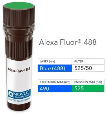
Product Image: TIGIT Antibody (TIGIT/3106) [Alexa Fluor® 488] [NBP2-79928AF488] - Vial of Alexa Fluor 488 conjugated antibody. Alexa Fluor 488 is optimally excited at 490 nm by the Blue laser (488 nm) and has an emission maximum of 525 nm.
Applications for TIGIT Antibody (TIGIT/3106) [Alexa Fluor® 488]
Application
Recommended Usage
ELISA
Optimal dilutions of this antibody should be experimentally determined.
Immunohistochemistry
Optimal dilutions of this antibody should be experimentally determined.
Immunohistochemistry-Paraffin
Optimal dilutions of this antibody should be experimentally determined.
Protein Array
Optimal dilutions of this antibody should be experimentally determined.
Application Notes
Optimal dilution of this antibody should be experimentally determined.
Formulation, Preparation, and Storage
Purification
Protein A or G purified
Formulation
50mM Sodium Borate
Preservative
0.05% Sodium Azide
Concentration
Please see the vial label for concentration. If unlisted please contact technical services.
Shipping
The product is shipped with polar packs. Upon receipt, store it immediately at the temperature recommended below.
Stability & Storage
Store at 4C in the dark.
Background: TIGIT
TIGIT is commonly used as a marker for T cell exhaustion and its expression correlates with disease progression. Furthermore, in viral infections including HIV and SIV, TIGIT serves as a target for immune restoration (6). In cancer detection, TIGIT is upregulated and coexpressed with programmed cell death protein 1 (PD-1) on the majority of circulating tumor antigen (TA)-specific CD8+ T cells (7). In addition to this, TIGIT can inhibit immune cells at multiple steps within the cancer immunity cycle. These include inhibiting NK cell effector function, suppressing dendritic cell costimulatory abilities, suppressing CD8+ T cell effector function by Tregs or polio virus receptor (PVR)-stimulated myeloid cells, and by directly inhibiting CD8+ T cells and preventing elimination of cancer cells (7).
References
1. Joller, N., & Kuchroo, V. K. (2017). Tim-3, Lag-3, and TIGIT. Current topics in microbiology and immunology, 410, 127-156. https://doi.org/10.1007/82_2017_62
2. Xu, Z., Jin, B. A novel interface consisting of homologous immunoglobulin superfamily members with multiple functions. Cell Mol Immunol 7, 11-19 (2010). https://doi.org/10.1038/cmi.2009.108
3. Uniprot(P86176
4. Khan, M., Arooj, S., & Wang, H. (2020). NK Cell-Based Immune Checkpoint Inhibition. Frontiers in immunology, 11, 167. https://doi.org/10.3389/fimmu.2020.00167
5. Harjunpaa, H., & Guillerey, C. (2020). TIGIT as an emerging immune checkpoint. Clinical and experimental immunology, 200(2), 108-119. https://doi.org/10.1111/cei.13407
6. Blake, S. J., Dougall, W. C., Miles, J. J., Teng, M. W., & Smyth, M. J. (2016). Molecular Pathways: Targeting CD96 and TIGIT for Cancer Immunotherapy. Clinical cancer research: an official journal of the American Association for Cancer Research, 22(21), 5183-5188. https://doi.org/10.1158/1078-0432.CCR-16-0933
7. Manieri, N. A., Chiang, E. Y., & Grogan, J. L. (2017). TIGIT: A Key Inhibitor of the Cancer Immunity Cycle. Trends in immunology, 38(1), 20-28. https://doi.org/10.1016/j.it.2016.10.002
Long Name
T Cell Immunoreceptor with Ig and ITIM Domains
Alternate Names
VSIG9, VSTM3, WUCAM
Gene Symbol
Tigit
Additional TIGIT Products
Product Documents for TIGIT Antibody (TIGIT/3106) [Alexa Fluor® 488]
Certificate of Analysis
To download a Certificate of Analysis, please enter a lot or batch number in the search box below.
Product Specific Notices for TIGIT Antibody (TIGIT/3106) [Alexa Fluor® 488]
Alexa Fluor (R) products are provided under an intellectual property license from Life Technologies Corporation. The purchase of this product conveys to the buyer the non-transferable right to use the purchased product and components of the product only in research conducted by the buyer (whether the buyer is an academic or for-profit entity). The sale of this product is expressly conditioned on the buyer not using the product or its components, or any materials made using the product or its components, in any activity to generate revenue, which may include, but is not limited to use of the product or its components: (i) in manufacturing; (ii) to provide a service, information, or data in return for payment; (iii) for therapeutic, diagnostic or prophylactic purposes; or (iv) for resale, regardless of whether they are resold for use in research. For information on purchasing a license to this product for purposes other than as described above, contact Life Technologies Corporation, 5791 Van Allen Way, Carlsbad, CA 92008 USA or outlicensing@lifetech.com. This conjugate is made on demand. Actual recovery may vary from the stated volume of this product. The volume will be greater than or equal to the unit size stated on the datasheet.
This product is for research use only and is not approved for use in humans or in clinical diagnosis. Primary Antibodies are guaranteed for 1 year from date of receipt.
Customer Reviews for TIGIT Antibody (TIGIT/3106) [Alexa Fluor® 488]
There are currently no reviews for this product. Be the first to review TIGIT Antibody (TIGIT/3106) [Alexa Fluor® 488] and earn rewards!
Have you used TIGIT Antibody (TIGIT/3106) [Alexa Fluor® 488]?
Submit a review and receive an Amazon gift card!
$25/€18/£15/$25CAN/¥2500 Yen for a review with an image
$10/€7/£6/$10CAN/¥1110 Yen for a review without an image
Submit a review
Protocols
Find general support by application which include: protocols, troubleshooting, illustrated assays, videos and webinars.
- Antigen Retrieval Protocol (PIER)
- Antigen Retrieval for Frozen Sections Protocol
- Appropriate Fixation of IHC/ICC Samples
- Cellular Response to Hypoxia Protocols
- Chromogenic IHC Staining of Formalin-Fixed Paraffin-Embedded (FFPE) Tissue Protocol
- Chromogenic Immunohistochemistry Staining of Frozen Tissue
- ClariTSA™ Fluorophore Kits
- Detection & Visualization of Antibody Binding
- ELISA Sample Preparation & Collection Guide
- ELISA Troubleshooting Guide
- Fluorescent IHC Staining of Frozen Tissue Protocol
- Graphic Protocol for Heat-induced Epitope Retrieval
- Graphic Protocol for the Preparation and Fluorescent IHC Staining of Frozen Tissue Sections
- Graphic Protocol for the Preparation and Fluorescent IHC Staining of Paraffin-embedded Tissue Sections
- Graphic Protocol for the Preparation of Gelatin-coated Slides for Histological Tissue Sections
- How to Run an R&D Systems DuoSet ELISA
- How to Run an R&D Systems Quantikine ELISA
- How to Run an R&D Systems Quantikine™ QuicKit™ ELISA
- IHC Sample Preparation (Frozen sections vs Paraffin)
- Immunofluorescent IHC Staining of Formalin-Fixed Paraffin-Embedded (FFPE) Tissue Protocol
- Immunohistochemistry (IHC) and Immunocytochemistry (ICC) Protocols
- Immunohistochemistry Frozen Troubleshooting
- Immunohistochemistry Paraffin Troubleshooting
- Preparing Samples for IHC/ICC Experiments
- Preventing Non-Specific Staining (Non-Specific Binding)
- Primary Antibody Selection & Optimization
- Protocol for Heat-Induced Epitope Retrieval (HIER)
- Protocol for Making a 4% Formaldehyde Solution in PBS
- Protocol for VisUCyte™ HRP Polymer Detection Reagent
- Protocol for the Preparation & Fixation of Cells on Coverslips
- Protocol for the Preparation and Chromogenic IHC Staining of Frozen Tissue Sections
- Protocol for the Preparation and Chromogenic IHC Staining of Frozen Tissue Sections - Graphic
- Protocol for the Preparation and Chromogenic IHC Staining of Paraffin-embedded Tissue Sections
- Protocol for the Preparation and Chromogenic IHC Staining of Paraffin-embedded Tissue Sections - Graphic
- Protocol for the Preparation and Fluorescent IHC Staining of Frozen Tissue Sections
- Protocol for the Preparation and Fluorescent IHC Staining of Paraffin-embedded Tissue Sections
- Protocol for the Preparation of Gelatin-coated Slides for Histological Tissue Sections
- Quantikine HS ELISA Kit Assay Principle, Alkaline Phosphatase
- Quantikine HS ELISA Kit Principle, Streptavidin-HRP Polymer
- Sandwich ELISA (Colorimetric) – Biotin/Streptavidin Detection Protocol
- Sandwich ELISA (Colorimetric) – Direct Detection Protocol
- TUNEL and Active Caspase-3 Detection by IHC/ICC Protocol
- The Importance of IHC/ICC Controls
- Troubleshooting Guide: ELISA
- Troubleshooting Guide: Immunohistochemistry
- View all Protocols, Troubleshooting, Illustrated assays and Webinars
Loading...
Associated Pathways
