
TLR (Cell Surface) Screening Antibody Pack
Novus Biologicals | Catalog # NBP2-25086

![Flow Cytometry: TLR (Cell Surface) Screening Antibody Pack [NBP2-25086]](https://resources.rndsystems.com/images/products/TLR-Cell-Surface-Screening-Antibody-Pack-Flow-Cytometry-NBP2-25086-img0007.jpg "Flow Cytometry: TLR (Cell Surface) Screening Antibody Pack [NBP2-25086]")
Loading...
Key Product Details
Species
Human, Mouse
Applications
Dot Blot, Flow Cytometry, Immunocytochemistry/ Immunofluorescence, Immunohistochemistry, Western Blot
Loading...
Product Specifications
Application Notes
See individual datasheets of components for their validated applications
Reactivity Notes
See individual datasheets of components for their validated species
Scientific Data Images for TLR (Cell Surface) Screening Antibody Pack
Flow Cytometry: TLR (Cell Surface) Screening Antibody Pack [NBP2-25086]
Flow Cytometry: TLR (Cell Surface) Screening Antibody Pack [NBP2-25086] - TLR1 and TLR2 expression pattern in primary BMNCs from 3 MM patients analyzed by flow cytometry. CD138-positive cells were gated from the total cell population. Staining for was compared with isotype-matched controls (NC). Image collected and cropped by CiteAb from the following publication (//doi.org/10.1371/journal.pone.0060671) licensed under a CC-BY license. TLR1 Antibody [NB100-56563] and TLR2 Antibody (TL2.1) [NB100-56722].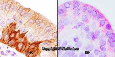
Dual RNAscope ISH-IHC: TLR (Cell Surface) Screening Antibody Pack [NBP2-25086] - Formalin-fixed paraffin-embedded tissue sections of human tonsil were probed for TLR4 mRNA (ACD RNAScope Probe, catalog #311281; Fast Red chromogen, ACD catalog # 322750). Adjacent tissue section was processed for immunohistochemistry using Mouse Monoclonal (Novus Biologicals catalog #NB100-56566) at 5ug/mL with 1 hour incubation at room temperature followed by incubation with anti-mouse IgG VisUCyte HRP Polymer Antibody (Catalog # VC001) and DAB chromogen (yellow-brown). Tissue was counterstained with hematoxylin (blue). Specific staining was localized to lymphocytes.
![Western Blot: TLR (Cell Surface) Screening Antibody Pack [NBP2-25086]](https://resources.rndsystems.com/images/products/TLR-Cell-Surface-Screening-Antibody-Pack-Western-Blot-NBP2-25086-img0006.jpg "Western Blot: TLR (Cell Surface) Screening Antibody Pack [NBP2-25086]")
Western Blot: TLR (Cell Surface) Screening Antibody Pack [NBP2-25086]
Western Blot: TLR (Cell Surface) Screening Antibody Pack [NBP2-25086] - Expression of TLR2 was determined by western blotting. The immunoreactivity of the anti-TLR2 was confirmed with human intestinal lysate. Data are representative for analysis of >=2 independent experiments. Image collected and cropped by CiteAb from the following publication (//doi.org/10.1371/journal.pone.0060671) licensed under a CC-BY license. TLR2 Antibody (TL2.1) [NB100-56722]![Immunohistochemistry-Paraffin: TLR (Cell Surface) Screening Antibody Pack [NBP2-25086]](https://resources.rndsystems.com/images/products/TLR-Cell-Surface-Screening-Antibody-Pack-Immunohistochemistry-Paraffin-NBP2-25086-img0010.jpg "Immunohistochemistry-Paraffin: TLR (Cell Surface) Screening Antibody Pack [NBP2-25086]")
Immunohistochemistry-Paraffin: TLR (Cell Surface) Screening Antibody Pack [NBP2-25086]
Immunohistochemistry-Paraffin: TLR (Cell Surface) Screening Antibody Pack [NBP2-25086] - Analysis of FFPE tissue section of normal human skin using 5 ug/ml concentration of TLR5 antibody (clone 19D759.2). Intense membrane staining of TLR5 was observed specifically in the pigmented basel cells of epidermal layer [10X Magnification]. TLR5 Antibody (19D759.2) [NBP2-24787]![Flow Cytometry: TLR (Cell Surface) Screening Antibody Pack [NBP2-25086]](https://resources.rndsystems.com/images/products/TLR-Cell-Surface-Screening-Antibody-Pack-Flow-Cytometry-NBP2-25086-img0011.jpg "Flow Cytometry: TLR (Cell Surface) Screening Antibody Pack [NBP2-25086]")
Flow Cytometry: TLR (Cell Surface) Screening Antibody Pack [NBP2-25086]
Flow Cytometry: TLR (Cell Surface) Screening Antibody Pack [NBP2-25086] - Detection of TLR10 on human PBMC using 10 ul (1 ug) of TLR10 antibody and 1 ug of CD19 antibody. Analysis using the Alexa Fluor (R) 647 conjugate of TLR10 antibody [NBP1-70343].![Immunohistochemistry: TLR (Cell Surface) Screening Antibody Pack [NBP2-25086]](https://resources.rndsystems.com/images/products/TLR-Cell-Surface-Screening-Antibody-Pack-Immunohistochemistry-NBP2-25086-img0009.jpg "Immunohistochemistry: TLR (Cell Surface) Screening Antibody Pack [NBP2-25086]")
Immunohistochemistry: TLR (Cell Surface) Screening Antibody Pack [NBP2-25086]
Immunohistochemistry: TLR (Cell Surface) Screening Antibody Pack [NBP2-25086] - Pericryptal Myofibroblasts are Responsible for Increased TLR4 Expression in a Subset of CRCs. Double-stained immunofluorescence for TLR4 (green) and vimentin (red) in normal (I), adenoma (II), and colon adenocarcinoma (III) (10x). In the stromal compartment of CRCs, immunofluorescent staining for TLR4 localized to the pericryptal myofibroblasts in a subset of samples. Image collected and cropped by CiteAb from the following publication (//www.jeccr.com/content/33/1/45), licensed under a CC-BY license. TLR4 Antibody (76B357.1) [NB100-56566]Kit Contents for TLR (Cell Surface) Screening Antibody Pack
- NB100-56536
- NB100-56563
- NB100-56566
- NB100-56722
- NBP1-70343
- NBP2-24787
- NL004
- NL007
Formulation, Preparation, and Storage
Concentration
Concentration of individual antibodies may be found on the vial label. If unlisted please contact technical services.
Shipping
The product is shipped with polar packs. Upon receipt, store it immediately at the temperature recommended below.
Storage
Store at 4C short term. Aliquot and store at -20C long term. Avoid freeze-thaw cycles.
Product Documents for TLR (Cell Surface) Screening Antibody Pack
Certificate of Analysis
To download a Certificate of Analysis, please enter a lot or batch number in the search box below.
Product Specific Notices for TLR (Cell Surface) Screening Antibody Pack
This product is for research use only and is not approved for use in humans or in clinical diagnosis. Antibody Packs are guaranteed for 1 year from date of receipt.
Citations for TLR (Cell Surface) Screening Antibody Pack
 Powered by Bioz
Customer Reviews for TLR (Cell Surface) Screening Antibody Pack
There are currently no reviews for this product. Be the first to review TLR (Cell Surface) Screening Antibody Pack and earn rewards!
Have you used TLR (Cell Surface) Screening Antibody Pack?
Submit a review and receive an Amazon gift card!
$25/€18/£15/$25CAN/¥2500 for a review with an image
$10/€7/£6/$10CAN/¥1110 for a review without an image
Submit a review
Protocols
Find general support by application which include: protocols, troubleshooting, illustrated assays, videos and webinars.
- 7-Amino Actinomycin D (7-AAD) Cell Viability Flow Cytometry Protocol
- Antigen Retrieval Protocol (PIER)
- Antigen Retrieval for Frozen Sections Protocol
- Appropriate Fixation of IHC/ICC Samples
- Cellular Response to Hypoxia Protocols
- Chromogenic IHC Staining of Formalin-Fixed Paraffin-Embedded (FFPE) Tissue Protocol
- Chromogenic Immunohistochemistry Staining of Frozen Tissue
- ClariTSA™ Fluorophore Kits
- Detection & Visualization of Antibody Binding
- Extracellular Membrane Flow Cytometry Protocol
- Flow Cytometry Protocol for Cell Surface Markers
- Flow Cytometry Protocol for Staining Membrane Associated Proteins
- Flow Cytometry Staining Protocols
- Flow Cytometry Troubleshooting Guide
- Fluorescent IHC Staining of Frozen Tissue Protocol
- Graphic Protocol for Heat-induced Epitope Retrieval
- Graphic Protocol for the Preparation and Fluorescent IHC Staining of Frozen Tissue Sections
- Graphic Protocol for the Preparation and Fluorescent IHC Staining of Paraffin-embedded Tissue Sections
- Graphic Protocol for the Preparation of Gelatin-coated Slides for Histological Tissue Sections
- ICC Cell Smear Protocol for Suspension Cells
- ICC Immunocytochemistry Protocol Videos
- ICC for Adherent Cells
- IHC Sample Preparation (Frozen sections vs Paraffin)
- Immunocytochemistry (ICC) Protocol
- Immunocytochemistry Troubleshooting
- Immunofluorescence of Organoids Embedded in Cultrex Basement Membrane Extract
- Immunofluorescent IHC Staining of Formalin-Fixed Paraffin-Embedded (FFPE) Tissue Protocol
- Immunohistochemistry (IHC) and Immunocytochemistry (ICC) Protocols
- Immunohistochemistry Frozen Troubleshooting
- Immunohistochemistry Paraffin Troubleshooting
- Intracellular Flow Cytometry Protocol Using Alcohol (Methanol)
- Intracellular Flow Cytometry Protocol Using Detergents
- Intracellular Nuclear Staining Flow Cytometry Protocol Using Detergents
- Intracellular Staining Flow Cytometry Protocol Using Alcohol Permeabilization
- Intracellular Staining Flow Cytometry Protocol Using Detergents to Permeabilize Cells
- Preparing Samples for IHC/ICC Experiments
- Preventing Non-Specific Staining (Non-Specific Binding)
- Primary Antibody Selection & Optimization
- Propidium Iodide Cell Viability Flow Cytometry Protocol
- Protocol for Heat-Induced Epitope Retrieval (HIER)
- Protocol for Liperfluo
- Protocol for Making a 4% Formaldehyde Solution in PBS
- Protocol for VisUCyte™ HRP Polymer Detection Reagent
- Protocol for the Characterization of Human Th22 Cells
- Protocol for the Characterization of Human Th9 Cells
- Protocol for the Fluorescent ICC Staining of Cell Smears - Graphic
- Protocol for the Fluorescent ICC Staining of Cultured Cells on Coverslips - Graphic
- Protocol for the Preparation & Fixation of Cells on Coverslips
- Protocol for the Preparation and Chromogenic IHC Staining of Frozen Tissue Sections
- Protocol for the Preparation and Chromogenic IHC Staining of Frozen Tissue Sections - Graphic
- Protocol for the Preparation and Chromogenic IHC Staining of Paraffin-embedded Tissue Sections
- Protocol for the Preparation and Chromogenic IHC Staining of Paraffin-embedded Tissue Sections - Graphic
- Protocol for the Preparation and Fluorescent ICC Staining of Cells on Coverslips
- Protocol for the Preparation and Fluorescent ICC Staining of Non-adherent Cells
- Protocol for the Preparation and Fluorescent ICC Staining of Stem Cells on Coverslips
- Protocol for the Preparation and Fluorescent IHC Staining of Frozen Tissue Sections
- Protocol for the Preparation and Fluorescent IHC Staining of Paraffin-embedded Tissue Sections
- Protocol for the Preparation of Gelatin-coated Slides for Histological Tissue Sections
- Protocol for the Preparation of a Cell Smear for Non-adherent Cell ICC - Graphic
- Protocol: Annexin V and PI Staining by Flow Cytometry
- Protocol: Annexin V and PI Staining for Apoptosis by Flow Cytometry
- R&D Systems Quality Control Western Blot Protocol
- TUNEL and Active Caspase-3 Detection by IHC/ICC Protocol
- The Importance of IHC/ICC Controls
- Troubleshooting Guide: Fluorokine Flow Cytometry Kits
- Troubleshooting Guide: Immunohistochemistry
- Troubleshooting Guide: Western Blot Figures
- Western Blot Conditions
- Western Blot Protocol
- Western Blot Protocol for Cell Lysates
- Western Blot Troubleshooting
- Western Blot Troubleshooting Guide
- View all Protocols, Troubleshooting, Illustrated assays and Webinars
Loading...