TNF RI/TNFRSF1A Antibody - BSA Free
Novus Biologicals | Catalog # NBP1-97453
![Western Blot: TNF RI/TNFRSF1A Antibody [NBP1-97453]](https://resources.rndsystems.com/images/products/TNF-RI-TNFRSF1A-Antibody-Western-Blot-NBP1-97453-img0005.jpg "Western Blot: TNF RI/TNFRSF1A Antibody [NBP1-97453]")
Key Product Details
Species Reactivity
Validated:
Human, Mouse, Rat, Porcine, Bovine, Canine, Drosophila, Goat, Hamster, Monkey, Rabbit, Sheep, Xenopus, Yeast
Cited:
Rabbit
Applications
Validated:
Immunohistochemistry, Immunohistochemistry-Paraffin, Western Blot, Flow Cytometry, Immunoprecipitation
Cited:
Western Blot, Immunoprecipitation
Label
Unconjugated
Antibody Source
Polyclonal Rabbit IgG
Format
BSA Free
Loading...
Product Specifications
Immunogen
Synthetic peptide corresponding to a portion of mouse TNF-R1. The sequence is completely conserved in rat and human.
Reactivity Notes
Yeast TNF-R1. Mouse and Goat reactivity reported from a verified customer review.
Clonality
Polyclonal
Host
Rabbit
Isotype
IgG
Theoretical MW
55 kDa.
Disclaimer note: The observed molecular weight of the protein may vary from the listed predicted molecular weight due to post translational modifications, post translation cleavages, relative charges, and other experimental factors.
Disclaimer note: The observed molecular weight of the protein may vary from the listed predicted molecular weight due to post translational modifications, post translation cleavages, relative charges, and other experimental factors.
Scientific Data Images for TNF RI/TNFRSF1A Antibody - BSA Free
Western Blot: TNF RI/TNFRSF1A Antibody [NBP1-97453]
Western Blot: TNF RI/TNFRSF1A Antibody [NBP1-97453] - TNF-R1 Antibody [NBP1-97453] - Analysis of TNF-R1: Lane 1: MW marker, Lane 2: Hela (HS), Lane 3: Jurkat.![Immunohistochemistry-Paraffin: TNF RI/TNFRSF1A Antibody [NBP1-97453]](https://resources.rndsystems.com/images/products/TNF-RI-TNFRSF1A-Antibody-Immunohistochemistry-Paraffin-NBP1-97453-img0004.jpg "Immunohistochemistry-Paraffin: TNF RI/TNFRSF1A Antibody [NBP1-97453]")
Immunohistochemistry-Paraffin: TNF RI/TNFRSF1A Antibody [NBP1-97453]
Immunohistochemistry-Paraffin: TNF RI/TNFRSF1A Antibody [NBP1-97453] - TNF-R1 Antibody [NBP1-97453] - Analysis of human spleen tissue stained with TNF receptor 1 pAb at 10ug/ml.![Flow Cytometry: TNF RI/TNFRSF1A Antibody [NBP1-97453]](https://resources.rndsystems.com/images/products/TNF-RI-TNFRSF1A-Antibody-Flow-Cytometry-NBP1-97453-img0006.jpg "Flow Cytometry: TNF RI/TNFRSF1A Antibody [NBP1-97453]")
Flow Cytometry: TNF RI/TNFRSF1A Antibody [NBP1-97453]
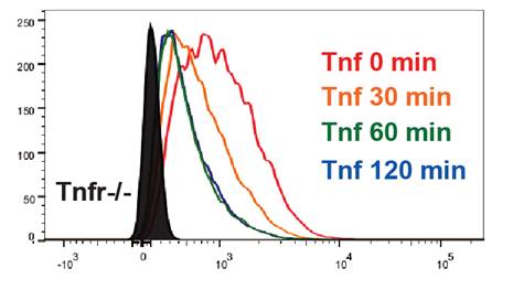
Flow Cytometry: TNF RI/TNFRSF1A Antibody [NBP1-97453] - TNF Receptor I Antibody [NBP1-97453] - MLL-AF9 transduced murine leukemia cells. Tnfr-/- cells were used as negative control. Image provided by verified customer.
Western Blot: Rabbit Polyclonal TNF RI/TNFRSF1A Antibody [NBP1-97453]
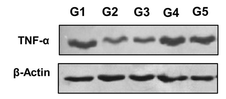
Western Blot: Rabbit Polyclonal TNF RI/TNFRSF1A Antibody [NBP1-97453] -Quantification of TNF alpha in bone graft in the tibia of intrailiac cellularized goats at different times compared with tibial autograft and untreated bone defect. NBP1-97453 was used at a dilution of 1:1000 and Beta-actin antibody (Catalog # NB600-501) was used at a dilution of 1:3000. Evaluation of pre-cellularized graft in iliac crest in the treatment of bone defects in the tibia of goats. G1: cellularized graft for 4 weeks applied to tibial bone defect, G2: cellularized graft for 6 weeks applied to tibial bone defect, G3: cellularized graft for 8 weeks applied to tibial bone defect, G4: tibial autograft and G5: untreated bone defect. All groups were evaluated 10 days after graft implantation. Image from a verified customer review.Applications for TNF RI/TNFRSF1A Antibody - BSA Free
Application
Recommended Usage
Flow Cytometry
1:20
Immunohistochemistry
1:10-1:500
Immunohistochemistry-Paraffin
1:10-1:500
Immunoprecipitation
12.5 ug/ml
Western Blot
1:1000
Application Notes
TNF RI/TNFRSF1A antibody validated for FLOW from a verified customer review.
Reviewed Applications
Read 2 reviews rated 5 using NBP1-97453 in the following applications:
Flow Cytometry Panel Builder
Bio-Techne Knows Flow Cytometry
Save time and reduce costly mistakes by quickly finding compatible reagents using the Panel Builder Tool.
Advanced Features
- Spectra Viewer - Custom analysis of spectra from multiple fluorochromes
- Spillover Popups - Visualize the spectra of individual fluorochromes
- Antigen Density Selector - Match fluorochrome brightness with antigen density

Formulation, Preparation, and Storage
Purification
Immunogen affinity purified
Formulation
PBS and 50% Glycerol
Format
BSA Free
Preservative
0.09% Sodium Azide
Concentration
1.0 mg/ml
Shipping
The product is shipped with polar packs. Upon receipt, store it immediately at the temperature recommended below.
Stability & Storage
Store at -20C. Avoid freeze-thaw cycles.
Background: TNF RI/TNFRSF1A
Long Name
Tumor Necrosis Factor Receptor I
Alternate Names
CD120a, TNFRI, TNFRSF1A
Entrez Gene IDs
7132 (Human)
Gene Symbol
TNFRSF1A
Additional TNF RI/TNFRSF1A Products
Product Documents for TNF RI/TNFRSF1A Antibody - BSA Free
Certificate of Analysis
To download a Certificate of Analysis, please enter a lot or batch number in the search box below.
Product Specific Notices for TNF RI/TNFRSF1A Antibody - BSA Free
This product is for research use only and is not approved for use in humans or in clinical diagnosis. Primary Antibodies are guaranteed for 1 year from date of receipt.
Citations for TNF RI/TNFRSF1A Antibody - BSA Free
 Powered by Bioz
Customer Reviews for TNF RI/TNFRSF1A Antibody - BSA Free (2)
5 out of 5
2 Customer Ratings
Have you used TNF RI/TNFRSF1A Antibody - BSA Free?
Submit a review and receive an Amazon gift card!
$25/€18/£15/$25CAN/¥2500 Yen for a review with an image
$10/€7/£6/$10CAN/¥1110 Yen for a review without an image
Submit a review
Customer Images
Showing
1
-
2 of
2 reviews
Showing All
Filter By:
-
Application: Western BlotSample Tested: Bone and Bone ExtractsSpecies: GoatVerified Customer | Posted 05/13/2025Quantification of TNF alpha in bone graft in the tibia of intrailiac cellularized goats at different times compared with tibial autograft and untreated bone defect. NBP1-97453 was used at a dilution of 1:1000.Evaluation of pre-cellularized graft in iliac crest in the treatment of bone defects in the tibia of goats. G1: cellularized graft for 4 weeks applied to tibial bone defect, G2: cellularized graft for 6 weeks applied to tibial bone defect, G3: cellularized graft for 8 weeks applied to tibial bone defect, G4: tibial autograft and G5: untreated bone defect. All groups were evaluated 10 days after graft implantation.
 Bio-Techne ResponseThis review reflects a new species or application tested on a primary antibody.
 -
Application: Flow CytometrySample Tested: Mouse MLL-AF9 transduced leukemia cellsSpecies: MouseVerified Customer | Posted 01/31/2014MLL-AF9 transduced murine leukemia cells. Tnfr-/- cells were used as negative control
There are no reviews that match your criteria.
Protocols
Find general support by application which include: protocols, troubleshooting, illustrated assays, videos and webinars.
- 7-Amino Actinomycin D (7-AAD) Cell Viability Flow Cytometry Protocol
- Antigen Retrieval Protocol (PIER)
- Antigen Retrieval for Frozen Sections Protocol
- Appropriate Fixation of IHC/ICC Samples
- Cellular Response to Hypoxia Protocols
- Chromogenic IHC Staining of Formalin-Fixed Paraffin-Embedded (FFPE) Tissue Protocol
- Chromogenic Immunohistochemistry Staining of Frozen Tissue
- ClariTSA™ Fluorophore Kits
- Detection & Visualization of Antibody Binding
- Extracellular Membrane Flow Cytometry Protocol
- Flow Cytometry Protocol for Cell Surface Markers
- Flow Cytometry Protocol for Staining Membrane Associated Proteins
- Flow Cytometry Staining Protocols
- Flow Cytometry Troubleshooting Guide
- Fluorescent IHC Staining of Frozen Tissue Protocol
- Graphic Protocol for Heat-induced Epitope Retrieval
- Graphic Protocol for the Preparation and Fluorescent IHC Staining of Frozen Tissue Sections
- Graphic Protocol for the Preparation and Fluorescent IHC Staining of Paraffin-embedded Tissue Sections
- Graphic Protocol for the Preparation of Gelatin-coated Slides for Histological Tissue Sections
- IHC Sample Preparation (Frozen sections vs Paraffin)
- Immunofluorescent IHC Staining of Formalin-Fixed Paraffin-Embedded (FFPE) Tissue Protocol
- Immunohistochemistry (IHC) and Immunocytochemistry (ICC) Protocols
- Immunohistochemistry Frozen Troubleshooting
- Immunohistochemistry Paraffin Troubleshooting
- Immunoprecipitation Protocol
- Intracellular Flow Cytometry Protocol Using Alcohol (Methanol)
- Intracellular Flow Cytometry Protocol Using Detergents
- Intracellular Nuclear Staining Flow Cytometry Protocol Using Detergents
- Intracellular Staining Flow Cytometry Protocol Using Alcohol Permeabilization
- Intracellular Staining Flow Cytometry Protocol Using Detergents to Permeabilize Cells
- Preparing Samples for IHC/ICC Experiments
- Preventing Non-Specific Staining (Non-Specific Binding)
- Primary Antibody Selection & Optimization
- Propidium Iodide Cell Viability Flow Cytometry Protocol
- Protocol for Heat-Induced Epitope Retrieval (HIER)
- Protocol for Liperfluo
- Protocol for Making a 4% Formaldehyde Solution in PBS
- Protocol for VisUCyte™ HRP Polymer Detection Reagent
- Protocol for the Characterization of Human Th22 Cells
- Protocol for the Characterization of Human Th9 Cells
- Protocol for the Preparation & Fixation of Cells on Coverslips
- Protocol for the Preparation and Chromogenic IHC Staining of Frozen Tissue Sections
- Protocol for the Preparation and Chromogenic IHC Staining of Frozen Tissue Sections - Graphic
- Protocol for the Preparation and Chromogenic IHC Staining of Paraffin-embedded Tissue Sections
- Protocol for the Preparation and Chromogenic IHC Staining of Paraffin-embedded Tissue Sections - Graphic
- Protocol for the Preparation and Fluorescent IHC Staining of Frozen Tissue Sections
- Protocol for the Preparation and Fluorescent IHC Staining of Paraffin-embedded Tissue Sections
- Protocol for the Preparation of Gelatin-coated Slides for Histological Tissue Sections
- Protocol: Annexin V and PI Staining by Flow Cytometry
- Protocol: Annexin V and PI Staining for Apoptosis by Flow Cytometry
- R&D Systems Quality Control Western Blot Protocol
- TUNEL and Active Caspase-3 Detection by IHC/ICC Protocol
- The Importance of IHC/ICC Controls
- Troubleshooting Guide: Fluorokine Flow Cytometry Kits
- Troubleshooting Guide: Immunohistochemistry
- Troubleshooting Guide: Western Blot Figures
- Western Blot Conditions
- Western Blot Protocol
- Western Blot Protocol for Cell Lysates
- Western Blot Troubleshooting
- Western Blot Troubleshooting Guide
- View all Protocols, Troubleshooting, Illustrated assays and Webinars
Loading...
Associated Pathways


