TOMM20 Antibody - BSA Free
Novus Biologicals | Catalog # NBP1-81556
![Western Blot: TOMM20 Antibody [NBP1-81556]](https://resources.rndsystems.com/images/products/TOMM20-Antibody-Western-Blot-NBP1-81556-img0019.jpg "Western Blot: TOMM20 Antibody [NBP1-81556]")
Loading...
Key Product Details
Validated by
Orthogonal Validation
Species Reactivity
Validated:
Human
Cited:
Human, Mouse, Rat
Predicted:
Mouse (99%), Rat (99%). Backed by our 100% Guarantee.
Applications
Validated:
Immunohistochemistry, Immunohistochemistry-Paraffin, Western Blot, Immunocytochemistry/ Immunofluorescence, Simple Western
Cited:
Western Blot, Immunocytochemistry/ Immunofluorescence, IF/IHC
Label
Unconjugated
Antibody Source
Polyclonal Rabbit IgG
Format
BSA Free
Loading...
Product Specifications
Immunogen
This antibody was developed against Recombinant Protein corresponding to amino acids: KRRSDPNFKNRLRERRKKQKLAKERAGLSKLPDLKDAEAVQKFFLEEIQLGEELLAQGEYEKGVDHLTNAIAVCGQPQQLLQVLQQTLPPPVFQMLLTKLPTISQRIVSAQSLAEDDV
Clonality
Polyclonal
Host
Rabbit
Isotype
IgG
Scientific Data Images for TOMM20 Antibody - BSA Free
![Western Blot: TOMM20 Antibody [NBP1-81556]](https://resources.rndsystems.com/images/products/TOMM20-Antibody-Western-Blot-NBP1-81556-img0022.jpg "Western Blot: TOMM20 Antibody [NBP1-81556]")
Western Blot: TOMM20 Antibody [NBP1-81556]
Western Blot: TOMM20 Antibody [NBP1-81556] - Analysis in mouse cell line NIH-3T3.![Immunohistochemistry-Paraffin: TOMM20 Antibody [NBP1-81556]](https://resources.rndsystems.com/images/products/TOMM20-Antibody-Immunohistochemistry-Paraffin-NBP1-81556-img0017.jpg "Immunohistochemistry-Paraffin: TOMM20 Antibody [NBP1-81556]")
Immunohistochemistry-Paraffin: TOMM20 Antibody [NBP1-81556]
Immunohistochemistry-Paraffin: TOMM20 Antibody [NBP1-81556] - Staining of human duodenum shows moderate to strong positivity in mitochondria in glandular cells.![Immunohistochemistry-Paraffin: TOMM20 Antibody [NBP1-81556]](https://resources.rndsystems.com/images/products/TOMM20-Antibody-Immunohistochemistry-Paraffin-NBP1-81556-img0013.jpg "Immunohistochemistry-Paraffin: TOMM20 Antibody [NBP1-81556]")
Immunohistochemistry-Paraffin: TOMM20 Antibody [NBP1-81556]
Immunohistochemistry-Paraffin: TOMM20 Antibody [NBP1-81556] - Staining of human endometrium shows moderate to strong positivity in mitochondria in glandular cells.![Immunohistochemistry-Paraffin: TOMM20 Antibody [NBP1-81556]](https://resources.rndsystems.com/images/products/TOMM20-Antibody-Immunohistochemistry-Paraffin-NBP1-81556-img0014.jpg "Immunohistochemistry-Paraffin: TOMM20 Antibody [NBP1-81556]")
Immunohistochemistry-Paraffin: TOMM20 Antibody [NBP1-81556]
Immunohistochemistry-Paraffin: TOMM20 Antibody [NBP1-81556] - Staining of human prostate shows moderate to strong positivity in mitochondria in glandular cells.![Immunohistochemistry-Paraffin: TOMM20 Antibody [NBP1-81556]](https://resources.rndsystems.com/images/products/TOMM20-Antibody-Immunohistochemistry-Paraffin-NBP1-81556-img0015.jpg "Immunohistochemistry-Paraffin: TOMM20 Antibody [NBP1-81556]")
Immunohistochemistry-Paraffin: TOMM20 Antibody [NBP1-81556]
Immunohistochemistry-Paraffin: TOMM20 Antibody [NBP1-81556] - Staining of human cerebral cortex shows moderate to strong positivity in mitochondria in neurons.![Simple Western: TOMM20 Antibody [NBP1-81556]](https://resources.rndsystems.com/images/products/TOMM20-Antibody-Simple-Western-NBP1-81556-img0006.jpg "Simple Western: TOMM20 Antibody [NBP1-81556]")
Simple Western: TOMM20 Antibody [NBP1-81556]
Simple Western: TOMM20 Antibody [NBP1-81556] - Simple Western lane view shows a specific band for TOMM20 in 0.2 mg/ml of RT-4 (Left) and U-251MG (Right) lysate. This experiment was performed under reducing conditions using the 12-230 kDa separation system.![Simple Western: TOMM20 Antibody [NBP1-81556]](https://resources.rndsystems.com/images/products/TOMM20-Antibody-Simple-Western-NBP1-81556-img0007.jpg "Simple Western: TOMM20 Antibody [NBP1-81556]")
Simple Western: TOMM20 Antibody [NBP1-81556]
Simple Western: TOMM20 Antibody [NBP1-81556] - Electropherogram image(s) of corresponding Simple Western lane view. TOMM20 antibody was used at 1:30 dilution on RT-4 and U-251MG lysate(s).
Western Blot: TOMM20 Antibody [NBP1-81556] -
Western Blot: TOMM20 Antibody [NBP1-81556] - Quantification of LC3B-II & TOM20 proteins in WT & Ts1Cje hippocampus. Hippocampal proteins from WT & Ts1Cje mice pairs were analyzed in Western blot with anti-LC3B or anti-TOM20 antibody. A LC3B western blot showing WT & Ts1Cje littermate pairs analyzed & total protein loaded. The ratio LC3B-II/LC3B-I is shown as the mean ± SEM (WT: 0.8714 ± 0.04154; Ts1Cje: 0.6500 ± 0.06802; p = 0.0152, Student’s t-test, n = 7 for WT & n = 6 for Ts1Cje). B TOM20 western blot showing WT & Ts1Cje littermate pairs analyzed & total protein loaded. The signals were normalized to the corresponding total protein loaded & the mean ± SEM values are shown (WT: 1.024 ± 0.08799; Ts1Cje: 1.412 ± 0.1452; p = 0.0414, Student’s t-test, n = 7 animals per genotype) Image collected & cropped by CiteAb from the following publication (https://pubmed.ncbi.nlm.nih.gov/34034796), licensed under a CC-BY license. Not internally tested by Novus Biologicals.![TOMM20 Antibody - BSA Free Immunocytochemistry/ Immunofluorescence: TOMM20 Antibody [NBP1-81556]](https://resources.rndsystems.com/images/products/nbp1-81556_-immunocytochemistry-immunofluorescence-639174076378446640.jpg "Immunocytochemistry/ Immunofluorescence: TOMM20 Antibody [NBP1-81556]")
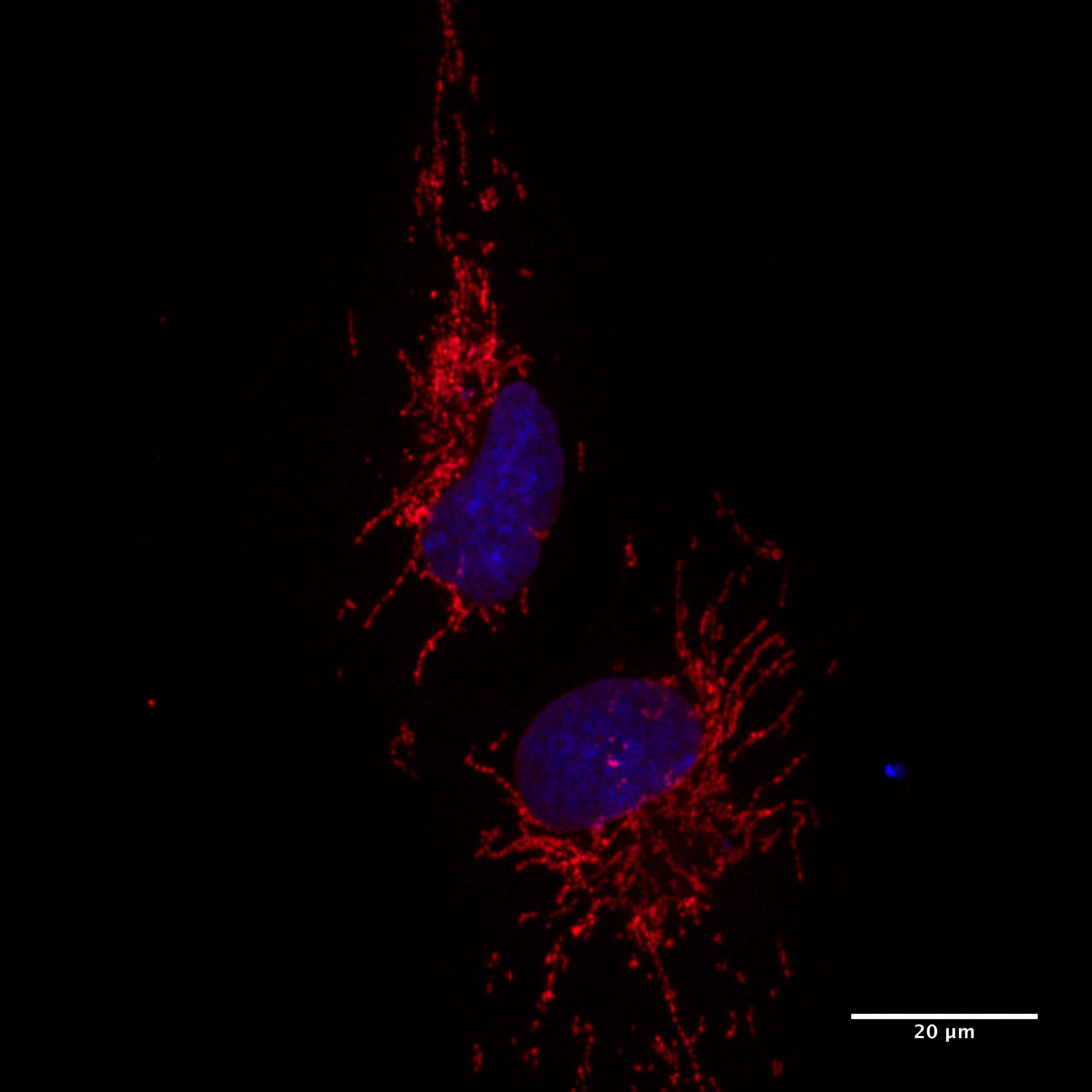
Immunocytochemistry/ Immunofluorescence: TOMM20 Antibody [NBP1-81556]
Staining of human cell line U-2 OS shows localization to mitochondria.Applications for TOMM20 Antibody - BSA Free
Application
Recommended Usage
Immunocytochemistry/ Immunofluorescence
0.25-2 ug/ml
Immunohistochemistry
1:500 - 1:1000
Immunohistochemistry-Paraffin
1:500 - 1:1000
Simple Western
1:30
Western Blot
0.04-0.4 ug/ml
Application Notes
For IHC-Paraffin, HIER pH 6 retrieval is recommended. ICC/IF Fixation Permeabilization: Use PFA/Triton X-100.
In Simple Western only 10 - 15 uL of the recommended dilution is used per data point.
See Simple Western Antibody Database for Simple Western validation: Tested in RT-4 and U-251MG, separated by Size, antibody dilution of 1:30, apparent MW was 18 kDa. Separated by Size-Wes, Sally Sue/Peggy Sue.
In Simple Western only 10 - 15 uL of the recommended dilution is used per data point.
See Simple Western Antibody Database for Simple Western validation: Tested in RT-4 and U-251MG, separated by Size, antibody dilution of 1:30, apparent MW was 18 kDa. Separated by Size-Wes, Sally Sue/Peggy Sue.
Reviewed Applications
Read 3 reviews rated 4.3 using NBP1-81556 in the following applications:
Formulation, Preparation, and Storage
Purification
Affinity purified
Formulation
PBS (pH 7.2) and 40% Glycerol
Format
BSA Free
Preservative
0.02% Sodium Azide
Concentration
Concentrations vary lot to lot. See vial label for concentration. If unlisted please contact technical services.
Shipping
The product is shipped with polar packs. Upon receipt, store it immediately at the temperature recommended below.
Stability & Storage
Store at 4C short term. Aliquot and store at -20C long term. Avoid freeze-thaw cycles.
Background: TOMM20
Long Name
TOMM20
Alternate Names
MAS20, mitochondrial 20 kDa outer membrane protein, mitochondrial import receptor subunit TOM20 homolo, MOM19, outer mitochondrial membrane receptor Tom20, TOM20, translocase of outer mitochondrial membrane 20 hom
Gene Symbol
TOMM20
Additional TOMM20 Products
Product Documents for TOMM20 Antibody - BSA Free
Certificate of Analysis
To download a Certificate of Analysis, please enter a lot or batch number in the search box below.
Product Specific Notices for TOMM20 Antibody - BSA Free
This product is for research use only and is not approved for use in humans or in clinical diagnosis. Primary Antibodies are guaranteed for 1 year from date of receipt.
Citations for TOMM20 Antibody - BSA Free
 Powered by Bioz
Customer Reviews for TOMM20 Antibody - BSA Free (3)
4.3 out of 5
3 Customer Ratings
Have you used TOMM20 Antibody - BSA Free?
Submit a review and receive an Amazon gift card!
$25/€18/£15/$25CAN/¥2500 Yen for a review with an image
$10/€7/£6/$10CAN/¥1110 Yen for a review without an image
Submit a review
Customer Images
Showing
1
-
3 of
3 reviews
Showing All
Filter By:
-
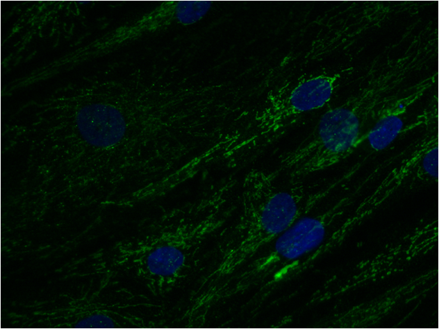
Application: ImmunocytochemistrySample Tested: Human primary fibroblastSpecies: HumanVerified Customer | Posted 10/31/2020Tomm20 antibody in human primary fibroblasts. 1:200 dilution. Secondary ab: Donkey anti-rabbit Alexa Fluor 488. Nuclear staining with DAPI.PFA 4% 15 min fixed. Ab incubation:1:200 dilution O/N 4ºC in 10% Donkey Serum in PBST (0.1% Triton). Secondary antibody Donkey anti-rabbit Alexa Fluor 488 1:500 2h RT in 10% Donkey Serum in PBST (0.1% Triton).

-
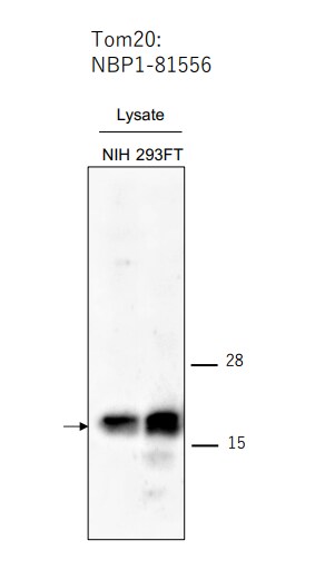
Application: Western BlotSample Tested: 293FT cell lysate and nih3t3 cell lysateSpecies: Human and MouseVerified Customer | Posted 06/29/2017
-
Application: ImmunocytochemistrySample Tested: Retinal Pigmented Epithelial CellsSpecies: HumanVerified Customer | Posted 04/10/2017RPE1 cells were fixed with 4% PFA, permeablized and blocked in 2.5% BSA with 0.2% Triton-X. Primary antibody was diluted 1:200 in PBS containing 2.5% BSA overnight at 4 deg C. Cells were incubated with anti-rabbit secondary antibody and counterstained with hoescht 33342. Image was collected using Zeiss 780 LSM.
There are no reviews that match your criteria.
Protocols
Find general support by application which include: protocols, troubleshooting, illustrated assays, videos and webinars.
- Antigen Retrieval Protocol (PIER)
- Antigen Retrieval for Frozen Sections Protocol
- Appropriate Fixation of IHC/ICC Samples
- Cellular Response to Hypoxia Protocols
- Chromogenic IHC Staining of Formalin-Fixed Paraffin-Embedded (FFPE) Tissue Protocol
- Chromogenic Immunohistochemistry Staining of Frozen Tissue
- ClariTSA™ Fluorophore Kits
- Detection & Visualization of Antibody Binding
- Fluorescent IHC Staining of Frozen Tissue Protocol
- Graphic Protocol for Heat-induced Epitope Retrieval
- Graphic Protocol for the Preparation and Fluorescent IHC Staining of Frozen Tissue Sections
- Graphic Protocol for the Preparation and Fluorescent IHC Staining of Paraffin-embedded Tissue Sections
- Graphic Protocol for the Preparation of Gelatin-coated Slides for Histological Tissue Sections
- ICC Cell Smear Protocol for Suspension Cells
- ICC Immunocytochemistry Protocol Videos
- ICC for Adherent Cells
- IHC Sample Preparation (Frozen sections vs Paraffin)
- Immunocytochemistry (ICC) Protocol
- Immunocytochemistry Troubleshooting
- Immunofluorescence of Organoids Embedded in Cultrex Basement Membrane Extract
- Immunofluorescent IHC Staining of Formalin-Fixed Paraffin-Embedded (FFPE) Tissue Protocol
- Immunohistochemistry (IHC) and Immunocytochemistry (ICC) Protocols
- Immunohistochemistry Frozen Troubleshooting
- Immunohistochemistry Paraffin Troubleshooting
- Preparing Samples for IHC/ICC Experiments
- Preventing Non-Specific Staining (Non-Specific Binding)
- Primary Antibody Selection & Optimization
- Protocol for Heat-Induced Epitope Retrieval (HIER)
- Protocol for Making a 4% Formaldehyde Solution in PBS
- Protocol for VisUCyte™ HRP Polymer Detection Reagent
- Protocol for the Fluorescent ICC Staining of Cell Smears - Graphic
- Protocol for the Fluorescent ICC Staining of Cultured Cells on Coverslips - Graphic
- Protocol for the Preparation & Fixation of Cells on Coverslips
- Protocol for the Preparation and Chromogenic IHC Staining of Frozen Tissue Sections
- Protocol for the Preparation and Chromogenic IHC Staining of Frozen Tissue Sections - Graphic
- Protocol for the Preparation and Chromogenic IHC Staining of Paraffin-embedded Tissue Sections
- Protocol for the Preparation and Chromogenic IHC Staining of Paraffin-embedded Tissue Sections - Graphic
- Protocol for the Preparation and Fluorescent ICC Staining of Cells on Coverslips
- Protocol for the Preparation and Fluorescent ICC Staining of Non-adherent Cells
- Protocol for the Preparation and Fluorescent ICC Staining of Stem Cells on Coverslips
- Protocol for the Preparation and Fluorescent IHC Staining of Frozen Tissue Sections
- Protocol for the Preparation and Fluorescent IHC Staining of Paraffin-embedded Tissue Sections
- Protocol for the Preparation of Gelatin-coated Slides for Histological Tissue Sections
- Protocol for the Preparation of a Cell Smear for Non-adherent Cell ICC - Graphic
- R&D Systems Quality Control Western Blot Protocol
- TUNEL and Active Caspase-3 Detection by IHC/ICC Protocol
- The Importance of IHC/ICC Controls
- Troubleshooting Guide: Immunohistochemistry
- Troubleshooting Guide: Western Blot Figures
- Western Blot Conditions
- Western Blot Protocol
- Western Blot Protocol for Cell Lysates
- Western Blot Troubleshooting
- Western Blot Troubleshooting Guide
- View all Protocols, Troubleshooting, Illustrated assays and Webinars
Loading...