TrkB [p Tyr707, p Tyr706] Antibody - BSA Free
Novus Biologicals | Catalog # NBP2-54764
Key Product Details
Species Reactivity
Validated:
Human, Mouse, Rat
Cited:
Rat
Applications
Validated:
Immunohistochemistry, Immunohistochemistry-Paraffin, Western Blot
Cited:
Western Blot
Label
Unconjugated
Antibody Source
Polyclonal Rabbit IgG
Format
BSA Free
Loading...
Product Specifications
Immunogen
Trk B (phospho Tyr706/Tyr707) antibody was raised against a peptide sequence around phosphorylation site of tyrosine 706/tyrosine 707 (T-D-Y (p)-Y (p)-R-V)derived from Human Trk B.
Modification
p Tyr706, p Tyr707
Specificity
The antibody detects endogenous levels of TrkB only when phosphorylated at tyrosine 706 and tyrosine 707.
Clonality
Polyclonal
Host
Rabbit
Isotype
IgG
Theoretical MW
91 kDa.
Disclaimer note: The observed molecular weight of the protein may vary from the listed predicted molecular weight due to post translational modifications, post translation cleavages, relative charges, and other experimental factors.
Disclaimer note: The observed molecular weight of the protein may vary from the listed predicted molecular weight due to post translational modifications, post translation cleavages, relative charges, and other experimental factors.
Scientific Data Images for TrkB [p Tyr707, p Tyr706] Antibody - BSA Free
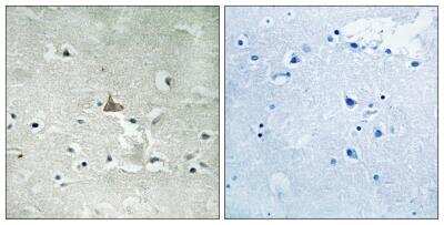
Immunohistochemistry-Paraffin: TrkB [p Tyr706] Antibody [NBP2-54764] - Human brain tissue using Trk B [P-Tyr706/Tyr707) antibody (left) or the same antibody preincubated with blocking peptide (right).
![TrkB [p Tyr707, p Tyr706] Antibody - BSA Free](https://resources.rndsystems.com/images/products/nbp2-54764_rabbit-polyclonal-trkb-p-tyr706-antibody-western-blot-132202615271916.jpg "Western Blot: TrkB [p Tyr707, p Tyr706] Antibody - BSA Free [NBP2-54764] -")
Western Blot: TrkB [p Tyr707, p Tyr706] Antibody - BSA Free [NBP2-54764] -
Effects of the ABA protocol on the protein levels of TrkB, the high affinity receptor for mBDNF, and on its downstream signaling effectors in the amygdala. Rats were exposed to the combination of food restriction and free access to the activity wheel and the analysis of phospho(p)TrkBY706 (A), TrkB receptor (B), pAktS473 (D), Akt (E), pERK2T185-Y187 (G), and ERK2 (H) were measured in the crude membrane fraction in the acute phase of the disease (PND42), and after a 7-day period of body weight recovery (PND49). Pearson’s product–moment correlation (r) analyses and linear regression analyses (R2) between body weight and pTrkBY706 (C), pAktS473 (F), and pERK2T185-Y187 (I) phosphorylation levels of CTRL and ABA rats at both time points are represented. In panel (L), representative immunoblots are shown for pTrkB Y706 (145 kDa), TrkB (145 kDa), pAktS473, Akt, pERK2T185-Y187, ERK2, and beta -Actin (43 kDa). Data are expressed as percentages of CTRL animals. Bar graphs represent the mean +/- SEM from five independent determinations for each experimental group. Unpaired Student’s T test. *p < 0.05, **p < 0.01, ***p < 0.05. CTRL or C, control; ABA or A, activity-based anorexia. Image collected and cropped by CiteAb from the following open publication (https://pubmed.ncbi.nlm.nih.gov/36570702), licensed under a CC-BY license. Not internally tested by Novus Biologicals.Applications for TrkB [p Tyr707, p Tyr706] Antibody - BSA Free
Application
Recommended Usage
Immunohistochemistry
1:10 - 1:200
Immunohistochemistry-Paraffin
1:10 - 1:500
Application Notes
This TrkB [p Tyr706] Antibody is validated for WB from a verified customer review.
Reviewed Applications
Read 1 review rated 3 using NBP2-54764 in the following applications:
Formulation, Preparation, and Storage
Purification
Epitope affinity purified
Formulation
PBS (pH 7.4) (without Mg2+ and Ca2+), 150mM NaCl, 50% Glycerol
Format
BSA Free
Preservative
0.02% Sodium Azide
Concentration
1 mg/ml
Shipping
The product is shipped with polar packs. Upon receipt, store it immediately at the temperature recommended below.
Stability & Storage
Store at -20 °C.
Background: TrkB
Long Name
Neurotrophic Tyrosine Kinase Receptor B
Alternate Names
NTRK2
Gene Symbol
NTRK2
Additional TrkB Products
Product Documents for TrkB [p Tyr707, p Tyr706] Antibody - BSA Free
Certificate of Analysis
To download a Certificate of Analysis, please enter a lot or batch number in the search box below.
Product Specific Notices for TrkB [p Tyr707, p Tyr706] Antibody - BSA Free
This product is for research use only and is not approved for use in humans or in clinical diagnosis. Primary Antibodies are guaranteed for 1 year from date of receipt.
Citations for TrkB [p Tyr707, p Tyr706] Antibody - BSA Free
 Powered by Bioz
Customer Reviews for TrkB [p Tyr707, p Tyr706] Antibody - BSA Free (1)
3 out of 5
1 Customer Rating
Have you used TrkB [p Tyr707, p Tyr706] Antibody - BSA Free?
Submit a review and receive an Amazon gift card!
$25/€18/£15/$25CAN/¥2500 Yen for a review with an image
$10/€7/£6/$10CAN/¥1110 Yen for a review without an image
Submit a review
Customer Images
![TrkB [p Tyr707, p Tyr706] Antibody - BSA Free NBP2-54764](https://resources.rndsystems.com/images/reviews/review_nbp2-54764_46356_0_0_0.jpg)
Showing
1
-
1 of
1 review
Showing All
Filter By:
-
Application: Western BlotSample Tested: Rat brain tissue, Rat brain synaptosomal (P2) fraction and Rat brain lysate (ventral hippocampus)Species: RatVerified Customer | Posted 02/19/2019F1 pTrkBTyr706 mPFC HOMO, P2. 145 kDa F2 pTrkBTyr706 95 kDa F3 total TrkB Primary TrkB ab #4603 Cell signalling F4 beta-Actin F5 pTrkBTyr706 Hipp HOMO 145kDa, 95kDa F6 total TrkB,145kDa and 95kDa F7 beta-ActinI’m writing to inform you about the Western Blot trial that I have done regarding the pTyr706 NBP2-54764; antibody that I bought Order n° 1334 15/11/2018 and that was untested for WB analysis. First of all, in the datasheet that you provide, bands of trkB are expected at 95kDa. However, we know from the literature that trkB receptor is present in two forms: the full length that weights 145 kDa and the truncated one that weights 95 kDa. As you can see from the blots attached, I effectively obtained with your antibody (against the phosphorilated form) and with an antibody against the total form of trkB (cell signaling #4603) two different bands at these specific molecular weights. The test was conducted on rat brain prefrontal cortex tissue extracts (Figure 1-4), and on rat brain hippocampus (Figure 5-6) obtained following the below-mentioned protocol. Prefrontal cortex (Pfc) tissues were processed following a classic protein extraction method done at room temperature, which allow us to obtain aliquots of whole homogenate (HOMO), nuclei (P1), cytosol (S2) and a crude membrane fractions (P2). 1. Pfc tissues were homogenized in a glass-glass potter in a homogenization buffer (0,32 M sucrose, 0,1 mM PMSF, 1 mM Hepes, 0,1 mM EGTA pH 7,4, 1X Complete (protease inhibitor cocktail), 1X phosphatase inhibitor cocktail). From that step we kept an aliquot of homogenate, we sonicated it and maintained at -20°C for following WB analysis. 2. The left homogenate was moved on through the extraction and centrifuged 10 minutes at 4°C at 1000g. 3. The obtained surnatant S1 was again centrifuged 15 minutes at 4°C at 9000g to obtain the P2 and the S2, while the pellet was the P1 fraction that was resuspended in a buffer solution (20 mM Hepes, 0,1 mM DTT (ditiotreitol), 0,1 mM, EGTA pH 7,4, 1X Complete, 1X phosphatase inhibitor cocktail). The obtained P2 was as for the P1 resuspended in the same previous buffer. Protein extracts were separated by polyacrylamide SDS/PAGE and transferred to nitrocellulose membranes. Blots were immunostained overnight at 4°C with the specific primary antibodies (see figures for specifics). After washing, the membranes were incubated for 1 h at room temperature with the appropriate secondary antibody (anti-mouse, anti-rabbit). After secondary antibody incubations, membranes were washed and finally incubated with ECL detection reagent. Western blot-1: - Figure1: blot of pTrkB Tyr706 measured in rat brain extracts of Prefrontal cortex in the whole homogenate (HOMO) and crude synaptosomal fraction (P2). We revealed one band at 145 kDa, molecular weight of Full length TrkB. - Figure2: blot of pTrkB Tyr706 measured in rat brain extracts of Prefrontal cortex in the whole homogenate (HOMO) and crude synaptosomal fraction (P2). We revealed one band at 95 kDa, molecular weight of truncated TrkB. - Figure3: blot of total TrkB measured in rat brain extracts of Prefrontal cortex in the whole homogenate (HOMO) and crude synaptosomal fraction (P2). Primary TrkB antibody (#4603) Cell signalling - Figure4: blot of beta-Actin (Molecular weight 43 kDa) - Prefrontal cortex Western blot-2: - Figure5: blot of pTrkB Tyr706 measured in rat brain extracts of hippocampus in the whole homogenate (HOMO). We revealed two bands: one at 145 kDa molecular weight of the TrkB full length and one at 95 kDa (truncated TrkB). - Figure6: blot of total TrkB measured in rat brain extracts of hippocampus in the whole homogenate (HOMO). We revealed two bands: one at 145 kDa molecular weight of the TrkB full length and one at 95 kDa (truncated TrkB). - Figure7: blot of beta-Actin (Molecular weight 43 kDa) - Hippocampus
![TrkB [p Tyr707, p Tyr706] Antibody - BSA Free NBP2-54764](data:image/png;base64,R0lGODlhAQABAAD/ACwAAAAAAQABAAACADs=)
There are no reviews that match your criteria.
Protocols
Find general support by application which include: protocols, troubleshooting, illustrated assays, videos and webinars.
- Antigen Retrieval Protocol (PIER)
- Antigen Retrieval for Frozen Sections Protocol
- Appropriate Fixation of IHC/ICC Samples
- Cellular Response to Hypoxia Protocols
- Chromogenic IHC Staining of Formalin-Fixed Paraffin-Embedded (FFPE) Tissue Protocol
- Chromogenic Immunohistochemistry Staining of Frozen Tissue
- ClariTSA™ Fluorophore Kits
- Detection & Visualization of Antibody Binding
- Fluorescent IHC Staining of Frozen Tissue Protocol
- Graphic Protocol for Heat-induced Epitope Retrieval
- Graphic Protocol for the Preparation and Fluorescent IHC Staining of Frozen Tissue Sections
- Graphic Protocol for the Preparation and Fluorescent IHC Staining of Paraffin-embedded Tissue Sections
- Graphic Protocol for the Preparation of Gelatin-coated Slides for Histological Tissue Sections
- IHC Sample Preparation (Frozen sections vs Paraffin)
- Immunofluorescent IHC Staining of Formalin-Fixed Paraffin-Embedded (FFPE) Tissue Protocol
- Immunohistochemistry (IHC) and Immunocytochemistry (ICC) Protocols
- Immunohistochemistry Frozen Troubleshooting
- Immunohistochemistry Paraffin Troubleshooting
- Preparing Samples for IHC/ICC Experiments
- Preventing Non-Specific Staining (Non-Specific Binding)
- Primary Antibody Selection & Optimization
- Protocol for Heat-Induced Epitope Retrieval (HIER)
- Protocol for Making a 4% Formaldehyde Solution in PBS
- Protocol for VisUCyte™ HRP Polymer Detection Reagent
- Protocol for the Preparation & Fixation of Cells on Coverslips
- Protocol for the Preparation and Chromogenic IHC Staining of Frozen Tissue Sections
- Protocol for the Preparation and Chromogenic IHC Staining of Frozen Tissue Sections - Graphic
- Protocol for the Preparation and Chromogenic IHC Staining of Paraffin-embedded Tissue Sections
- Protocol for the Preparation and Chromogenic IHC Staining of Paraffin-embedded Tissue Sections - Graphic
- Protocol for the Preparation and Fluorescent IHC Staining of Frozen Tissue Sections
- Protocol for the Preparation and Fluorescent IHC Staining of Paraffin-embedded Tissue Sections
- Protocol for the Preparation of Gelatin-coated Slides for Histological Tissue Sections
- R&D Systems Quality Control Western Blot Protocol
- TUNEL and Active Caspase-3 Detection by IHC/ICC Protocol
- The Importance of IHC/ICC Controls
- Troubleshooting Guide: Immunohistochemistry
- Troubleshooting Guide: Western Blot Figures
- Western Blot Conditions
- Western Blot Protocol
- Western Blot Protocol for Cell Lysates
- Western Blot Troubleshooting
- Western Blot Troubleshooting Guide
- View all Protocols, Troubleshooting, Illustrated assays and Webinars
Loading...
Associated Pathways

