TSG101 Antibody - BSA Free
Novus Biologicals | Catalog # NBP1-80659
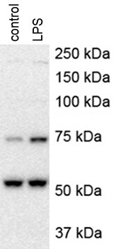
![Western Blot: TSG101 Antibody [NBP1-80659]](https://resources.rndsystems.com/images/products/TSG101-Antibody-Western-Blot-NBP1-80659-img0011.jpg "Western Blot: TSG101 Antibody [NBP1-80659]")
Loading...
Key Product Details
Validated by
Knockout/Knockdown
Species Reactivity
Validated:
Human, Mouse, Rat
Cited:
Human, Mouse, Rat
Applications
Validated:
Immunohistochemistry, Immunohistochemistry-Paraffin, Western Blot, Immunocytochemistry/ Immunofluorescence, Knockdown Validated
Cited:
Western Blot, Immunocytochemistry/ Immunofluorescence
Label
Unconjugated
Antibody Source
Polyclonal Rabbit IgG
Format
BSA Free
Loading...
Product Specifications
Immunogen
This antibody was developed against Recombinant Protein corresponding to amino acids: RMKEEMDRAQAELNALKRTEEDLKKGHQKLEEMVTRLDQEVAEVDKNIELLKKKDEELSSALEKMENQSENNDIDEVIIPTAPLYKQILNLYAEENAIEDTIFYLGEALRRGVIDLDVFLKHVRLLS
Marker
Exosome Marker
Clonality
Polyclonal
Host
Rabbit
Isotype
IgG
Scientific Data Images for TSG101 Antibody - BSA Free
![Immunocytochemistry/ Immunofluorescence: TSG101 Antibody [NBP1-80659]](https://resources.rndsystems.com/images/products/TSG101-Antibody-Immunocytochemistry-Immunofluorescence-NBP1-80659-img0009.jpg "Immunocytochemistry/ Immunofluorescence: TSG101 Antibody [NBP1-80659]")
Immunocytochemistry/ Immunofluorescence: TSG101 Antibody [NBP1-80659]
Immunocytochemistry/Immunofluorescence: TSG101 Antibody [NBP1-80659] - Staining of human cell line A-431 shows localization to plasma membrane & cytosol. Antibody staining is shown in green.![Immunohistochemistry-Paraffin: TSG101 Antibody [NBP1-80659]](https://resources.rndsystems.com/images/products/TSG101-Antibody-Immunohistochemistry-Paraffin-NBP1-80659-img0015.jpg "Immunohistochemistry-Paraffin: TSG101 Antibody [NBP1-80659]")
Immunohistochemistry-Paraffin: TSG101 Antibody [NBP1-80659]
Immunohistochemistry-Paraffin: TSG101 Antibody [NBP1-80659] - Staining of human fallopian tube shows strong cytoplasmic positivity in glandular cells.![Western Blot: TSG101 Antibody [NBP1-80659]](https://resources.rndsystems.com/images/products/TSG101-Antibody-Western-Blot-NBP1-80659-img0008.jpg "Western Blot: TSG101 Antibody [NBP1-80659]")
Western Blot: TSG101 Antibody [NBP1-80659]
Western Blot: TSG101 Antibody [NBP1-80659] - Analysis in mouse cell line NIH-3T3 and rat cell line NBT-II.![Immunohistochemistry-Paraffin: TSG101 Antibody [NBP1-80659]](https://resources.rndsystems.com/images/products/TSG101-Antibody-Immunohistochemistry-Paraffin-NBP1-80659-img0012.jpg "Immunohistochemistry-Paraffin: TSG101 Antibody [NBP1-80659]")
Immunohistochemistry-Paraffin: TSG101 Antibody [NBP1-80659]
Immunohistochemistry-Paraffin: TSG101 Antibody [NBP1-80659] - Staining of human prostate shows moderate cytoplasmic positivity in glandular and in smooth muscle cells.![Immunohistochemistry-Paraffin: TSG101 Antibody [NBP1-80659]](https://resources.rndsystems.com/images/products/TSG101-Antibody-Immunohistochemistry-Paraffin-NBP1-80659-img0013.jpg "Immunohistochemistry-Paraffin: TSG101 Antibody [NBP1-80659]")
Immunohistochemistry-Paraffin: TSG101 Antibody [NBP1-80659]
Immunohistochemistry-Paraffin: TSG101 Antibody [NBP1-80659] - Staining of human cerebral cortex shows moderate cytoplasmic positivity in neurons.![Immunohistochemistry-Paraffin: TSG101 Antibody [NBP1-80659]](https://resources.rndsystems.com/images/products/TSG101-Antibody-Immunohistochemistry-Paraffin-NBP1-80659-img0014.jpg "Immunohistochemistry-Paraffin: TSG101 Antibody [NBP1-80659]")
Immunohistochemistry-Paraffin: TSG101 Antibody [NBP1-80659]
Immunohistochemistry-Paraffin: TSG101 Antibody [NBP1-80659] - Staining of human small intestine shows strong cytoplasmic positivity in glandular cells.Applications for TSG101 Antibody - BSA Free
Application
Recommended Usage
Immunocytochemistry/ Immunofluorescence
0.25-2 ug/ml
Immunohistochemistry
1:200 - 1:500
Immunohistochemistry-Paraffin
1:200 - 1:500
Western Blot
0.04-0.4 ug/ml
Application Notes
IHC-Paraffin, HIER pH 6 retrieval is recommended. ICC/IF, Fixation Permeabilization, Use PFA/Triton X-100.
Reviewed Applications
Read 2 reviews rated 4 using NBP1-80659 in the following applications:
Formulation, Preparation, and Storage
Purification
Affinity purified
Formulation
PBS (pH 7.2) and 40% Glycerol
Format
BSA Free
Preservative
0.02% Sodium Azide
Concentration
Concentrations vary lot to lot. See vial label for concentration. If unlisted please contact technical services.
Shipping
The product is shipped with polar packs. Upon receipt, store it immediately at the temperature recommended below.
Stability & Storage
Store at 4C short term. Aliquot and store at -20C long term. Avoid freeze-thaw cycles.
Background: TSG101
Upon its initial discovery, TSG101 was recognized as a tumor suppressor protein due to the identification of deletions within the TSG101 gene in human breast carcinomas. However, re-examination of the initial findings argued against this function and supported that TSG101 promotes tumorigenesis (2). In agreement with this role, TSG101 expression is upregulated in several types of cancer including breast, ovarian, and colorectal carcinoma.
References
1. Schmidt, O., & Teis, D. (2012). The ESCRT machinery. Current Biology. https://doi.org/10.1016/j.cub.2012.01.028
2. Jiang, Y., Ou, Y., & Cheng, X. (2013). Role of TSG101 in cancer. Frontiers in Bioscience. https://doi.org/10.2741/4099
3. Balut, C. M., Gao, Y., Murray, S. A., Thibodeau, P. H., & Devor, D. C. (2010). ESCRT-dependent targeting of plasma membrane localized KCa3.1 to the lysosomes. American Journal of Physiology - Cell Physiology. https://doi.org/10.1152/ajpcell.00120.2010
4. Su, V., & Lau, A. F. (2014). Connexins: Mechanisms regulating protein levels and intercellular communication. FEBS Letters. https://doi.org/10.1016/j.febslet.2014.01.013
Long Name
Tumor Susceptibility 101
Alternate Names
TSG10, VPS23
Entrez Gene IDs
7251 (Human)
Gene Symbol
TSG101
UniProt
Additional TSG101 Products
Product Documents for TSG101 Antibody - BSA Free
Certificate of Analysis
To download a Certificate of Analysis, please enter a lot or batch number in the search box below.
Product Specific Notices for TSG101 Antibody - BSA Free
This product is for research use only and is not approved for use in humans or in clinical diagnosis. Primary Antibodies are guaranteed for 1 year from date of receipt.
Citations for TSG101 Antibody - BSA Free
 Powered by Bioz
Customer Reviews for TSG101 Antibody - BSA Free (2)
4 out of 5
2 Customer Ratings
Have you used TSG101 Antibody - BSA Free?
Submit a review and receive an Amazon gift card!
$25/€18/£15/$25CAN/¥2500 Yen for a review with an image
$10/€7/£6/$10CAN/¥1110 Yen for a review without an image
Submit a review
Customer Images

Showing
1
-
2 of
2 reviews
Showing All
Filter By:
-
Application: Western BlotSample Tested: Rat astrocytesSpecies: RatVerified Customer | Posted 01/17/2018

-
Application: Western BlotSample Tested: Primary mouse hepatocytesSpecies: MouseVerified Customer | Posted 12/08/2017mouse hepatocytes were treated with 100 ng/ml LPS for 4 hoursmouse hepatocytes were treated with 100 ng/ml LPS for 4 hours
There are no reviews that match your criteria.
Protocols
Find general support by application which include: protocols, troubleshooting, illustrated assays, videos and webinars.
- Antigen Retrieval Protocol (PIER)
- Antigen Retrieval for Frozen Sections Protocol
- Appropriate Fixation of IHC/ICC Samples
- Cellular Response to Hypoxia Protocols
- Chromogenic IHC Staining of Formalin-Fixed Paraffin-Embedded (FFPE) Tissue Protocol
- Chromogenic Immunohistochemistry Staining of Frozen Tissue
- ClariTSA™ Fluorophore Kits
- Detection & Visualization of Antibody Binding
- Fluorescent IHC Staining of Frozen Tissue Protocol
- Graphic Protocol for Heat-induced Epitope Retrieval
- Graphic Protocol for the Preparation and Fluorescent IHC Staining of Frozen Tissue Sections
- Graphic Protocol for the Preparation and Fluorescent IHC Staining of Paraffin-embedded Tissue Sections
- Graphic Protocol for the Preparation of Gelatin-coated Slides for Histological Tissue Sections
- ICC Cell Smear Protocol for Suspension Cells
- ICC Immunocytochemistry Protocol Videos
- ICC for Adherent Cells
- IHC Sample Preparation (Frozen sections vs Paraffin)
- Immunocytochemistry (ICC) Protocol
- Immunocytochemistry Troubleshooting
- Immunofluorescence of Organoids Embedded in Cultrex Basement Membrane Extract
- Immunofluorescent IHC Staining of Formalin-Fixed Paraffin-Embedded (FFPE) Tissue Protocol
- Immunohistochemistry (IHC) and Immunocytochemistry (ICC) Protocols
- Immunohistochemistry Frozen Troubleshooting
- Immunohistochemistry Paraffin Troubleshooting
- Preparing Samples for IHC/ICC Experiments
- Preventing Non-Specific Staining (Non-Specific Binding)
- Primary Antibody Selection & Optimization
- Protocol for Heat-Induced Epitope Retrieval (HIER)
- Protocol for Making a 4% Formaldehyde Solution in PBS
- Protocol for VisUCyte™ HRP Polymer Detection Reagent
- Protocol for the Fluorescent ICC Staining of Cell Smears - Graphic
- Protocol for the Fluorescent ICC Staining of Cultured Cells on Coverslips - Graphic
- Protocol for the Preparation & Fixation of Cells on Coverslips
- Protocol for the Preparation and Chromogenic IHC Staining of Frozen Tissue Sections
- Protocol for the Preparation and Chromogenic IHC Staining of Frozen Tissue Sections - Graphic
- Protocol for the Preparation and Chromogenic IHC Staining of Paraffin-embedded Tissue Sections
- Protocol for the Preparation and Chromogenic IHC Staining of Paraffin-embedded Tissue Sections - Graphic
- Protocol for the Preparation and Fluorescent ICC Staining of Cells on Coverslips
- Protocol for the Preparation and Fluorescent ICC Staining of Non-adherent Cells
- Protocol for the Preparation and Fluorescent ICC Staining of Stem Cells on Coverslips
- Protocol for the Preparation and Fluorescent IHC Staining of Frozen Tissue Sections
- Protocol for the Preparation and Fluorescent IHC Staining of Paraffin-embedded Tissue Sections
- Protocol for the Preparation of Gelatin-coated Slides for Histological Tissue Sections
- Protocol for the Preparation of a Cell Smear for Non-adherent Cell ICC - Graphic
- R&D Systems Quality Control Western Blot Protocol
- TUNEL and Active Caspase-3 Detection by IHC/ICC Protocol
- The Importance of IHC/ICC Controls
- Troubleshooting Guide: Immunohistochemistry
- Troubleshooting Guide: Western Blot Figures
- Western Blot Conditions
- Western Blot Protocol
- Western Blot Protocol for Cell Lysates
- Western Blot Troubleshooting
- Western Blot Troubleshooting Guide
- View all Protocols, Troubleshooting, Illustrated assays and Webinars
Loading...