TXNIP Antibody (JY2) - Azide and BSA Free
Novus Biologicals | Catalog # NBP1-54578
Key Product Details
Validated by
Biological Validation
Species Reactivity
Validated:
Human, Mouse, Rat
Cited:
Human, Mouse, Rat, Amphibian
Applications
Validated:
Knockout Validated, Immunohistochemistry, Immunohistochemistry-Paraffin, Immunohistochemistry-Frozen, Western Blot, Flow Cytometry, Simple Western, Immunoprecipitation, Proximity Ligation Assay, CyTOF-ready
Cited:
Knockout Validated, Immunohistochemistry-Paraffin, Western Blot, Simple Western, Immunoprecipitation, Proximity Ligation Assay, IF/IHC, Knockdown Validated
Label
Unconjugated
Antibody Source
Monoclonal Mouse IgG1 Clone # JY2
Format
Azide and BSA Free
Loading...
Product Specifications
Immunogen
Human recombinant TXNIP
Reactivity Notes
Use in Mouse reported in scientific literature (PMID:34576095). Please note that this antibody is reactive to Mouse and derived from the same host, Mouse. Additional Mouse on Mouse blocking steps may be required for IHC and ICC experiments. Please contact Technical Support for more information.
Clonality
Monoclonal
Host
Mouse
Isotype
IgG1
Scientific Data Images for TXNIP Antibody (JY2) - Azide and BSA Free
![Immunohistochemistry-Frozen: TXNIP Antibody (JY2) - Azide and BSA Free [NBP1-54578]](https://resources.rndsystems.com/images/products/TXNIP-Antibody-JY2-Immunohistochemistry-Frozen-NBP1-54578-img0007.jpg "Immunohistochemistry-Frozen: TXNIP Antibody (JY2) - Azide and BSA Free [NBP1-54578]")
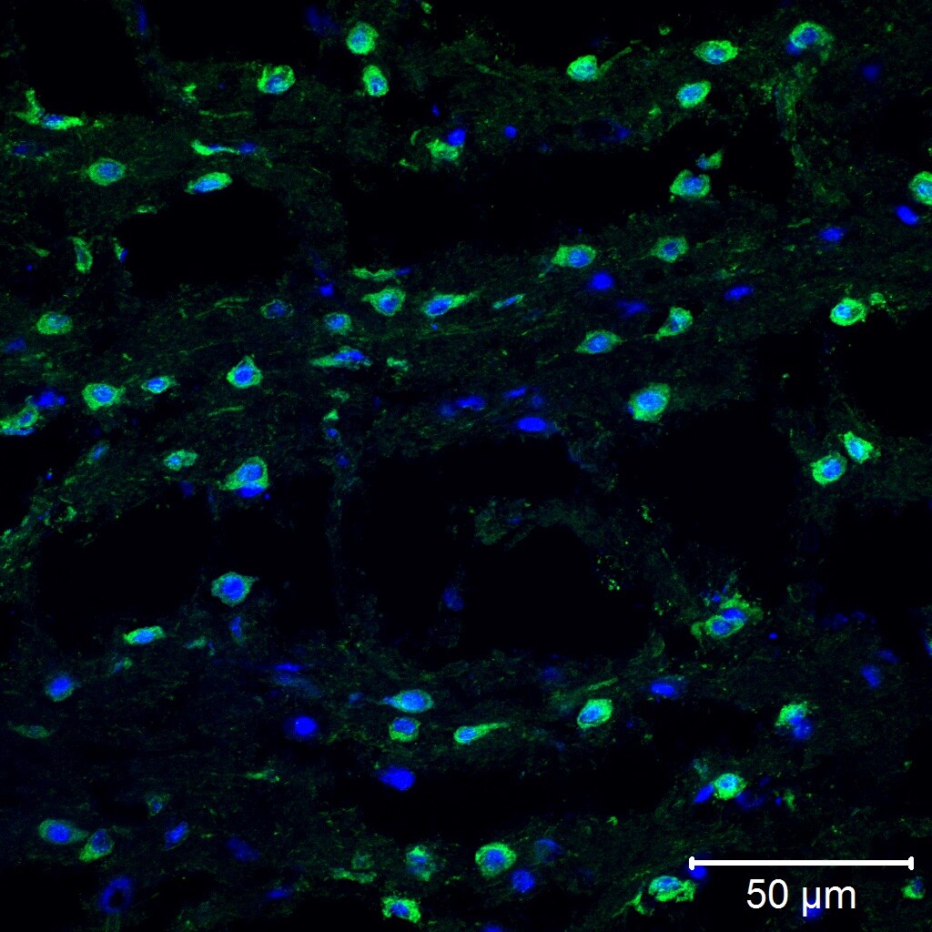
Immunohistochemistry-Frozen: TXNIP Antibody (JY2) - Azide and BSA Free [NBP1-54578]
Immunohistochemistry-Frozen: TXNIP Antibody (JY2) [NBP1-54578] - TXNIP in mouse brain. Antibody dilution: 1:100, incubated overnight in 5%BSA + 0.2% Triton X 100. Image from verified customer review.![Western Blot: TXNIP Antibody (JY2)Azide and BSA Free [NBP1-54578]](https://resources.rndsystems.com/images/products/TXNIP-Antibody-JY2-Western-Blot-NBP1-54578-img0001.jpg "Western Blot: TXNIP Antibody (JY2)Azide and BSA Free [NBP1-54578]")
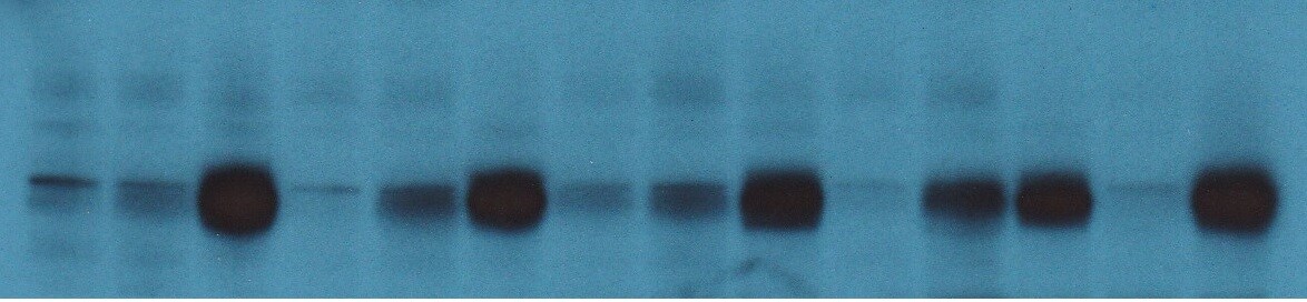
Western Blot: TXNIP Antibody (JY2)Azide and BSA Free [NBP1-54578]
Western Blot: TXNIP Antibody (JY2) [NBP1-54578] - TXNIP expression in Raji (1), K562 (2), KG1 (3), MRC5 (4), IC2Tr (5), HEL (6), P19 (7) and WR19L (8).![Western Blot: TXNIP Antibody (JY2)Azide and BSA Free [NBP1-54578]](https://resources.rndsystems.com/images/products/TXNIP-Antibody-JY2-Azide-and-BSA-Free-Western-Blot-NBP1-54578-img0011.jpg "Western Blot: TXNIP Antibody (JY2)Azide and BSA Free [NBP1-54578]")
![Flow Cytometry: TXNIP Antibody (JY2) - Azide and BSA Free [NBP1-54578]](https://resources.rndsystems.com/images/products/TXNIP-Antibody-JY2-Azide-and-BSA-Free-Flow-Cytometry-NBP1-54578-img0010.jpg "Flow Cytometry: TXNIP Antibody (JY2) - Azide and BSA Free [NBP1-54578]")
Flow Cytometry: TXNIP Antibody (JY2) - Azide and BSA Free [NBP1-54578]
Flow Cytometry: TXNIP Antibody (JY2) - Azide and BSA Free [NBP1-54578] - An intracellular stain was performed on THP-1 cells with TXNIP Antibody (JY2) NBP1-54578AF488 (blue) and a matched isotype control (orange). Cells were fixed with 4% PFA and then permeabilized with 0.1% saponin. Cells were incubated in an antibody dilution of 5 ug/mL for 30 minutes at room temperature. Both antibodies were conjugated to Alexa Fluor 488.![Western Blot: TXNIP Antibody (JY2)Azide and BSA Free [NBP1-54578]](https://resources.rndsystems.com/images/products/TXNIP-Antibody-JY2-Western-Blot-NBP1-54578-img0006.jpg "Western Blot: TXNIP Antibody (JY2)Azide and BSA Free [NBP1-54578]")
Western Blot: TXNIP Antibody (JY2)Azide and BSA Free [NBP1-54578]
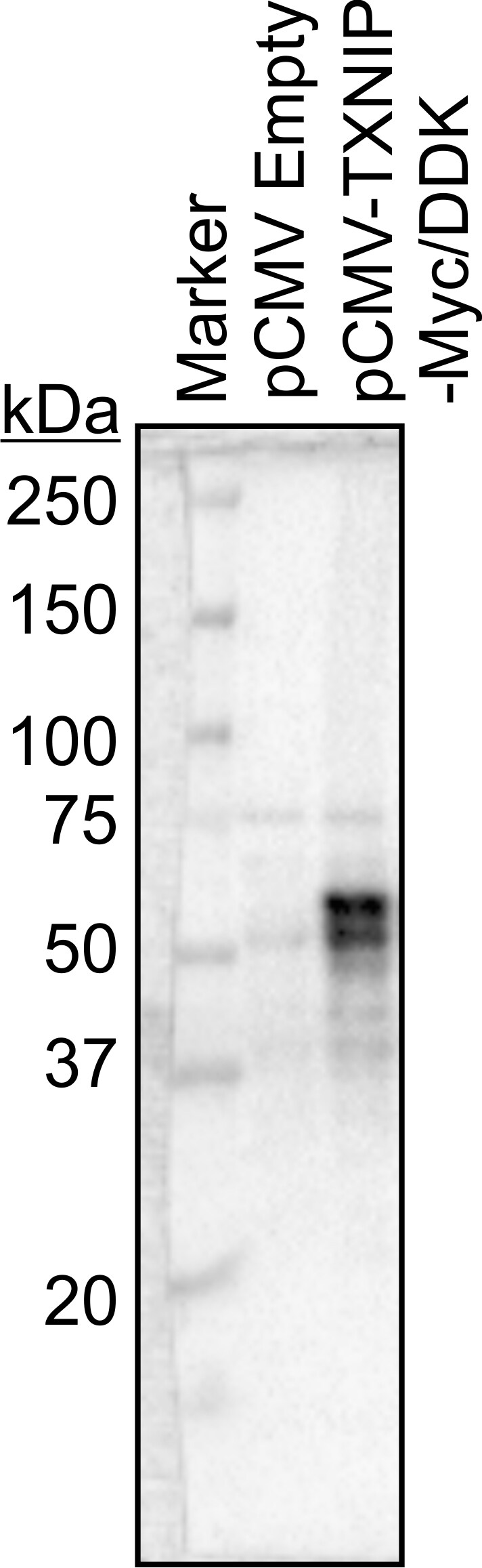
Western Blot: TXNIP Antibody (JY2) [NBP1-54578] - Hepa1 lysates probed with anti-TXNIP (JY2) at 1:2K. Cells were transfected with either empty vector or mouse TXNIP expression vector (MYC/DDK tagged). 30ug protein per lane. Image from verified customer review.![Western Blot: TXNIP Antibody (JY2)Azide and BSA Free [NBP1-54578]](https://resources.rndsystems.com/images/products/TXNIP-Antibody-JY2-Western-Blot-NBP1-54578-img0009.jpg "Western Blot: TXNIP Antibody (JY2)Azide and BSA Free [NBP1-54578]")
Western Blot: TXNIP Antibody (JY2)Azide and BSA Free [NBP1-54578]
Western Blot: TXNIP Antibody (JY2) [NBP1-54578] - AMPK mediates anti-pyroptotic effects of Exendin-4. (a) ROS determination by FACS. The left arrow indicated the ROS-negative population and the right pointed the positive. (b) Transcription activity of TXNIP in cardiomyocytes. (c) Western blot. (d) RNA silencing of TXNIP in cardiomyocytes. (e) IL-1 ELISA with TXNIP RNAi. (f) Caspase-1 activity assay with TXNIP RNAi. (g) Quantification of pAMPK/AMPK with CC treatment. (h) IL-1 ELISA with CC treatment. (i) Caspase-1 activity assay with CC treatment. Values are the mean SEM of 3 samples per group.0.05,0.01,0.005, and0.001. Exendin-4 Protects against Hyperglycemia-Induced Cardiomyocyte Pyroptosis via the AMPK-TXNIP Pathway. J Diabetes Res (2019)![Immunohistochemistry-Paraffin: TXNIP Antibody (JY2) - Azide and BSA Free [NBP1-54578]](https://resources.rndsystems.com/images/products/TXNIP-Antibody-JY2-Immunohistochemistry-Paraffin-NBP1-54578-img0003.jpg "Immunohistochemistry-Paraffin: TXNIP Antibody (JY2) - Azide and BSA Free [NBP1-54578]")
Immunohistochemistry-Paraffin: TXNIP Antibody (JY2) - Azide and BSA Free [NBP1-54578]
Immunohistochemistry-Paraffin: TXNIP Antibody (JY2) [NBP1-54578] - Formalin fixed and paraffin embedded tissue section of mouse kidney using TXNIP antibody (clone JY2) at 1:250 dilution. The signal was developed using HRP-conjugated secondary antibody and DAB reagent which followed counterstaining of the cell nuclei with hematoxylin. This TXNIP antibody generated a diffused cytoplasmic signal in the cells of various tubules and glomeruli.![Immunohistochemistry-Paraffin: TXNIP Antibody (JY2) - Azide and BSA Free [NBP1-54578]](https://resources.rndsystems.com/images/products/TXNIP-Antibody-JY2-Immunohistochemistry-Paraffin-NBP1-54578-img0005.jpg "Immunohistochemistry-Paraffin: TXNIP Antibody (JY2) - Azide and BSA Free [NBP1-54578]")
Immunohistochemistry-Paraffin: TXNIP Antibody (JY2) - Azide and BSA Free [NBP1-54578]
Immunohistochemistry-Paraffin: TXNIP Antibody (JY2) [NBP1-54578] - Formalin fixed and paraffin embedded tissue section of mouse kidney using TXNIP antibody (clone JY2) at 1:100 dilution. The signal was developed using HRP-conjugated secondary antibody and DAB reagent which followed counterstaining of the cell nuclei with hematoxylin. This TXNIP antibody generated a diffused cytoplasmic signal in the cells of various tubules with negligible staining in glomeruli.![Flow Cytometry: TXNIP Antibody (JY2) - Azide and BSA Free [NBP1-54578]](https://resources.rndsystems.com/images/products/TXNIP-Antibody-JY2-Flow-Cytometry-NBP1-54578-img0008.jpg "Flow Cytometry: TXNIP Antibody (JY2) - Azide and BSA Free [NBP1-54578]")
Flow Cytometry: TXNIP Antibody (JY2) - Azide and BSA Free [NBP1-54578]
Flow Cytometry: TXNIP Antibody (JY2) [NBP1-54578] - An intracellular stain was performed on RH-30 cells with TXNIP (JY2) Antibody NBP1-54578F (blue) and a matched isotype control (orange). Cells were fixed with 4% PFA and then permeabilized with 0.1% saponin. Cells were incubated in an antibody dilution of 5 ug/mL for 30 minutes at room temperature. Both antibodies were conjugated to FITC.![Immunoprecipitation: TXNIP Antibody (JY2) - Azide and BSA Free [NBP1-54578]](https://resources.rndsystems.com/images/products/TXNIP-Antibody-JY2-Immunoprecipitation-NBP1-54578-img0002.jpg "Immunoprecipitation: TXNIP Antibody (JY2) - Azide and BSA Free [NBP1-54578]")
Immunoprecipitation: TXNIP Antibody (JY2) - Azide and BSA Free [NBP1-54578]
Immunoprecipitation: TXNIP Antibody (JY2) [NBP1-54578] - Txnip/VDUP1 from Raji with NBP1-54578 (1) or mouse IgG1 (2). - Azide and BSA Free [NBP1-54578] -")
Western Blot: TXNIP Antibody (JY2) - Azide and BSA Free [NBP1-54578] -
Western Blot: TXNIP Antibody (JY2) - Azide and BSA Free [NBP1-54578] - AMPK mediates anti-pyroptotic effects of Exendin-4. (a) ROS determination by FACS. The left arrow indicated the ROS-negative population & the right pointed the positive. (b) Transcription activity of TXNIP in cardiomyocytes. (c) Western blot. (d) RNA silencing of TXNIP in cardiomyocytes. (e) IL-1 beta ELISA with TXNIP RNAi. (f) Caspase-1 activity assay with TXNIP RNAi. (g) Quantification of pAMPK/AMPK with CC treatment. (h) IL-1 beta ELISA with CC treatment. (i) Caspase-1 activity assay with CC treatment. Values are the mean ± SEM of 3 samples per group. ∗p < 0.05, ∗∗p < 0.01, ∗∗∗p < 0.005, & ∗∗∗∗p < 0.001. Image collected & cropped by CiteAb from the following publication (https://pubmed.ncbi.nlm.nih.gov/31886288), licensed under a CC-BY license. Not internally tested by Novus Biologicals. - Azide and BSA Free [NBP1-54578] -")
Simple Western: TXNIP Antibody (JY2) - Azide and BSA Free [NBP1-54578] -
Proinflammatory cytokine (IL-6) and TXNIP expression and TUNEL assay for STZ-induced mice treated with Verapamil (V) and R-Vera (RV).(A) STZ-induced mice administered 100 mg/kg/day racemic or R-Vera exhibited significantly reduced serum IL-6 expression compared to vehicle mice whereas those administered 50 mg/kg/day R-Vera did not. **P < 0.01 indicates significant difference from vehicle using one-way ANOVA. N = 8 in each group. (B) STZ-induced mice showed highly upregulated islet tissue TXNIP compared to normal C57BL/6J mice. TXNIP was downregulated after racemic or R-Vera administration compared to vehicle. #P < 0.05 indicated significant difference between normal and vehicle; *P < 0.05 indicated significant difference to vehicle using one-way ANOVA. N = 8 in each group. (C) beta -cell apoptosis assessed using TUNEL staining. Deep staining spots in images are apoptotic beta -cells (200×). (D) TUNEL-positive cell counts show that STZ-induced mice administered 100 mg/kg/day R-Vera had significantly reduce beta -cell apoptosis. *P < 0.05 indicated a significant difference between vehicle and RV-100 using one-way ANOVA. The sample numbers of 5, 7, 5, and 9 indicate the section numbers from the Vehicle, V-100, RV-100, and RV-50 groups, respectively (rather than the number of mice). Each data point represents the percentage of TUNEL–positive cells per slide in each group. Image collected and cropped by CiteAb from the following open publication (https://pubmed.ncbi.nlm.nih.gov/34358247), licensed under a CC-BY license. Not internally tested by Novus Biologicals.Applications for TXNIP Antibody (JY2) - Azide and BSA Free
Application
Recommended Usage
Flow Cytometry
2-5 ug/0.1x10^6 cells
Immunohistochemistry-Frozen
reported by customer review
Immunohistochemistry-Paraffin
1:100-1:500
Immunoprecipitation
2ug/200ul of cell extract from 5x10^6 cells
Knockout Validated
reported in scientific literature (Brocker et al)
Simple Western
1:500
Western Blot
1 ug/ml
Reviewed Applications
Read 3 reviews rated 4.3 using NBP1-54578 in the following applications:
Flow Cytometry Panel Builder
Bio-Techne Knows Flow Cytometry
Save time and reduce costly mistakes by quickly finding compatible reagents using the Panel Builder Tool.
Advanced Features
- Spectra Viewer - Custom analysis of spectra from multiple fluorochromes
- Spillover Popups - Visualize the spectra of individual fluorochromes
- Antigen Density Selector - Match fluorochrome brightness with antigen density

Formulation, Preparation, and Storage
Purification
Protein A or G purified
Formulation
PBS
Format
Azide and BSA Free
Preservative
No Preservative
Concentration
1.0 mg/ml
Shipping
The product is shipped with polar packs. Upon receipt, store it immediately at the temperature recommended below.
Stability & Storage
Store at -20C. Avoid freeze-thaw cycles.
Background: TXNIP
Alternate Names
HHCPA78, THIF, thioredoxin binding protein 2, thioredoxin interacting protein, Thioredoxin-binding protein 2, thioredoxin-interacting protein, upregulated by 1,25-dihydroxyvitamin D-3, VDUP1EST01027, Vitamin D3 up-regulated protein 1
Gene Symbol
TXNIP
UniProt
Additional TXNIP Products
Product Documents for TXNIP Antibody (JY2) - Azide and BSA Free
Certificate of Analysis
To download a Certificate of Analysis, please enter a lot or batch number in the search box below.
Product Specific Notices for TXNIP Antibody (JY2) - Azide and BSA Free
This product is for research use only and is not approved for use in humans or in clinical diagnosis. Primary Antibodies are guaranteed for 1 year from date of receipt.
Citations for TXNIP Antibody (JY2) - Azide and BSA Free
 Powered by Bioz
Customer Reviews for TXNIP Antibody (JY2) - Azide and BSA Free (3)
4.3 out of 5
3 Customer Ratings
Have you used TXNIP Antibody (JY2) - Azide and BSA Free?
Submit a review and receive an Amazon gift card!
$25/€18/£15/$25CAN/¥2500 Yen for a review with an image
$10/€7/£6/$10CAN/¥1110 Yen for a review without an image
Submit a review
Customer Images
Showing
1
-
3 of
3 reviews
Showing All
Filter By:
-
Application: Immunohistochemistry-FrozenSample Tested: Mouse brainSpecies: MouseVerified Customer | Posted 02/28/2019TXNIP in mouse brainAntibody dilution: 1:100, incubated overnight in 5%BSA + 0.2% Triton X 100.

-
Application: Western BlotSample Tested: Mouse brain cortex whole lysate and Mouse brainSpecies: mouse brain and MouseVerified Customer | Posted 08/22/2018TXNIP expression in mouse brain
-
Application: Western BlotSample Tested: Hepa1 cells transfected with mouse TXNIP expression vector and Hepa1 whole cell lysatesSpecies: MouseVerified Customer | Posted 05/22/2018Hepa1 lysates probed with anti-TXNIP (JY2) @ 1:2K. Cells were transfected with either empty vector or mouse TXNIP expression vector (MYC/DDK tagged).Hepa1 lysates probed with anti-TXNIP (JY2) @ 1:2K. Cells were transfected with either empty vector or mouse TXNIP expression vector (MYC/DDK tagged). Loaded 30ug protein per lane.
There are no reviews that match your criteria.
Protocols
Find general support by application which include: protocols, troubleshooting, illustrated assays, videos and webinars.
- 7-Amino Actinomycin D (7-AAD) Cell Viability Flow Cytometry Protocol
- Antigen Retrieval Protocol (PIER)
- Antigen Retrieval for Frozen Sections Protocol
- Appropriate Fixation of IHC/ICC Samples
- Cellular Response to Hypoxia Protocols
- Chromogenic IHC Staining of Formalin-Fixed Paraffin-Embedded (FFPE) Tissue Protocol
- Chromogenic Immunohistochemistry Staining of Frozen Tissue
- ClariTSA™ Fluorophore Kits
- Detection & Visualization of Antibody Binding
- Extracellular Membrane Flow Cytometry Protocol
- Flow Cytometry Protocol for Cell Surface Markers
- Flow Cytometry Protocol for Staining Membrane Associated Proteins
- Flow Cytometry Staining Protocols
- Flow Cytometry Troubleshooting Guide
- Fluorescent IHC Staining of Frozen Tissue Protocol
- Graphic Protocol for Heat-induced Epitope Retrieval
- Graphic Protocol for the Preparation and Fluorescent IHC Staining of Frozen Tissue Sections
- Graphic Protocol for the Preparation and Fluorescent IHC Staining of Paraffin-embedded Tissue Sections
- Graphic Protocol for the Preparation of Gelatin-coated Slides for Histological Tissue Sections
- IHC Sample Preparation (Frozen sections vs Paraffin)
- Immunofluorescent IHC Staining of Formalin-Fixed Paraffin-Embedded (FFPE) Tissue Protocol
- Immunohistochemistry (IHC) and Immunocytochemistry (ICC) Protocols
- Immunohistochemistry Frozen Troubleshooting
- Immunohistochemistry Paraffin Troubleshooting
- Immunoprecipitation Protocol
- Intracellular Flow Cytometry Protocol Using Alcohol (Methanol)
- Intracellular Flow Cytometry Protocol Using Detergents
- Intracellular Nuclear Staining Flow Cytometry Protocol Using Detergents
- Intracellular Staining Flow Cytometry Protocol Using Alcohol Permeabilization
- Intracellular Staining Flow Cytometry Protocol Using Detergents to Permeabilize Cells
- Preparing Samples for IHC/ICC Experiments
- Preventing Non-Specific Staining (Non-Specific Binding)
- Primary Antibody Selection & Optimization
- Propidium Iodide Cell Viability Flow Cytometry Protocol
- Protocol for Heat-Induced Epitope Retrieval (HIER)
- Protocol for Liperfluo
- Protocol for Making a 4% Formaldehyde Solution in PBS
- Protocol for VisUCyte™ HRP Polymer Detection Reagent
- Protocol for the Characterization of Human Th22 Cells
- Protocol for the Characterization of Human Th9 Cells
- Protocol for the Preparation & Fixation of Cells on Coverslips
- Protocol for the Preparation and Chromogenic IHC Staining of Frozen Tissue Sections
- Protocol for the Preparation and Chromogenic IHC Staining of Frozen Tissue Sections - Graphic
- Protocol for the Preparation and Chromogenic IHC Staining of Paraffin-embedded Tissue Sections
- Protocol for the Preparation and Chromogenic IHC Staining of Paraffin-embedded Tissue Sections - Graphic
- Protocol for the Preparation and Fluorescent IHC Staining of Frozen Tissue Sections
- Protocol for the Preparation and Fluorescent IHC Staining of Paraffin-embedded Tissue Sections
- Protocol for the Preparation of Gelatin-coated Slides for Histological Tissue Sections
- Protocol: Annexin V and PI Staining by Flow Cytometry
- Protocol: Annexin V and PI Staining for Apoptosis by Flow Cytometry
- R&D Systems Quality Control Western Blot Protocol
- TUNEL and Active Caspase-3 Detection by IHC/ICC Protocol
- The Importance of IHC/ICC Controls
- Troubleshooting Guide: Fluorokine Flow Cytometry Kits
- Troubleshooting Guide: Immunohistochemistry
- Troubleshooting Guide: Western Blot Figures
- Western Blot Conditions
- Western Blot Protocol
- Western Blot Protocol for Cell Lysates
- Western Blot Troubleshooting
- Western Blot Troubleshooting Guide
- View all Protocols, Troubleshooting, Illustrated assays and Webinars
Loading...