
Ubiquilin 2 Antibody (5F5) - Azide and BSA Free
Novus Biologicals | Catalog # H00029978-M03

Scientific images may be resized, cropped, or adjusted for brightness or contrast for presentation purposes. These updates are limited to presentation and do not affect the underlying scientific interpretation of the data. Where available, additional source data may be provided upon request.
Key Product Details
Validated by
Species Reactivity
Validated:
Cited:
Applications
Validated:
Cited:
Label
Antibody Source
Format
Product Specifications
Immunogen
Specificity
Clonality
Host
Isotype
Scientific Data Images for Ubiquilin 2 Antibody (5F5) - Azide and BSA Free
Western Blot: Ubiquilin 2 Antibody (5F5) [H00029978-M03]
Ubiquilin-2-Antibody-5F5-Western-Blot-H00029978-M03-img0014.jpg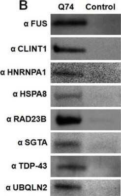
Western Blot: Ubiquilin 2 Antibody (5F5) [H00029978-M03]
Ubiquilin-2-Antibody-5F5-Western-Blot-H00029978-M03-img0015.jpg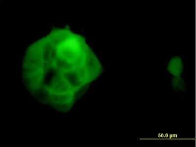
Immunocytochemistry/ Immunofluorescence: Ubiquilin 2 Antibody (5F5) [H00029978-M03]
Immunocytochemistry/Immunofluorescence: Ubiquilin 2 Antibody (5F5) [H00029978-M03] - Analysis of monoclonal antibody to UBQLN2 on A-431 cell. Antibody concentration 10 ug/ml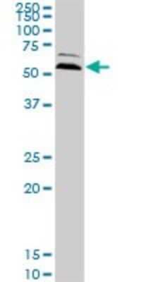
Western Blot: Ubiquilin 2 Antibody (5F5) [H00029978-M03]
Western Blot: Ubiquilin 2 Antibody (5F5) [H00029978-M03] - Analysis of UBQLN2 expression in PC-12 (Cat # L012V1).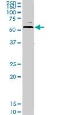
Western Blot: Ubiquilin 2 Antibody (5F5) [H00029978-M03]
Western Blot: Ubiquilin 2 Antibody (5F5) [H00029978-M03] - Analysis of UBQLN2 expression in Raw 264.7 (Cat # L024V1).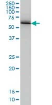
Western Blot: Ubiquilin 2 Antibody (5F5) [H00029978-M03]
Western Blot: Ubiquilin 2 Antibody (5F5) [H00029978-M03] - Analysis of UBQLN2 expression in A-431 (Cat # L015V1).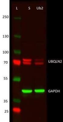
Western Blot: Ubiquilin 2 Antibody (5F5) [H00029978-M03]
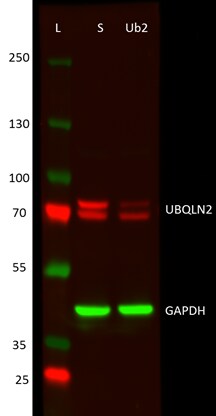
Western Blot: Ubiquilin 2 Antibody (5F5) [H00029978-M03] - UBQLN2 shRNA knockdown in SH-SY5Y cells. First lane is the ladder, then treatment with scrambled shRNA and third lane is UBQLN2 knockdown. 30 ug of the protein sample was loaded on the gel and transferred to the nitrocellulose membrane. Overnight incubation in primary antibodies at 4 degrees C in 5 % milk in TBST. Then incubated in secondary antibodies (Goat Anti-mouse DyLight 550) at RT for 2 h, and then imaged. Knockdown of the upper band with shRNA for UBQLN2 was about 82 %, and of the lower band only about 8 %, suggesting a high specificity of the used shRNA. This image was submitted via customer Review.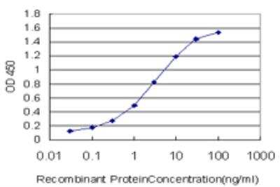
ELISA: Ubiquilin 2 Antibody (5F5) [H00029978-M03]
ELISA: Ubiquilin 2 Antibody (5F5) [H00029978-M03] - Detection limit for recombinant GST tagged UBQLN2 is approximately 0.03ng/ml as a capture antibody.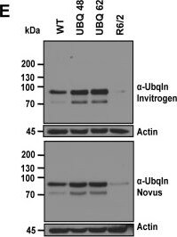
Western Blot: Ubiquilin 2 Antibody (5F5) [H00029978-M03] -
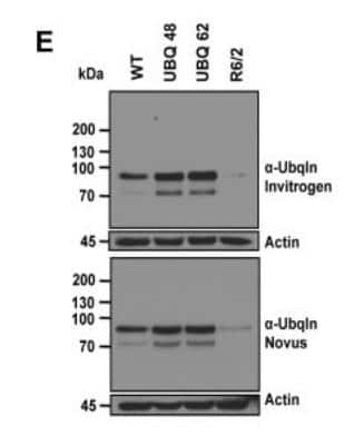
Western Blot: Ubiquilin 2 Antibody (5F5) [H00029978-M03] - Generation of transgenic mice that overexpress human ubiquilin-1.(A) Schematic of the Thy1.2 expression construct used to generate ubiquilin-1 transgenic mice. Human ubiquilin-1 with an N-terminal FLAG tag was cloned in the appropriate orientation between the XhoI site of the Thy1.2 expression cassette. (B) Southern Blot of the first generation offspring of two founder mice (48 & 62). (C) Validation of a PCR genotyping protocol. Amplification of the transgene was only observed in mice that Southern blotting revealed to be positive. (D) Immunoblots of brain cortical lysates with an anti-FLAG antibody & for tubulin indicated that line 62 offspring express higher levels of FLAG-ubiquilin-1 than line 48. (E) Immunoblots of equal amounts of total brain lysates from 12 month-old WT mouse, 12 month-old Ubqln-1 48 transgenic mouse, 12 month-old Ubqln-1 62 transgenic mouse & end stage 15 week-old R6/2 transgenic mouse. The top panel was probed with a monoclonal anti-ubiquilin antibody (Invitrogen antibody clone 3D5E2) & the lower panel with a different monoclonal anti-ubiquilin antibody (Novus antibody clone 5F5). Note two immunoreactive ubiquilin bands are seen at ∼70 kDa & at ∼90 kDa, which we presume is a modified form of ubiquilin. Both blots were also probed for actin to ensure equal loading. (F) Cryostat sections of a Ubqln-1 62 transgenic mouse brain (a–f) & WT mouse brain (g-i) showing anti-FLAG antibody staining (Alexa 594, left panels) & corresponding DAPI staining (center panels) & the result of merging the fluorescent & DAPI signals (right hand panels). The brain sections shown are of the hippocampus (a–c & g–i) & cerebellum (d–f). Identical exposure settings were used for the left hand panels. Image collected & cropped by CiteAb from the following publication (https://pubmed.ncbi.nlm.nih.gov/24475300), licensed under a CC-BY license. Not internally tested by Novus Biologicals.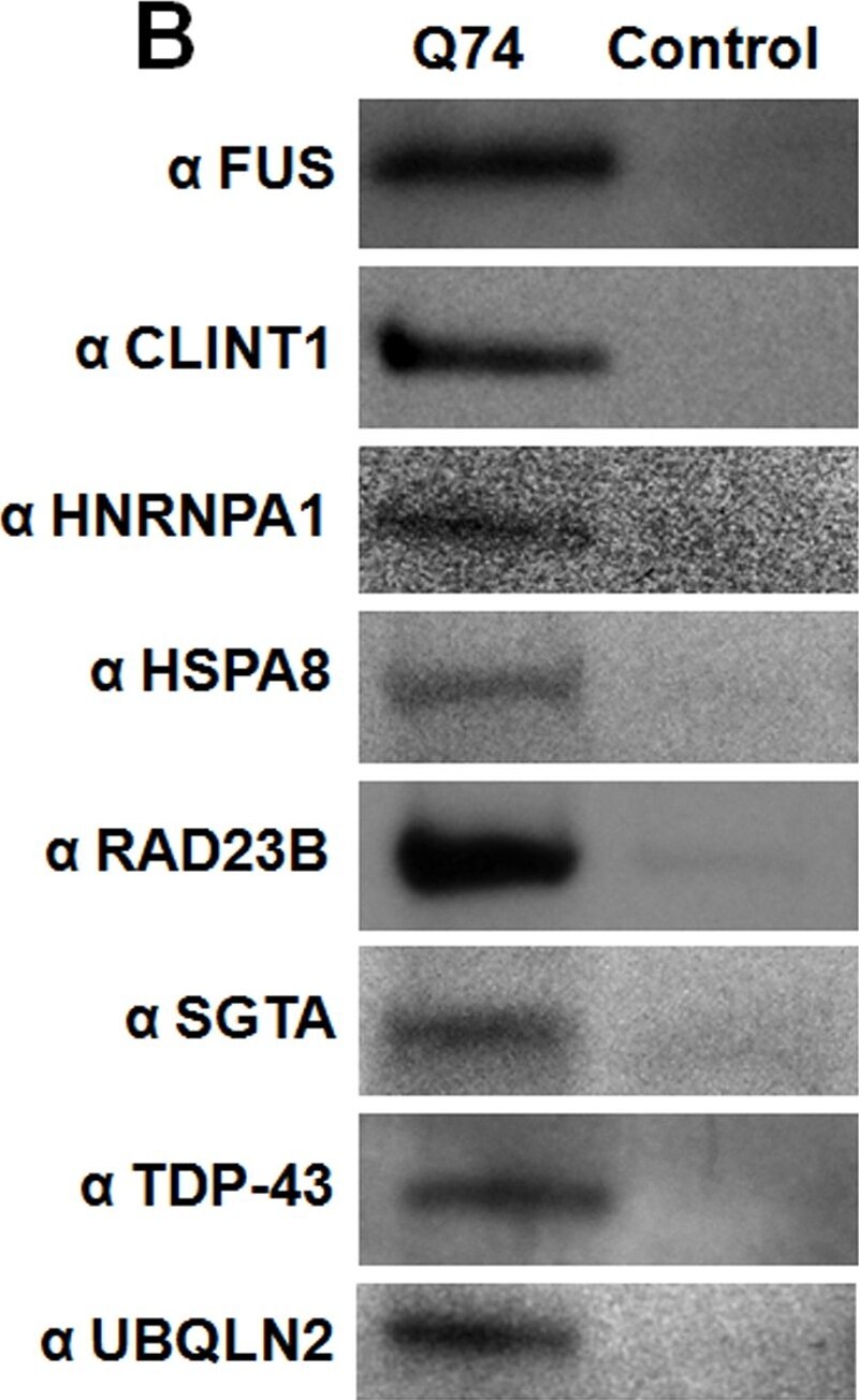
Western Blot: Ubiquilin 2 Antibody (5F5) [H00029978-M03] -
Western Blot: Ubiquilin 2 Antibody (5F5) [H00029978-M03] - Confirmation of polyQ-associated proteins from PC-12 cells identified by TAPI.(A) Western blotting shows that the addition of doxycycline to the PC-12 cell model induces the expression of HttQ74-GFP, resulting in aggregates that can be purified by TAPI. The kinase ERK is probed as a negative control; ERK was never identified by mass spectrometry, so is not expected to co-fractionate with polyQ aggregates. (B) Western blot analysis of TAPI-purified polyQ aggregates from PC-12 cells confirms the presence of several disease-associated proteins only in the Htt-Q74 samples. All proteins migrated near their predicted molecular weights. For control, the TAPI procedure was conducted in parallel on the induced Htt-Q23 cell line (FUS, TDP-43, UBQLN2, HNRNPA1) or the un-induced Htt-Q74 cell line (CLINT1, HSPA8, RAD23B, SGTA). (C) Confocal microscopy shows localization of identified proteins to Htt-Q74 aggregates in PC12 cells. (left) RAD23B, nominally a DNA repair protein, localizes to nuclear Htt-Q74 inclusions but not cytoplasmic inclusions. (middle) FUS, an RNA-binding protein localizes to nuclear & cytoplasmic Htt-Q74 inclusions. (right) CLINT1, a clatherin-interacting protein, is observed in cytoplasmic Htt-Q74 aggregates. Arrows indicate foci with co-localized proteins. Green = GFP; Magenta = CLINT1, FUS or RAD23B in merge. Image collected & cropped by CiteAb from the following publication (https://dx.plos.org/10.1371/journal.pone.0136362), licensed under a CC0-1.0 license. Not internally tested by Novus Biologicals.Applications for Ubiquilin 2 Antibody (5F5) - Azide and BSA Free
ELISA
Immunocytochemistry/ Immunofluorescence
Immunohistochemistry
Immunohistochemistry-Frozen
Immunohistochemistry-Paraffin
Reviewed Applications
Read 1 review rated 3 using H00029978-M03 in the following applications:
Formulation, Preparation, and Storage
Purification
Formulation
Format
Preservative
Concentration
Shipping
Stability & Storage
Background: Ubiquilin 2
Alternate Names
Entrez Gene IDs
Gene Symbol
OMIM
UniProt
Additional Ubiquilin 2 Products
Product Documents for Ubiquilin 2 Antibody (5F5) - Azide and BSA Free
Certificate of Analysis
To download a Certificate of Analysis, please enter a lot or batch number in the search box below.
Product Specific Notices for Ubiquilin 2 Antibody (5F5) - Azide and BSA Free
This product is produced by and distributed for Abnova, a company based in Taiwan.
This product is for research use only and is not approved for use in humans or in clinical diagnosis. Primary Antibodies are guaranteed for 1 year from date of receipt.
Citations for Ubiquilin 2 Antibody (5F5) - Azide and BSA Free
 Powered by Bioz
Customer Reviews for Ubiquilin 2 Antibody (5F5) - Azide and BSA Free (1)
Have you used Ubiquilin 2 Antibody (5F5) - Azide and BSA Free?
Submit a review and receive an Amazon gift card!
$25/€18/£15/$25CAN/¥2500 for a review with an image
$10/€7/£6/$10CAN/¥1110 for a review without an image
Submit a review
Customer Images
-
Application: Western BlotSample Tested: SH-SY5Y cell lysateSpecies: HumanVerified Customer | Posted 02/24/2017Western blot of UBQLN2 shRNA knockdown in SH-SY5Y cells. First lane is the ladder, then treatment with scrambled shRNA and third lane is UBQLN2 knockdown.The samples were lysed in Ripa buffer, followed by sonication. 30 ug of the protein sample was loaded on the gel and transferred to the nitrocellulose membrane. Blocked for 45 min at RT in 5 % milk in TBST, overnight incubation in primary antibodies at 4 °C in 5 % milk in TBST, washed with TBST 3x for 10 min, incubated in secondary antibodies (Goat Anti-mouse DyLight 550) at RT for 2 h and then imaged using BioRad ChemiDoc. Images were quantified using ImageLab Software (BioRad). Knockdown of the upper band with shRNA for UBQLN2 was about 82 %, and of the lower band only about 8 %, suggesting a high specificity of the used shRNA, yet if comparing to the data provided in the antibody datasheet, my shRNA wouldn’t really be targeting UBQLN2, but some undefined band. It is possible that the antibody is not able to differentiate between UBQLN2 and the member of the same family, UBQLN1. UBQLN1 is slightly smaller than UBQLN2, but they both share a significant similarity at the C-terminus, from which the immunogen for the UBQLN2 antibody was raised.

There are no reviews that match your criteria.
Protocols
Find general support by application which include: protocols, troubleshooting, illustrated assays, videos and webinars.
- Antigen Retrieval Protocol (PIER)
- Antigen Retrieval for Frozen Sections Protocol
- Appropriate Fixation of IHC/ICC Samples
- Cellular Response to Hypoxia Protocols
- Chromogenic IHC Staining of Formalin-Fixed Paraffin-Embedded (FFPE) Tissue Protocol
- Chromogenic Immunohistochemistry Staining of Frozen Tissue
- ClariTSA™ Fluorophore Kits
- Detection & Visualization of Antibody Binding
- ELISA Sample Preparation & Collection Guide
- ELISA Troubleshooting Guide
- Fluorescent IHC Staining of Frozen Tissue Protocol
- Graphic Protocol for Heat-induced Epitope Retrieval
- Graphic Protocol for the Preparation and Fluorescent IHC Staining of Frozen Tissue Sections
- Graphic Protocol for the Preparation and Fluorescent IHC Staining of Paraffin-embedded Tissue Sections
- Graphic Protocol for the Preparation of Gelatin-coated Slides for Histological Tissue Sections
- How to Run an R&D Systems DuoSet ELISA
- How to Run an R&D Systems Quantikine ELISA
- How to Run an R&D Systems Quantikine™ QuicKit™ ELISA
- ICC Cell Smear Protocol for Suspension Cells
- ICC Immunocytochemistry Protocol Videos
- ICC for Adherent Cells
- IHC Sample Preparation (Frozen sections vs Paraffin)
- Immunocytochemistry (ICC) Protocol
- Immunocytochemistry Troubleshooting
- Immunofluorescence of Organoids Embedded in Cultrex Basement Membrane Extract
- Immunofluorescent IHC Staining of Formalin-Fixed Paraffin-Embedded (FFPE) Tissue Protocol
- Immunohistochemistry (IHC) and Immunocytochemistry (ICC) Protocols
- Immunohistochemistry Frozen Troubleshooting
- Immunohistochemistry Paraffin Troubleshooting
- Preparing Samples for IHC/ICC Experiments
- Preventing Non-Specific Staining (Non-Specific Binding)
- Primary Antibody Selection & Optimization
- Protocol for Heat-Induced Epitope Retrieval (HIER)
- Protocol for Making a 4% Formaldehyde Solution in PBS
- Protocol for VisUCyte™ HRP Polymer Detection Reagent
- Protocol for the Fluorescent ICC Staining of Cell Smears - Graphic
- Protocol for the Fluorescent ICC Staining of Cultured Cells on Coverslips - Graphic
- Protocol for the Preparation & Fixation of Cells on Coverslips
- Protocol for the Preparation and Chromogenic IHC Staining of Frozen Tissue Sections
- Protocol for the Preparation and Chromogenic IHC Staining of Frozen Tissue Sections - Graphic
- Protocol for the Preparation and Chromogenic IHC Staining of Paraffin-embedded Tissue Sections
- Protocol for the Preparation and Chromogenic IHC Staining of Paraffin-embedded Tissue Sections - Graphic
- Protocol for the Preparation and Fluorescent ICC Staining of Cells on Coverslips
- Protocol for the Preparation and Fluorescent ICC Staining of Non-adherent Cells
- Protocol for the Preparation and Fluorescent ICC Staining of Stem Cells on Coverslips
- Protocol for the Preparation and Fluorescent IHC Staining of Frozen Tissue Sections
- Protocol for the Preparation and Fluorescent IHC Staining of Paraffin-embedded Tissue Sections
- Protocol for the Preparation of Gelatin-coated Slides for Histological Tissue Sections
- Protocol for the Preparation of a Cell Smear for Non-adherent Cell ICC - Graphic
- Quantikine HS ELISA Kit Assay Principle, Alkaline Phosphatase
- Quantikine HS ELISA Kit Principle, Streptavidin-HRP Polymer
- R&D Systems Quality Control Western Blot Protocol
- Sandwich ELISA (Colorimetric) – Biotin/Streptavidin Detection Protocol
- Sandwich ELISA (Colorimetric) – Direct Detection Protocol
- TUNEL and Active Caspase-3 Detection by IHC/ICC Protocol
- The Importance of IHC/ICC Controls
- Troubleshooting Guide: ELISA
- Troubleshooting Guide: Immunohistochemistry
- Troubleshooting Guide: Western Blot Figures
- Western Blot Conditions
- Western Blot Protocol
- Western Blot Protocol for Cell Lysates
- Western Blot Troubleshooting
- Western Blot Troubleshooting Guide
- View all Protocols, Troubleshooting, Illustrated assays and Webinars
FAQs for Ubiquilin 2 Antibody (5F5) - Azide and BSA Free
-
Q: Do you know if the mAb detects ubiquilin 1?
A: Based on the sequence homology, it looks like there could be some cross-reactivity with Ubiquilin 1. The immunogen sequence has 92% homology with bovine Ubiquilin 1.