Ubiquitin Antibody (Ubi-1) - BSA Free
Novus Biologicals | Catalog # NB300-130
![Western Blot: Ubiquitin Antibody (Ubi-1) [NB300-130]](https://resources.rndsystems.com/images/products/Ubiquitin-Antibody-Ubi-1-Western-Blot-NB300-130-img0014.jpg "Western Blot: Ubiquitin Antibody (Ubi-1) [NB300-130]")
Key Product Details
Validated by
Species Reactivity
Validated:
Cited:
Applications
Validated:
Cited:
Label
Antibody Source
Format
Product Specifications
Immunogen
Reactivity Notes
Specificity
Clonality
Host
Isotype
Theoretical MW
Disclaimer note: The observed molecular weight of the protein may vary from the listed predicted molecular weight due to post translational modifications, post translation cleavages, relative charges, and other experimental factors.
Scientific Data Images for Ubiquitin Antibody (Ubi-1) - BSA Free
![Immunocytochemistry/ Immunofluorescence: Ubiquitin Antibody (Ubi-1) [NB300-130]](https://resources.rndsystems.com/images/products/Ubiquitin-Antibody-Ubi-1-Immunocytochemistry-Immunofluorescence-NB300-130-img0013.jpg "Immunocytochemistry/ Immunofluorescence: Ubiquitin Antibody (Ubi-1) [NB300-130]")
Immunocytochemistry/ Immunofluorescence: Ubiquitin Antibody (Ubi-1) [NB300-130]
Ubiquitin-Antibody-Ubi-1-Immunocytochemistry-Immunofluorescence-NB300-130-img0013.jpg![Western Blot: Ubiquitin Antibody (Ubi-1) [NB300-130]](https://resources.rndsystems.com/images/products/Ubiquitin-Antibody-Ubi-1-Western-Blot-NB300-130-img0012.jpg "Western Blot: Ubiquitin Antibody (Ubi-1) [NB300-130]")
Western Blot: Ubiquitin Antibody (Ubi-1) [NB300-130]
Western Blot: Ubiquitin Antibody (Ubi-1) [NB300-130] - Analysis of HEK293 cell lysates using mouse mAb Ubiquitin, dilution 1:1000 (Green). [1] protein standard (Red), [2] cells maintained in normal medium, [3] cells treated with 10uM of proteasome inhibitor lactacystin (Lc) for 16hrs. Lysed cells were electrophoresed on 4-20% SDS-PAGE and transferred to PVDF membranes. The smear detected above the 200kDa standard represent accumulation of ubiquitinated proteins in proteasome inhibitor-Lc treated cells. The prominent band at 8kDa corresponds to monoubiquitin. Rabbit pAb to HSP60, dilution 1:5000 (Red) was used as a loading control.![Immunohistochemistry-Paraffin: Ubiquitin Antibody (Ubi-1) [NB300-130]](https://resources.rndsystems.com/images/products/Ubiquitin-Antibody-Ubi-1-Immunohistochemistry-Paraffin-NB300-130-img0011.jpg "Immunohistochemistry-Paraffin: Ubiquitin Antibody (Ubi-1) [NB300-130]")
Immunohistochemistry-Paraffin: Ubiquitin Antibody (Ubi-1) [NB300-130]
Immunohistochemistry-Paraffin: Ubiquitin Antibody (Ubi-1) [NB300-130] - FFPE section of cerebral cortex of an Alzheimer patient processed with Ubiquitin antibody using HRP/DAB (Brown) and also stained with haemotoxylin (Blue). A typical flame shaped tangle is seen in a pyramidal neuron in the center and is surrounded by some dystrophic neurites, also strongly ubiquitin positive. Both are commonly seen in cortical and hippocampal Alzheimer brain sections and are typical for this disease, but are rare or absent in healthy brain.![Immunohistochemistry-Paraffin: Ubiquitin Antibody (Ubi-1) [NB300-130]](https://resources.rndsystems.com/images/products/Ubiquitin-Antibody-Ubi-1-Immunohistochemistry-Paraffin-NB300-130-img0008.jpg "Immunohistochemistry-Paraffin: Ubiquitin Antibody (Ubi-1) [NB300-130]")
Immunohistochemistry-Paraffin: Ubiquitin Antibody (Ubi-1) [NB300-130]
Immunohistochemistry-Paraffin: Ubiquitin Antibody (Ubi-1) [NB300-130] - Staining of Ubiquitin in hippocampal tissue from an Alzheimer patient.![Western Blot: Ubiquitin Antibody (Ubi-1) [NB300-130]](https://resources.rndsystems.com/images/products/Ubiquitin-Antibody-Ubi-1-Western-Blot-NB300-130-img0015.jpg "Western Blot: Ubiquitin Antibody (Ubi-1) [NB300-130]")
![Western Blot: Ubiquitin Antibody (Ubi-1) [NB300-130]](https://resources.rndsystems.com/images/products/Ubiquitin-Antibody-Ubi-1-Western-Blot-NB300-130-img0009.jpg "Western Blot: Ubiquitin Antibody (Ubi-1) [NB300-130]")
Western Blot: Ubiquitin Antibody (Ubi-1) [NB300-130]
Western Blot: Ubiquitin Antibody (Ubi-1) [NB300-130] - Western blot of Ubiquitin expression on (Lane 1) poly-ubiquitin (lys63 linked), (Lane 2) pure ubiquitin and crude homogenates of adult rat, (Lane 3) cortex, (Lane 4) cerebellum and (Lane 5) brain stem.![Immunohistochemistry-Paraffin: Ubiquitin Antibody (Ubi-1) [NB300-130]](https://resources.rndsystems.com/images/products/Ubiquitin-Antibody-Ubi-1-Immunohistochemistry-Paraffin-NB300-130-img0010.jpg "Immunohistochemistry-Paraffin: Ubiquitin Antibody (Ubi-1) [NB300-130]")
Immunohistochemistry-Paraffin: Ubiquitin Antibody (Ubi-1) [NB300-130]
Immunohistochemistry-Paraffin: Ubiquitin Antibody (Ubi-1) [NB300-130] - IHC staining of Ubiquitin in human rectal cancer using DAB with hematoxylin counterstain. [NB300-130] -")
Western Blot: Ubiquitin Antibody (Ubi-1) [NB300-130] -
Western Blot: Ubiquitin Antibody (Ubi-1) [NB300-130] - In Vivo Applications of Affimers(A) HA-NleL 293 T-Rex cells were induced with 1 μg/mL doxycycline for 12 hr or left untreated. Whole-cell lysate (WCL) blots are shown for actin, HA(-NleL), Ub, & K6 chains. Western blots with the K6 affimer are also shown after Ub enrichment using TUBEs.(B) TUBE-PD of HEK293 cells after 1 hr of MG132 (10 μM) without further treatment (−) or with additional UV (40 J/m2) or CCCP treatment (10 μM for 1 hr) & subsequently blotted with the K6 affimer. Input controls are shown for total Ub & actin & gamma H2AX. The relative signal increase from two experiments is shown below the respective lanes.(C) Expression of WT or catalytically inactive (C431S) Parkin in HeLa Flp-In cells was induced with 0.2 μg/mL doxycycline for 16 hr. Mitochondria were depolarized with O/A for 2 hr. WCL inputs are shown for total Ub, expressed Parkin, TOM20, & actin. The TUBE-PD was also blotted using the K6 affimer & total Ub.(D) Confocal fluorescence microscopy images of cells as in (B) stained with K6 affimer (green), TOM20 (red), & DAPI (blue). Cells & a magnified area are outlined in white. Scale bars correspond to 20 μm.See also Figure S5. Image collected & cropped by CiteAb from the following publication (https://pubmed.ncbi.nlm.nih.gov/28943312), licensed under a CC-BY license. Not internally tested by Novus Biologicals. [NB300-130] -")
Western Blot: Ubiquitin Antibody (Ubi-1) [NB300-130] -
Western Blot: Ubiquitin Antibody (Ubi-1) [NB300-130] - HUWE1 Assembles K6 Chains In Vitro & In Vivo(A) Table summarizing proteins identified with the corresponding number peptide-spectrum matches (PSMs) in three replicates of DUB-treated K6 affimer pull-downs.(B) In vitro assembly reaction of the HECT E3 HUWE1 with Ub WT, Ub K6R, Ub K11R, & Ub K48R on Coomassie. Arrows indicate K6 diUb.(C) Linkage composition of HUWE1-generated diUb after 1 hr as determined by AQUA MS.(D) AQUA-derived total cellular chain composition of HUWE1−/− HeLa & parental cells after TUBE-based enrichment. Error bars indicate mean ± SD from n = 3. ∗p < 0.05, according to a two-tailed Student’s t test. N.S., not significant.(E) TUBE-PD from a doxycycline-inducible HUWE1 shRNA Ls174T cell line blotted with the K6-specific affimer, with input controls for actin, total Ub, & HUWE1.(F) K6 affimer pull-down in doxycycline-inducible HUWE1 shRNA Ls174T cells blotted against Mfn2. Cells were left untreated or treated with 10 μg/mL MG132 for 4 hr and/or 1 μg/mL doxycycline for 72 hr. Pull-downs were incubated with 250 nM USP21 as indicated, with a K48 blot to show completeness of the deubiquitination reaction. Input controls are shown for Mfn2, actin, & HUWE1.See also Figure S7. Image collected & cropped by CiteAb from the following publication (https://pubmed.ncbi.nlm.nih.gov/28943312), licensed under a CC-BY license. Not internally tested by Novus Biologicals. [NB300-130] -")
Western Blot: Ubiquitin Antibody (Ubi-1) [NB300-130] -
Western Blot: Ubiquitin Antibody (Ubi-1) [NB300-130] - In Vivo Applications of Affimers(A) HA-NleL 293 T-Rex cells were induced with 1 μg/mL doxycycline for 12 hr or left untreated. Whole-cell lysate (WCL) blots are shown for actin, HA(-NleL), Ub, & K6 chains. Western blots with the K6 affimer are also shown after Ub enrichment using TUBEs.(B) TUBE-PD of HEK293 cells after 1 hr of MG132 (10 μM) without further treatment (−) or with additional UV (40 J/m2) or CCCP treatment (10 μM for 1 hr) & subsequently blotted with the K6 affimer. Input controls are shown for total Ub & actin & gamma H2AX. The relative signal increase from two experiments is shown below the respective lanes.(C) Expression of WT or catalytically inactive (C431S) Parkin in HeLa Flp-In cells was induced with 0.2 μg/mL doxycycline for 16 hr. Mitochondria were depolarized with O/A for 2 hr. WCL inputs are shown for total Ub, expressed Parkin, TOM20, & actin. The TUBE-PD was also blotted using the K6 affimer & total Ub.(D) Confocal fluorescence microscopy images of cells as in (B) stained with K6 affimer (green), TOM20 (red), & DAPI (blue). Cells & a magnified area are outlined in white. Scale bars correspond to 20 μm.See also Figure S5. Image collected & cropped by CiteAb from the following publication (https://pubmed.ncbi.nlm.nih.gov/28943312), licensed under a CC-BY license. Not internally tested by Novus Biologicals.Applications for Ubiquitin Antibody (Ubi-1) - BSA Free
Immunocytochemistry/ Immunofluorescence
Immunohistochemistry
Immunohistochemistry-Frozen
Immunohistochemistry-Paraffin
Western Blot
Reviewed Applications
Read 4 reviews rated 4.3 using NB300-130 in the following applications:
Formulation, Preparation, and Storage
Purification
Formulation
Format
Preservative
Concentration
Shipping
Stability & Storage
Background: Ubiquitin
Alternate Names
Gene Symbol
UniProt
Additional Ubiquitin Products
Product Documents for Ubiquitin Antibody (Ubi-1) - BSA Free
Certificate of Analysis
To download a Certificate of Analysis, please enter a lot or batch number in the search box below.
Product Specific Notices for Ubiquitin Antibody (Ubi-1) - BSA Free
This product is for research use only and is not approved for use in humans or in clinical diagnosis. Primary Antibodies are guaranteed for 1 year from date of receipt.
Related Research Areas
Citations for Ubiquitin Antibody (Ubi-1) - BSA Free
 Powered by Bioz
Customer Reviews for Ubiquitin Antibody (Ubi-1) - BSA Free (4)
Have you used Ubiquitin Antibody (Ubi-1) - BSA Free?
Submit a review and receive an Amazon gift card!
$25/€18/£15/$25CAN/¥2500 Yen for a review with an image
$10/€7/£6/$10CAN/¥1110 Yen for a review without an image
Submit a review
Customer Images
-
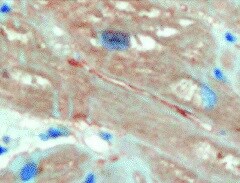
Application: Immunohistochemistry-ParaffinSample Tested: Adult heartSpecies: HumanVerified Customer | Posted 08/25/2021Ubiquitin Antibody in heart sample.

-
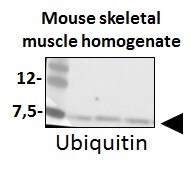
Application: Western BlotSample Tested: Mouse skeletal muscle homogenateSpecies: MouseVerified Customer | Posted 03/27/2018Western Blot: Ubiquitin Antibody (Ubi-1) [NB300-130SS] - Total protein from Mouse skeletal muscle homogenate, separated on a 4-12% gel by SDS-PAGE, transferred to nitrocellulose membrane and blocked in 5% non-fat milk for 1h at room temperature. The membrane was probed with anti-Myogenin 1:1000 in non-fat milk.
-
Application: Western BlotSample Tested: 293 whole cell lysate, Hela whole cell lysate, HCT116 whole cell lysate, MEF whole cell lysate, mouse liver lysate and mouse bone marrow-derived macrophage cellsSpecies: Human and MouseVerified Customer | Posted 08/04/2017Works well for WB on total cell lysates (boiling the membrane (nitrocellulose) can increase signal drastically if it is low, e.g. when blotting on primary cells/tissues), and it works really well for WB on Ubiquitin pulldowns, e.g. after a TUBE (tandem ubiquitin-binding entity) pulldown.
-
Application: ImmunofluorescenceSample Tested: primary mouse neuronal cultureSpecies: MouseVerified Customer | Posted 05/27/2009
There are no reviews that match your criteria.
Protocols
View specific protocols for Ubiquitin Antibody (Ubi-1) - BSA Free (NB300-130):
Materials
1) 1 Phosphate buffered saline (pH 7.6): NaCl 137mmol/L, KCl 2.7mmol/L, Na2HPO4 4.3mmol/L, KH2PO4 1.4 mmol/L
2) Citrate buffer, 0.01 M, pH6.0, Sodium Citrate 3g, Citric acid 0.4g
3) 3% Hydrogen peroxide
4) Primary antibody
5) Blocking serum (normal serum)
6) Biotinylated secondary antibody
7) DAB staining kit
Methods
1. Dewax and hydration of slides using xylene and EtOH:
Dry slides for 20 min in a 60 C oven
Add Xylene, 2 x 10 min
100%, 95%, 80%, and 70% EtOH, 5 min each EtOH concentration
Rinse in PBS, 5'
2 Antigen retrieval method (only for paraffin slides)
1a. High-pressure antigen retrieval procedure (recommended method)
Place slides in a glass slide holder (ensure that the slide holder is completely filled with slides, slides without sections if necessary, to ensure even heating. The entire slide holder is immersed in 1000 ml of Citrate buffer (0.01M, pH6.0) within a pressure cooker
Once steam is produced, and ONLY when steam is visible, from the pressure cooker (usually 15-20 min), the required high-pressure will have been reached, and slides will be incubated for 2 min.
Turn off heat, and allow buffer and slides to cool to room temperature
Slides are then rinsed in PBS for 5 minutes
2. Add 3% hydrogen peroxide solution, 10'at RT, then PBS, 3X5'
3. Normal blocking serum, 20'at RT
4. Incubate with Primary Ab, 4C overnight or 1.5 hours at 37C
5. Rinse with PBS, 3 X 5' each rinse
6. Add Biotin-conjugated second antibody, 10'at RT
7. Rinse with PBS, 3 X 5' each rinse
8. Add Streptavidin-Peroxidase, 10'at RT
9. Rinse with PBS, 3 X 5' each rinse
10. Staining with DAB solution, 2-5'under microscope
11. Stop the reaction by washing in tap water
12. Counterstain in Haematoxylin for 3-5 minutes
13. 75%, 80%, 95% and 100% ethanol, 5x2', xylene 2 x 10'
Find general support by application which include: protocols, troubleshooting, illustrated assays, videos and webinars.
- Antigen Retrieval Protocol (PIER)
- Antigen Retrieval for Frozen Sections Protocol
- Appropriate Fixation of IHC/ICC Samples
- Cellular Response to Hypoxia Protocols
- Chromogenic IHC Staining of Formalin-Fixed Paraffin-Embedded (FFPE) Tissue Protocol
- Chromogenic Immunohistochemistry Staining of Frozen Tissue
- ClariTSA™ Fluorophore Kits
- Detection & Visualization of Antibody Binding
- Fluorescent IHC Staining of Frozen Tissue Protocol
- Graphic Protocol for Heat-induced Epitope Retrieval
- Graphic Protocol for the Preparation and Fluorescent IHC Staining of Frozen Tissue Sections
- Graphic Protocol for the Preparation and Fluorescent IHC Staining of Paraffin-embedded Tissue Sections
- Graphic Protocol for the Preparation of Gelatin-coated Slides for Histological Tissue Sections
- ICC Cell Smear Protocol for Suspension Cells
- ICC Immunocytochemistry Protocol Videos
- ICC for Adherent Cells
- IHC Sample Preparation (Frozen sections vs Paraffin)
- Immunocytochemistry (ICC) Protocol
- Immunocytochemistry Troubleshooting
- Immunofluorescence of Organoids Embedded in Cultrex Basement Membrane Extract
- Immunofluorescent IHC Staining of Formalin-Fixed Paraffin-Embedded (FFPE) Tissue Protocol
- Immunohistochemistry (IHC) and Immunocytochemistry (ICC) Protocols
- Immunohistochemistry Frozen Troubleshooting
- Immunohistochemistry Paraffin Troubleshooting
- Preparing Samples for IHC/ICC Experiments
- Preventing Non-Specific Staining (Non-Specific Binding)
- Primary Antibody Selection & Optimization
- Protocol for Heat-Induced Epitope Retrieval (HIER)
- Protocol for Making a 4% Formaldehyde Solution in PBS
- Protocol for VisUCyte™ HRP Polymer Detection Reagent
- Protocol for the Fluorescent ICC Staining of Cell Smears - Graphic
- Protocol for the Fluorescent ICC Staining of Cultured Cells on Coverslips - Graphic
- Protocol for the Preparation & Fixation of Cells on Coverslips
- Protocol for the Preparation and Chromogenic IHC Staining of Frozen Tissue Sections
- Protocol for the Preparation and Chromogenic IHC Staining of Frozen Tissue Sections - Graphic
- Protocol for the Preparation and Chromogenic IHC Staining of Paraffin-embedded Tissue Sections
- Protocol for the Preparation and Chromogenic IHC Staining of Paraffin-embedded Tissue Sections - Graphic
- Protocol for the Preparation and Fluorescent ICC Staining of Cells on Coverslips
- Protocol for the Preparation and Fluorescent ICC Staining of Non-adherent Cells
- Protocol for the Preparation and Fluorescent ICC Staining of Stem Cells on Coverslips
- Protocol for the Preparation and Fluorescent IHC Staining of Frozen Tissue Sections
- Protocol for the Preparation and Fluorescent IHC Staining of Paraffin-embedded Tissue Sections
- Protocol for the Preparation of Gelatin-coated Slides for Histological Tissue Sections
- Protocol for the Preparation of a Cell Smear for Non-adherent Cell ICC - Graphic
- R&D Systems Quality Control Western Blot Protocol
- TUNEL and Active Caspase-3 Detection by IHC/ICC Protocol
- The Importance of IHC/ICC Controls
- Troubleshooting Guide: Immunohistochemistry
- Troubleshooting Guide: Western Blot Figures
- Western Blot Conditions
- Western Blot Protocol
- Western Blot Protocol for Cell Lysates
- Western Blot Troubleshooting
- Western Blot Troubleshooting Guide
- View all Protocols, Troubleshooting, Illustrated assays and Webinars
FAQs for Ubiquitin Antibody (Ubi-1) - BSA Free
-
Q: Regarding the WB image on datasheet of NCX1 antibody (6H2) (NB300-127), can you confirm if lane 2 is a rabbit heart sample? In addition, can you provide the dilution fold and loading mount used to obtain this image? Lastly, would you happen to know if t
A: A protein stabilizer has not been added to the antibody NB300-130. However, note that this antibody is supplied as ascites fluid, not as a purified antibody. If you are not familiar with this format, the following descriptions will be useful: Ascites fluid is a complex solution containing multiple proteins and background antibodies as well as the antibody of interest. Ascites contains the antibody produced by the hybridoma cells as well as many other proteins, including background antibodies produced by the mice and albumin. Antibody concentrations in ascites are typically high, but an accurate concentration of the target antibody cannot be readily determined. Therefore, unfortunately you cannot determine the concentration of the ubiquitin antibody within NB300-130 as sold. This is why this product's pack sizes are volumes rather than masses, and why we state on our datasheet: 'This product is unpurified. The exact concentration of antibody is not quantifiable.' All I can suggest is that you carry out antigen affinity chromatography of the antibody, and then determine its concentration.