![Western Blot: UCP2 Antibody [NB100-59742]](https://resources.rndsystems.com/images/products/UCP2-Antibody-Western-Blot-NB100-59742-img0001.jpg "Western Blot: UCP2 Antibody [NB100-59742]")
Key Product Details
Species Reactivity
Validated:
Cited:
Predicted:
Applications
Validated:
Cited:
Label
Antibody Source
Product Specifications
Immunogen
Reactivity Notes
Clonality
Host
Isotype
Theoretical MW
Disclaimer note: The observed molecular weight of the protein may vary from the listed predicted molecular weight due to post translational modifications, post translation cleavages, relative charges, and other experimental factors.
Scientific Data Images for UCP2 Antibody
Western Blot: UCP2 Antibody [NB100-59742]
Western Blot: UCP2 Antibody [NB100-59742] - Staining of rat adipose (A) and mouse spleen (B) lysate (35 ug protein in RIPA buffer). Antibody at 1 ug/mL. Detected by chemiluminescence. Approx 37 kDa band observed in rat adipose and 28-30 kDa in mouse spleen lysates (calculated MW of 33.4kDa according to Mouse NP_035801.3 and 33.4kDa according to Rat NP_062227.2).![Immunocytochemistry/ Immunofluorescence: UCP2 Antibody [NB100-59742]](https://resources.rndsystems.com/images/products/UCP2-Antibody-Immunocytochemistry-Immunofluorescence-NB100-59742-img0002.jpg "Immunocytochemistry/ Immunofluorescence: UCP2 Antibody [NB100-59742]")
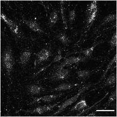
Immunocytochemistry/ Immunofluorescence: UCP2 Antibody [NB100-59742]
Immunocytochemistry/Immunofluorescence: UCP2 Antibody [NB100-59742] - Analysis of paraformaldehyde fixed MCF7 cells, permeabilized with 0.15% Triton. Primary incubation 1hr (10 ug/mL) followed by Alexa Fluor 488 secondary antibody (2 ug/mL), showing Mitochondrial staining. The nuclear stain is DAPI (blue). Negative control: Unimmunized goat IgG (10 ug/mL) followed by Alexa Fluor 488 secondary antibody (2 ug/mL).Applications for UCP2 Antibody
Immunocytochemistry/ Immunofluorescence
Immunohistochemistry
Peptide ELISA
Western Blot
Reviewed Applications
Read 2 reviews rated 3 using NB100-59742 in the following applications:
Formulation, Preparation, and Storage
Purification
Formulation
Preservative
Concentration
Shipping
Stability & Storage
Background: UCP2
Long Name
Alternate Names
Gene Symbol
UniProt
Additional UCP2 Products
Product Documents for UCP2 Antibody
Certificate of Analysis
To download a Certificate of Analysis, please enter a lot or batch number in the search box below.
Product Specific Notices for UCP2 Antibody
This product is for research use only and is not approved for use in humans or in clinical diagnosis. Primary Antibodies are guaranteed for 1 year from date of receipt.
Related Research Areas
Citations for UCP2 Antibody
 Powered by Bioz
Customer Reviews for UCP2 Antibody (2)
Have you used UCP2 Antibody?
Submit a review and receive an Amazon gift card!
$25/€18/£15/$25CAN/¥2500 Yen for a review with an image
$10/€7/£6/$10CAN/¥1110 Yen for a review without an image
Submit a review
Customer Images
-
Application: ImmunocytochemistrySample Tested: Human primary fibroblastSpecies: HumanVerified Customer | Posted 10/24/2020UCP2 in human primary fibroblasts PFA fixed. Donkey anti-Goat Alexa Fluor 647 detection.Fixation: PFA 4% 15 RT. Blocking/permeabilization: 1h in 10% DS in PBST (0.1% Triton). UCP2 antibody incubation O/N 4ºC 1/100 dilution in 10% DS in PBST (0.1%Triton). Secondary antibody: Donkey anti-Goat Alexa Fluor 647 1:500 1h RT.
 Bio-Techne ResponseThank you for reviewing our product. We are sorry to that that this antibody did not perform as expected. We have been in touch with the customer to resolve this issue according to our Product Guarantee and to the customer’s satisfaction.
 -
Application: Western BlotSample Tested: Liver mitochondrial protein preparationsSpecies: RatVerified Customer | Posted 12/19/2014
There are no reviews that match your criteria.
Protocols
Find general support by application which include: protocols, troubleshooting, illustrated assays, videos and webinars.
- Antigen Retrieval Protocol (PIER)
- Antigen Retrieval for Frozen Sections Protocol
- Appropriate Fixation of IHC/ICC Samples
- Cellular Response to Hypoxia Protocols
- Chromogenic IHC Staining of Formalin-Fixed Paraffin-Embedded (FFPE) Tissue Protocol
- Chromogenic Immunohistochemistry Staining of Frozen Tissue
- ClariTSA™ Fluorophore Kits
- Detection & Visualization of Antibody Binding
- ELISA Sample Preparation & Collection Guide
- ELISA Troubleshooting Guide
- Fluorescent IHC Staining of Frozen Tissue Protocol
- Graphic Protocol for Heat-induced Epitope Retrieval
- Graphic Protocol for the Preparation and Fluorescent IHC Staining of Frozen Tissue Sections
- Graphic Protocol for the Preparation and Fluorescent IHC Staining of Paraffin-embedded Tissue Sections
- Graphic Protocol for the Preparation of Gelatin-coated Slides for Histological Tissue Sections
- How to Run an R&D Systems DuoSet ELISA
- How to Run an R&D Systems Quantikine ELISA
- How to Run an R&D Systems Quantikine™ QuicKit™ ELISA
- ICC Cell Smear Protocol for Suspension Cells
- ICC Immunocytochemistry Protocol Videos
- ICC for Adherent Cells
- IHC Sample Preparation (Frozen sections vs Paraffin)
- Immunocytochemistry (ICC) Protocol
- Immunocytochemistry Troubleshooting
- Immunofluorescence of Organoids Embedded in Cultrex Basement Membrane Extract
- Immunofluorescent IHC Staining of Formalin-Fixed Paraffin-Embedded (FFPE) Tissue Protocol
- Immunohistochemistry (IHC) and Immunocytochemistry (ICC) Protocols
- Immunohistochemistry Frozen Troubleshooting
- Immunohistochemistry Paraffin Troubleshooting
- Preparing Samples for IHC/ICC Experiments
- Preventing Non-Specific Staining (Non-Specific Binding)
- Primary Antibody Selection & Optimization
- Protocol for Heat-Induced Epitope Retrieval (HIER)
- Protocol for Making a 4% Formaldehyde Solution in PBS
- Protocol for VisUCyte™ HRP Polymer Detection Reagent
- Protocol for the Fluorescent ICC Staining of Cell Smears - Graphic
- Protocol for the Fluorescent ICC Staining of Cultured Cells on Coverslips - Graphic
- Protocol for the Preparation & Fixation of Cells on Coverslips
- Protocol for the Preparation and Chromogenic IHC Staining of Frozen Tissue Sections
- Protocol for the Preparation and Chromogenic IHC Staining of Frozen Tissue Sections - Graphic
- Protocol for the Preparation and Chromogenic IHC Staining of Paraffin-embedded Tissue Sections
- Protocol for the Preparation and Chromogenic IHC Staining of Paraffin-embedded Tissue Sections - Graphic
- Protocol for the Preparation and Fluorescent ICC Staining of Cells on Coverslips
- Protocol for the Preparation and Fluorescent ICC Staining of Non-adherent Cells
- Protocol for the Preparation and Fluorescent ICC Staining of Stem Cells on Coverslips
- Protocol for the Preparation and Fluorescent IHC Staining of Frozen Tissue Sections
- Protocol for the Preparation and Fluorescent IHC Staining of Paraffin-embedded Tissue Sections
- Protocol for the Preparation of Gelatin-coated Slides for Histological Tissue Sections
- Protocol for the Preparation of a Cell Smear for Non-adherent Cell ICC - Graphic
- Quantikine HS ELISA Kit Assay Principle, Alkaline Phosphatase
- Quantikine HS ELISA Kit Principle, Streptavidin-HRP Polymer
- R&D Systems Quality Control Western Blot Protocol
- Sandwich ELISA (Colorimetric) – Biotin/Streptavidin Detection Protocol
- Sandwich ELISA (Colorimetric) – Direct Detection Protocol
- TUNEL and Active Caspase-3 Detection by IHC/ICC Protocol
- The Importance of IHC/ICC Controls
- Troubleshooting Guide: ELISA
- Troubleshooting Guide: Immunohistochemistry
- Troubleshooting Guide: Western Blot Figures
- Western Blot Conditions
- Western Blot Protocol
- Western Blot Protocol for Cell Lysates
- Western Blot Troubleshooting
- Western Blot Troubleshooting Guide
- View all Protocols, Troubleshooting, Illustrated assays and Webinars
FAQs for UCP2 Antibody
-
Q: Hi, I'd like to measure UCP2 levels in various mouse tissues by western blot. I was wondering which of your antibodies would be best for this purpose. In the literature I have seen some papers where they have isolated mitochondria to be able to see UCP2 and others where they have not. We would like to use frozen tissue samples, so isolating mitochondria will not be an option. We would like to use liver, muscle, pancreas, kidney, brown fat, subcutaneous fat, and gonadal fat. I'd appreciate any advice you might have.
A: Regarding your question on our UCP2 antibodies, we do have a product (catalog # NB100-78377), but our lab confirmed that it has only been validated in mouse liver and brain and there is no guarantee on other tissues.