VEGF-C Antibody - Azide and BSA Free
Novus Biologicals | Catalog # NB110-61022
![Immunocytochemistry/ Immunofluorescence: VEGF-C Antibody [NB110-61022]](https://resources.rndsystems.com/images/products/VEGF-C-Antibody-Immunocytochemistry-Immunofluorescence-NB110-61022-img0002.jpg "Immunocytochemistry/ Immunofluorescence: VEGF-C Antibody [NB110-61022]")
Key Product Details
Validated by
Species Reactivity
Validated:
Cited:
Applications
Validated:
Cited:
Label
Antibody Source
Format
Product Specifications
Immunogen
Specificity
Clonality
Host
Isotype
Scientific Data Images for VEGF-C Antibody - Azide and BSA Free
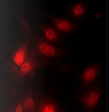
Immunocytochemistry/ Immunofluorescence: VEGF-C Antibody [NB110-61022]
Immunocytochemistry/Immunofluorescence: VEGF-C Antibody [NB110-61022] - bEnd.3 mouse brain endothelioma cell line stained with Vegf-c antibody at 1:500. ICC/IF image submitted by a verified customer review.![Immunohistochemistry-Paraffin: VEGF-C Antibody [NB110-61022]](https://resources.rndsystems.com/images/products/VEGF-C-Antibody-Immunohistochemistry-Paraffin-NB110-61022-img0003.jpg "Immunohistochemistry-Paraffin: VEGF-C Antibody [NB110-61022]")
![Immunohistochemistry: VEGF-C Antibody [NB110-61022]](https://resources.rndsystems.com/images/products/VEGF-C-Antibody-Immunohistochemistry-NB110-61022-img0001.jpg "Immunohistochemistry: VEGF-C Antibody [NB110-61022]")
Immunohistochemistry: VEGF-C Antibody [NB110-61022]
Immunohistochemistry: VEGF-C Antibody [NB110-61022] - VEGFC Antibody [NB110-61022] - 4% PFA fixed and paraffin embedded mouse Lung tissue section was subjected to IHC staining of mouse VEGF-C.
Western Blot: VEGF-C Antibody [NB110-61022] -
Western Blot: VEGF-C Antibody [NB110-61022] - Kallikrein-related peptidase 3 (KLK3)/Prostate specific antigen (PSA) activates VEGF-C.(A, B) Cleavage of pro-VEGF-C by KLK3 (PSA). Pro-VEGF-C was incubated with or without KLK3, with & without the monoclonal antibody against KLK3 (5C7). Detection of VEGF-C in Western blots probed with antiserum 6 & 3/4, resulting in the detection of pro-VEGF-C (29/31 kDa) & activated, mature VEGF-C (21/23 kDa). The band marked by the asterisk likely represents the N-terminal propeptide (~15 kDa) which is detected by the antiserum 6. Note that for the image shown for antiserum 6, two different exposures of the same blot were merged (n = 3). (C, D) VEGF-C processed by KLK3 is biologically active in Ba/F3 cell assays, which translate activation of a hybrid VEGFR/EpoR receptor into cell survival (n = 2). Error bars indicate SD.10.7554/eLife.44478.004Figure 1—source data 1.Ba/F3 assay showing the activity of KLK3-cleaved VEGF-C.Ba/F3 assay showing the activity of KLK3-cleaved VEGF-C. Image collected & cropped by CiteAb from the following publication (https://pubmed.ncbi.nlm.nih.gov/31099754), licensed under a CC-BY license. Not internally tested by Novus Biologicals.Applications for VEGF-C Antibody - Azide and BSA Free
Immunohistochemistry
Immunohistochemistry-Frozen
Immunohistochemistry-Paraffin
Western Blot
Reviewed Applications
Read 1 review rated 5 using NB110-61022 in the following applications:
Formulation, Preparation, and Storage
Purification
Reconstitution
Formulation
Format
Preservative
Concentration
Shipping
Stability & Storage
Calculators
Background: VEGF-C
Long Name
Alternate Names
Gene Symbol
UniProt
Additional VEGF-C Products
Product Documents for VEGF-C Antibody - Azide and BSA Free
Certificate of Analysis
To download a Certificate of Analysis, please enter a lot or batch number in the search box below.
Product Specific Notices for VEGF-C Antibody - Azide and BSA Free
This product is for research use only and is not approved for use in humans or in clinical diagnosis. Primary Antibodies are guaranteed for 1 year from date of receipt.
Citations for VEGF-C Antibody - Azide and BSA Free
 Powered by Bioz
Customer Reviews for VEGF-C Antibody - Azide and BSA Free (1)
Have you used VEGF-C Antibody - Azide and BSA Free?
Submit a review and receive an Amazon gift card!
$25/€18/£15/$25CAN/¥2500 Yen for a review with an image
$10/€7/£6/$10CAN/¥1110 Yen for a review without an image
Submit a review
Customer Images
-
Application: ImmunocytochemistrySample Tested: bEnd.3 mouse endothelioma cell lineSpecies: MouseVerified Customer | Posted 07/23/2020bend.3 stained with Vegf-c concentration 1:500
 Bio-Techne ResponseThis review was submitted through the legacy Novus Innovators Program, reflecting a new species or application tested on a primary antibody.
There are no reviews that match your criteria.
Protocols
Find general support by application which include: protocols, troubleshooting, illustrated assays, videos and webinars.
- Antigen Retrieval Protocol (PIER)
- Antigen Retrieval for Frozen Sections Protocol
- Appropriate Fixation of IHC/ICC Samples
- Cellular Response to Hypoxia Protocols
- Chromogenic IHC Staining of Formalin-Fixed Paraffin-Embedded (FFPE) Tissue Protocol
- Chromogenic Immunohistochemistry Staining of Frozen Tissue
- ClariTSA™ Fluorophore Kits
- Detection & Visualization of Antibody Binding
- Fluorescent IHC Staining of Frozen Tissue Protocol
- Graphic Protocol for Heat-induced Epitope Retrieval
- Graphic Protocol for the Preparation and Fluorescent IHC Staining of Frozen Tissue Sections
- Graphic Protocol for the Preparation and Fluorescent IHC Staining of Paraffin-embedded Tissue Sections
- Graphic Protocol for the Preparation of Gelatin-coated Slides for Histological Tissue Sections
- ICC Cell Smear Protocol for Suspension Cells
- ICC Immunocytochemistry Protocol Videos
- ICC for Adherent Cells
- IHC Sample Preparation (Frozen sections vs Paraffin)
- Immunocytochemistry (ICC) Protocol
- Immunocytochemistry Troubleshooting
- Immunofluorescence of Organoids Embedded in Cultrex Basement Membrane Extract
- Immunofluorescent IHC Staining of Formalin-Fixed Paraffin-Embedded (FFPE) Tissue Protocol
- Immunohistochemistry (IHC) and Immunocytochemistry (ICC) Protocols
- Immunohistochemistry Frozen Troubleshooting
- Immunohistochemistry Paraffin Troubleshooting
- Preparing Samples for IHC/ICC Experiments
- Preventing Non-Specific Staining (Non-Specific Binding)
- Primary Antibody Selection & Optimization
- Protocol for Heat-Induced Epitope Retrieval (HIER)
- Protocol for Making a 4% Formaldehyde Solution in PBS
- Protocol for VisUCyte™ HRP Polymer Detection Reagent
- Protocol for the Fluorescent ICC Staining of Cell Smears - Graphic
- Protocol for the Fluorescent ICC Staining of Cultured Cells on Coverslips - Graphic
- Protocol for the Preparation & Fixation of Cells on Coverslips
- Protocol for the Preparation and Chromogenic IHC Staining of Frozen Tissue Sections
- Protocol for the Preparation and Chromogenic IHC Staining of Frozen Tissue Sections - Graphic
- Protocol for the Preparation and Chromogenic IHC Staining of Paraffin-embedded Tissue Sections
- Protocol for the Preparation and Chromogenic IHC Staining of Paraffin-embedded Tissue Sections - Graphic
- Protocol for the Preparation and Fluorescent ICC Staining of Cells on Coverslips
- Protocol for the Preparation and Fluorescent ICC Staining of Non-adherent Cells
- Protocol for the Preparation and Fluorescent ICC Staining of Stem Cells on Coverslips
- Protocol for the Preparation and Fluorescent IHC Staining of Frozen Tissue Sections
- Protocol for the Preparation and Fluorescent IHC Staining of Paraffin-embedded Tissue Sections
- Protocol for the Preparation of Gelatin-coated Slides for Histological Tissue Sections
- Protocol for the Preparation of a Cell Smear for Non-adherent Cell ICC - Graphic
- R&D Systems Quality Control Western Blot Protocol
- TUNEL and Active Caspase-3 Detection by IHC/ICC Protocol
- The Importance of IHC/ICC Controls
- Troubleshooting Guide: Immunohistochemistry
- Troubleshooting Guide: Western Blot Figures
- Western Blot Conditions
- Western Blot Protocol
- Western Blot Protocol for Cell Lysates
- Western Blot Troubleshooting
- Western Blot Troubleshooting Guide
- View all Protocols, Troubleshooting, Illustrated assays and Webinars
FAQs for VEGF-C Antibody - Azide and BSA Free
-
Q: Do you have any data which show the molecular weight of the VEGFC antibody (NB110-61022) by western blotting?
A: We do not currently have any images of NB110-61022 being used in Western blot, although it is guaranteed for this use. NBP1-45924 and NBP1-97842 show a band around 16 kDa. This is the predicted molecular weight of VEGF-C (UniProt P49767) and this is the weight the band using NB110-61022 should show up at.
-
Q: I was wondering if you know of any blocking antibodies against IL-6 or VEGF-C that can be used in cell culture to quench these cytokines?
A:
We have no antibodies that are currently known to show blocking antibodies to VEGF-C. Please see this link to our IL-6 antibodies with blocking/neutralizing activity can be found at this link.
-
Q: What is the molecular weight for VEGFC Antibody (NB110-61022) on Western blot?
A: The expected molecular weight of VEGFC is about 47KDa according to UniProt.
-
Q: Do you have any data which show the molecular weight of the VEGFC antibody (NB110-61022) by western blotting?
A: We do not currently have any images of NB110-61022 being used in Western blot, although it is guaranteed for this use. NBP1-45924 and NBP1-97842 show a band around 16 kDa. This is the predicted molecular weight of VEGF-C (UniProt P49767) and this is the weight the band using NB110-61022 should show up at.
-
Q: I was wondering if you know of any blocking antibodies against IL-6 or VEGF-C that can be used in cell culture to quench these cytokines?
A:
We have no antibodies that are currently known to show blocking antibodies to VEGF-C. Please see this link to our IL-6 antibodies with blocking/neutralizing activity can be found at this link.
-
Q: What is the molecular weight for VEGFC Antibody (NB110-61022) on Western blot?
A: The expected molecular weight of VEGFC is about 47KDa according to UniProt.