VEGFR1/Flt-1 Antibody - BSA Free
Novus Biologicals | Catalog # NB100-527
![Western Blot: VEGFR1/Flt-1 Antibody [NB100-527]](https://resources.rndsystems.com/images/products/VEGF-R1-Flt-1-Antibody-Western-Blot-NB100-527-img0005.jpg "Western Blot: VEGFR1/Flt-1 Antibody [NB100-527]")
Key Product Details
Species Reactivity
Validated:
Cited:
Predicted:
Applications
Validated:
Cited:
Label
Antibody Source
Format
Product Specifications
Immunogen
Localization
Marker
Specificity
Clonality
Host
Isotype
Scientific Data Images for VEGFR1/Flt-1 Antibody - BSA Free
Western Blot: VEGFR1/Flt-1 Antibody [NB100-527]
Western Blot: VEGF R1/Flt-1 Antibody [NB100-527] - Chimeric CSF-1R/VEGFR-2 detection in transfected lysates.![Immunocytochemistry/ Immunofluorescence: VEGFR1/Flt-1 Antibody [NB100-527]](https://resources.rndsystems.com/images/products/VEGF-R1-Flt-1-Antibody-Immunocytochemistry-Immunofluorescence-NB100-527-img0006.jpg "Immunocytochemistry/ Immunofluorescence: VEGFR1/Flt-1 Antibody [NB100-527]")
Immunocytochemistry/ Immunofluorescence: VEGFR1/Flt-1 Antibody [NB100-527]
Immunocytochemistry/Immunofluorescence: VEGFR1/Flt-1 Antibody [NB100-527] - VEGF R1/Flt-1 antibody was tested in HeLa cells with DyLight 488 (green). Nuclei and alpha-tubulin were counterstained with DAPI (blue) and DyLight 550 (red).![Immunohistochemistry-Paraffin: VEGFR1/Flt-1 Antibody [NB100-527]](https://resources.rndsystems.com/images/products/VEGFR1-Flt-1-Antibody-Immunohistochemistry-Paraffin-NB100-527-img0009.jpg "Immunohistochemistry-Paraffin: VEGFR1/Flt-1 Antibody [NB100-527]")
Immunohistochemistry-Paraffin: VEGFR1/Flt-1 Antibody [NB100-527]
Immunohistochemistry-Paraffin: VEGFR1/Flt-1 Antibody [NB100-527] - Analysis of FFPE human breast carcinoma tissue section using 1:500 dilution of VEGFR1/Flt-1 antibody on a Bond Rx autostainer (Leica Biosystems). The assay involved 20 minutes of heat induced antigen retrieval (HIER) with 10mM sodium citrate buffer (pH 6.0) and endogenous peroxidase quenching using peroxide block. The sections were incubated with primary antibody for 30 minutes. Bond Polymer Refine Detection (Leica Biosystems) and DAB were used for signal detection which followed counterstaining with hematoxylin. Whole slide scanning and capturing of representative images (20X) were performed using Aperio AT2 (Leica Biosystems). This VEGFR1/Flt-1 antibody generated an expected membrane cytoplasmic staining of VEGFR1 protein in the cancer cells (punctate appearance typical of receptors). The tumor stroma/stromal cells did not show VEGFR1/Flt-1immunopositivity.![Immunohistochemistry-Paraffin: VEGFR1/Flt-1 Antibody [NB100-527]](https://resources.rndsystems.com/images/products/VEGFR1-Flt-1-Antibody-Immunohistochemistry-Paraffin-NB100-527-img0008.jpg "Immunohistochemistry-Paraffin: VEGFR1/Flt-1 Antibody [NB100-527]")
Immunohistochemistry-Paraffin: VEGFR1/Flt-1 Antibody [NB100-527]
Immunohistochemistry-Paraffin: VEGFR1/Flt-1 Antibody [NB100-527] - Analysis of a FFPE human breast carcinoma tissue section using 1:500 dilution of VEGFR1/Flt-1 antibody on a Bond Rx autostainer (Leica Biosystems). The assay involved 20 minutes of heat induced antigen retrieval (HIER) with 10mM sodium citrate buffer (pH 6.0) and endogenous peroxidase quenching using peroxide block. The sections were incubated with primary antibody for 30 minutes. Bond Polymer Refine Detection (Leica Biosystems) and DAB were used for signal detection which followed counterstaining with hematoxylin. Whole slide scanning and capturing of representative images (20X) were performed using Aperio AT2 (Leica Biosystems). This VEGFR1/Flt-1 antibody generated an expected membrane cytoplasmic staining of VEGFR1 protein in the cancer cells. The tumor stroma/stromal cells did not show VEGFR1/Flt-1immunopositivity. Staining was performed by Histowiz.![VEGFR1/Flt-1 Antibody - BSA Free Western Blot: Rabbit Polyclonal VEGFR1/Flt-1 Antibody [NB100-527]](https://resources.rndsystems.com/images/products/antibody/nb100-527_rabbit-polyclonal-vegfr1-flt-1-antibody-western-blot-2362025152643..png "Western Blot: Rabbit Polyclonal VEGFR1/Flt-1 Antibody [NB100-527]")
Western Blot: Rabbit Polyclonal VEGFR1/Flt-1 Antibody [NB100-527]
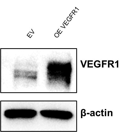
VEGFR1 overexpression in K562 cells. Image from a verified customer review.Applications for VEGFR1/Flt-1 Antibody - BSA Free
Immunocytochemistry/ Immunofluorescence
Immunohistochemistry
Immunohistochemistry-Paraffin
Western Blot
Reviewed Applications
Read 1 review rated 5 using NB100-527 in the following applications:
Formulation, Preparation, and Storage
Purification
Formulation
Format
Preservative
Concentration
Shipping
Stability & Storage
Background: VEGFR1/Flt-1
Long Name
Alternate Names
Gene Symbol
Additional VEGFR1/Flt-1 Products
Product Documents for VEGFR1/Flt-1 Antibody - BSA Free
Certificate of Analysis
To download a Certificate of Analysis, please enter a lot or batch number in the search box below.
Product Specific Notices for VEGFR1/Flt-1 Antibody - BSA Free
This product is for research use only and is not approved for use in humans or in clinical diagnosis. Primary Antibodies are guaranteed for 1 year from date of receipt.
Citations for VEGFR1/Flt-1 Antibody - BSA Free
 Powered by Bioz
Customer Reviews for VEGFR1/Flt-1 Antibody - BSA Free (1)
Have you used VEGFR1/Flt-1 Antibody - BSA Free?
Submit a review and receive an Amazon gift card!
$25/€18/£15/$25CAN/¥2500 Yen for a review with an image
$10/€7/£6/$10CAN/¥1110 Yen for a review without an image
Submit a review
Customer Images
-
Application: Western BlotSample Tested: k562 cellsSpecies: HumanVerified Customer | Posted 06/19/2025VEGFR1 overexpression in K562 cells

There are no reviews that match your criteria.
Protocols
View specific protocols for VEGFR1/Flt-1 Antibody - BSA Free (NB100-527):
Culture cells to appropriate density in 35 mm culture dishes or 6-well plates.
1. Remove culture medium and wash the cells briefly in PBS. Add 10% formalin to the dish and fix at room temperature for 10 minutes.
2. Remove the formalin and wash the cells in PBS.
3. Permeablize the cells with 0.1% Triton X100 or other suitable detergent for 10 min.
4. Remove the permeablization buffer and wash three times for 10 minutes each in PBS. Be sure to not let the specimen dry out.
5. To block nonspecific antibody binding, incubate in 10% normal goat serum from 1 hour to overnight at room temperature.
6. Add primary antibody at appropriate dilution and incubate overnight at 4C.
7. Remove primary antibody and replace with PBS. Wash three times for 10 minutes each.
8. Add secondary antibody at appropriate dilution. Incubate for 1 hour at room temperature.
9. Remove secondary antibody and replace with PBS. Wash three times for 10 minutes each.
10. Counter stain DNA with DAPi if required.
Antigen Unmasking:
Bring slides to a boil in 10 mM sodium citrate buffer (pH 6.0) then maintain at a sub-boiling temperature for 10 minutes. Cool slides on bench-top for 30 minutes (keep slides in the sodium citrate buffer at all times).
Staining:
1. Wash sections in deionized water three times for 5 minutes each.
2. Wash sections in PBS for 5 minutes.
3. Block each section with 100-400 ul blocking solution (1% BSA in PBS) for 1 hour at room temperature.
4. Remove blocking solution and add 100-400 ul diluted primary antibody. Incubate overnight at 4 C.
5. Remove antibody solution and wash sections in wash buffer three times for 5 minutes each.
6. Add 100-400 ul HRP polymer conjugated secondary antibody. Incubate 30 minutes at room temperature.
7. Wash sections three times in wash buffer for 5 minutes each.
8. Add 100-400 ul DAB substrate to each section and monitor staining closely.
9. As soon as the sections develop, immerse slides in deionized water.
10. Counterstain sections in hematoxylin.
11. Wash sections in deionized water two times for 5 minutes each.
12. Dehydrate sections.
13. Mount coverslips.
1. Perform SDS-PAGE on samples to be analyzed, loading 10-25 ug of total protein per lane.
2. Transfer proteins to PVDF membrane according to the instructions provided by the manufacturer of the membrane and transfer apparatus.
3. Stain the membrane with Ponceau S (or similar product) to assess transfer success, and mark molecular weight standards where appropriate.
4. Rinse the blot TBS -0.05% Tween 20 (TBST).
5. Block the membrane in 5% Non-fat milk in TBST (blocking buffer) for at least 1 hour.
6. Wash the membrane in TBST three times for 10 minutes each.
7. Dilute primary antibody in blocking buffer and incubate overnight at 4C with gentle rocking.
8. Wash the membrane in TBST three times for 10 minutes each.
9. Incubate the membrane in diluted HRP conjugated secondary antibody in blocking buffer (as per manufacturer's instructions) for 1 hour at room temperature.
10. Wash the blot in TBST three times for 10 minutes each (this step can be repeated as required to reduce background).
11. Apply the detection reagent of choice in accordance with the manufacturer's instructions.
Find general support by application which include: protocols, troubleshooting, illustrated assays, videos and webinars.
- Antigen Retrieval Protocol (PIER)
- Antigen Retrieval for Frozen Sections Protocol
- Appropriate Fixation of IHC/ICC Samples
- Cellular Response to Hypoxia Protocols
- Chromogenic IHC Staining of Formalin-Fixed Paraffin-Embedded (FFPE) Tissue Protocol
- Chromogenic Immunohistochemistry Staining of Frozen Tissue
- ClariTSA™ Fluorophore Kits
- Detection & Visualization of Antibody Binding
- Fluorescent IHC Staining of Frozen Tissue Protocol
- Graphic Protocol for Heat-induced Epitope Retrieval
- Graphic Protocol for the Preparation and Fluorescent IHC Staining of Frozen Tissue Sections
- Graphic Protocol for the Preparation and Fluorescent IHC Staining of Paraffin-embedded Tissue Sections
- Graphic Protocol for the Preparation of Gelatin-coated Slides for Histological Tissue Sections
- ICC Cell Smear Protocol for Suspension Cells
- ICC Immunocytochemistry Protocol Videos
- ICC for Adherent Cells
- IHC Sample Preparation (Frozen sections vs Paraffin)
- Immunocytochemistry (ICC) Protocol
- Immunocytochemistry Troubleshooting
- Immunofluorescence of Organoids Embedded in Cultrex Basement Membrane Extract
- Immunofluorescent IHC Staining of Formalin-Fixed Paraffin-Embedded (FFPE) Tissue Protocol
- Immunohistochemistry (IHC) and Immunocytochemistry (ICC) Protocols
- Immunohistochemistry Frozen Troubleshooting
- Immunohistochemistry Paraffin Troubleshooting
- Preparing Samples for IHC/ICC Experiments
- Preventing Non-Specific Staining (Non-Specific Binding)
- Primary Antibody Selection & Optimization
- Protocol for Heat-Induced Epitope Retrieval (HIER)
- Protocol for Making a 4% Formaldehyde Solution in PBS
- Protocol for VisUCyte™ HRP Polymer Detection Reagent
- Protocol for the Fluorescent ICC Staining of Cell Smears - Graphic
- Protocol for the Fluorescent ICC Staining of Cultured Cells on Coverslips - Graphic
- Protocol for the Preparation & Fixation of Cells on Coverslips
- Protocol for the Preparation and Chromogenic IHC Staining of Frozen Tissue Sections
- Protocol for the Preparation and Chromogenic IHC Staining of Frozen Tissue Sections - Graphic
- Protocol for the Preparation and Chromogenic IHC Staining of Paraffin-embedded Tissue Sections
- Protocol for the Preparation and Chromogenic IHC Staining of Paraffin-embedded Tissue Sections - Graphic
- Protocol for the Preparation and Fluorescent ICC Staining of Cells on Coverslips
- Protocol for the Preparation and Fluorescent ICC Staining of Non-adherent Cells
- Protocol for the Preparation and Fluorescent ICC Staining of Stem Cells on Coverslips
- Protocol for the Preparation and Fluorescent IHC Staining of Frozen Tissue Sections
- Protocol for the Preparation and Fluorescent IHC Staining of Paraffin-embedded Tissue Sections
- Protocol for the Preparation of Gelatin-coated Slides for Histological Tissue Sections
- Protocol for the Preparation of a Cell Smear for Non-adherent Cell ICC - Graphic
- R&D Systems Quality Control Western Blot Protocol
- TUNEL and Active Caspase-3 Detection by IHC/ICC Protocol
- The Importance of IHC/ICC Controls
- Troubleshooting Guide: Immunohistochemistry
- Troubleshooting Guide: Western Blot Figures
- Western Blot Conditions
- Western Blot Protocol
- Western Blot Protocol for Cell Lysates
- Western Blot Troubleshooting
- Western Blot Troubleshooting Guide
- View all Protocols, Troubleshooting, Illustrated assays and Webinars
FAQs for VEGFR1/Flt-1 Antibody - BSA Free
-
Q: I am studying the regulation of PLCg during fertilization in the starfish. I am interested in trying your anti-FLT1 polyclonal antibody. Your information sheet says that the immunogen for this antibody is "A synthetic peptide corresponding to a sequence in the middle region of human FLT1". Since I will be using this antibody in starfish eggs, I compared the starfish protein sequence to the human protein sequence and found areas of high identity between the two and I want to use an antibody that is expected to bind to amino acids common to both. Therefore, I would like some more information to match the immunogen to the starfish protein. Can you tell me specifically which amino acids of human FLT1 were used to make the antibody?
A: The immunogen sequence to this product is proprietary. I can tell you it falls between amino acids 800-900 of the mouse FLT1 protein [UniProt# P35969].
-
Q: Would you be able to tell me the concentration of the current lot of NB100-527 - VEGFR1/VEGFR2 Ab?
A:
The concentration of NB100-527 is 1mg/mL
-
Q: I am studying the regulation of PLCg during fertilization in the starfish. I am interested in trying your anti-FLT1 polyclonal antibody. Your information sheet says that the immunogen for this antibody is "A synthetic peptide corresponding to a sequence in the middle region of human FLT1". Since I will be using this antibody in starfish eggs, I compared the starfish protein sequence to the human protein sequence and found areas of high identity between the two and I want to use an antibody that is expected to bind to amino acids common to both. Therefore, I would like some more information to match the immunogen to the starfish protein. Can you tell me specifically which amino acids of human FLT1 were used to make the antibody?
A: The immunogen sequence to this product is proprietary. I can tell you it falls between amino acids 800-900 of the mouse FLT1 protein [UniProt# P35969].
-
Q: Would you be able to tell me the concentration of the current lot of NB100-527 - VEGFR1/VEGFR2 Ab?
A:
The concentration of NB100-527 is 1mg/mL
Associated Pathways


