Vinculin Antibody (hVIN-1)
Novus Biologicals | Catalog # NB600-1293
![Western Blot: Vinculin Antibody (hVIN-1) [NB600-1293]](https://resources.rndsystems.com/images/products/Vinculin-Antibody-hVIN-1-Western-Blot-NB600-1293-img0003.jpg "Western Blot: Vinculin Antibody (hVIN-1) [NB600-1293]")
Key Product Details
Validated by
Species Reactivity
Validated:
Cited:
Applications
Validated:
Cited:
Label
Antibody Source
Product Specifications
Immunogen
Reactivity Notes
Localization
Marker
Specificity
Clonality
Host
Isotype
Theoretical MW
Disclaimer note: The observed molecular weight of the protein may vary from the listed predicted molecular weight due to post translational modifications, post translation cleavages, relative charges, and other experimental factors.
Scientific Data Images for Vinculin Antibody (hVIN-1)
Western Blot: Vinculin Antibody (hVIN-1) [NB600-1293]
Western Blot: Vinculin Antibody (hVIN-1) [NB600-1293] - Cell line lysates were separated on SDS-PAGE and probed with 1:200 Monoclonal Anti-Vinculin Clone: hVIN-1. The antibody was developed using Goat Anti-Mouse IgG-Peroxidase and a chemiluminescent substrate.Lanes:1.HeLa2.COS73.NIH-3T34.RAT25.CHO6.MDBK7.MDCK![Immunocytochemistry/ Immunofluorescence: Vinculin Antibody (hVIN-1) [NB600-1293]](https://resources.rndsystems.com/images/products/Vinculin-Antibody-hVIN-1-Immunocytochemistry-Immunofluorescence-NB600-1293-img0004.jpg "Immunocytochemistry/ Immunofluorescence: Vinculin Antibody (hVIN-1) [NB600-1293]")
![Immunohistochemistry: Vinculin Antibody (hVIN-1) [NB600-1293]](https://resources.rndsystems.com/images/products/Vinculin-Antibody-hVIN-1-Immunohistochemistry-NB600-1293-img0001.jpg "Immunohistochemistry: Vinculin Antibody (hVIN-1) [NB600-1293]")
Immunohistochemistry: Vinculin Antibody (hVIN-1) [NB600-1293]
Immunohistochemistry: Vinculin Antibody (hVIN-1) [NB600-1293] - Enhanced Validation-By Independent Antibodies:Immunohistochemistry. Formalin-fixed, paraffin-embedded Rat Heart sections stained with 15 ug/mL Anti-Vinculin antibody produced in Rabbit (Cat. No. V4139) (A). The antibody was developed using Anti-Rabbit IgG (whole molecule)-FITC antibody produced in Goat (Cat. No. F9887), and 15 ug/mL Monoclonal Anti-Vinculin antibody produced in Mouse, Clone: hVIN1 (Cat. No. V9131) (B). The antibody was developed using Rabbit Anti-Mouse IgG-Cy3 conjugate antibody.Results:Two Anti-Vinculin antibodies, V4139 (A) and V9131 (B), target different regions of Vinculin show similar staining profiles between the two antibodies, demonstrating Independent Antibody Verification. [NB600-1293] -")
Western Blot: Vinculin Antibody (hVIN-1) [NB600-1293] -
A1B decreases TLR4 protein content in microglia chronically treated with LPS. (A) N9 cells seeded at 1 × 10⁵ cells per well on poly-d-lysine-coated glass cover slides in 6-well plates were pre-treated for 4 h with DMSO or with 0.5 uM K-604, then exposed with or without 200 ng/mL LPS for 24 h. Double immunofluorescence staining for TLR4 and for the plasma membrane marker N-cadherin was then performed. (i) Timeline of the experiment. (ii) Representative images demonstrating TLR4 distribution in N9 cells. (iii) Quantification of total TLR4 relative fluorescence intensity per cell, n = 15 cells/treatment group. (iv) Quantification of IM/PM TLR4 fluorescence intensity ratio. A total of 15 cells per group were analyzed. The TLR4 signals overlapping with that of N-cadherin are considered as TLR4 at the PM, while those not overlapping are considered as TLR4 at the IM. (B) N9 microglial cells were seeded at 2 × 10⁵ cells per well onto 6-well plates in RPMI-1640 with 10% serum. N9 cells were treated with DMSO (control group) or 0.5 uM K-604 for 4 h, then treated with or without 200 ng/mL LPS for 24 and 48 h. At different time points, cells were harvested for protein isolation and TLR4 Western blot analyses. n = 3 replicates. Vinculin was used as the protein loading control. (i) Timeline of experiment. (ii) Western blot. (iii) Quantitation of Western blot. * p < 0.05; ** p < 0.01; **** p < 0.0001. Image collected and cropped by CiteAb from the following open publication (https://www.mdpi.com/1422-0067/24/6/5616), licensed under a CC-BY license. Not internally tested by Novus Biologicals. [NB600-1293] -")
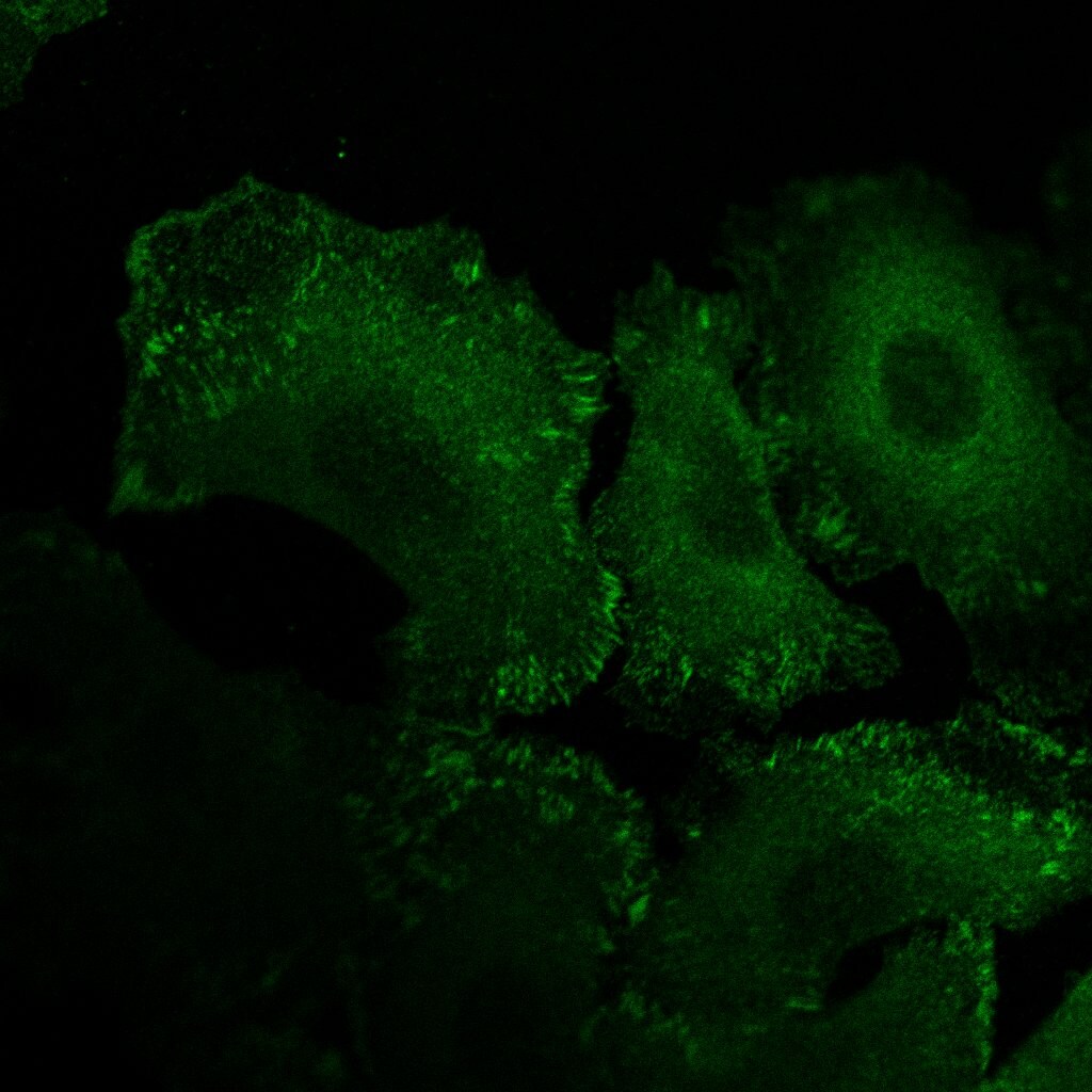
Immunocytochemistry/ Immunofluorescence: Vinculin Antibody (hVIN-1) [NB600-1293] -
Reduction of directionality and focal adhesion turnover by low eribulin concentrations(A) Effect of eribulin on MTOC directionality during wound healing. Percentage of cells with MTOC facing the wound (bottom). Values are mean +/- SEM (n = 3) **P < 0.01. (B) immunofluorescence images of LM8 cells treated with 0 nM, 1 nM, or 10 nM eribulin and stained for vinculin (red) and nucleus (blue) (left). Dotted line shows the cell shape. Scale bar: 10 μm. Quantitative analysis of the area of vinculin staining (right). Values are mean +/- SEM (≥30 cells per group). **P < 0.01. (C) Immunofluorescence images of LM8 cells treated with 0 nM, 1 nM, or 10 nM eribulin and stained for Tyr397-phosphorylated FAK (green), actin (red), and nucleus (blue) (left). Dotted line shows the cell shape. Scale bar: 10 μm. Quantitative analysis of the area of Tyr397-phosphorylated FAK staining (right). Values are mean +/- SEM (≥30 cells per group). *P < 0.05. Eribulin treatment shrank the Tyr397-phosphorylated FAK staining area in a dose-dependent manner. (D) Western blot of Tyr397-phosphorylated FAK in LM8 cells treated with eribulin (top). Quantitative densitometric analysis of the ratio of Tyr397-phosphorylated FAK to total FAK (bottom). Values are mean +/- SEM (n = 3). *P < 0.05. (E) Immunofluorescence images of LM8 cells treated with 0 nM or 10 nM eribulin and stained for APC (red), alpha -tubulin (green), and nucleus (blue). Scale bar: 10 μm. Image collected and cropped by CiteAb from the following open publication (https://pubmed.ncbi.nlm.nih.gov/30719211), licensed under a CC-BY license. Not internally tested by Novus Biologicals.Applications for Vinculin Antibody (hVIN-1)
Immunohistochemistry
Immunohistochemistry-Frozen
Single Cell Western
Western Blot
Reviewed Applications
Read 1 review rated 5 using NB600-1293 in the following applications:
Formulation, Preparation, and Storage
Purification
Formulation
Preservative
Concentration
Shipping
Stability & Storage
Background: Vinculin
Long Name
Alternate Names
Gene Symbol
UniProt
Additional Vinculin Products
Product Documents for Vinculin Antibody (hVIN-1)
Certificate of Analysis
To download a Certificate of Analysis, please enter a lot or batch number in the search box below.
Product Specific Notices for Vinculin Antibody (hVIN-1)
This product is for research use only and is not approved for use in humans or in clinical diagnosis. Primary Antibodies are guaranteed for 1 year from date of receipt.
Related Research Areas
Citations for Vinculin Antibody (hVIN-1)
 Powered by Bioz
Customer Reviews for Vinculin Antibody (hVIN-1) (1)
Have you used Vinculin Antibody (hVIN-1)?
Submit a review and receive an Amazon gift card!
$25/€18/£15/$25CAN/¥2500 Yen for a review with an image
$10/€7/£6/$10CAN/¥1110 Yen for a review without an image
Submit a review
Customer Images
-
Application: ImmunocytochemistrySample Tested: skin keratinocytesSpecies: MouseVerified Customer | Posted 04/26/2017Mouse primary keratinocytes stained with the clone hVIN-1

There are no reviews that match your criteria.
Protocols
Find general support by application which include: protocols, troubleshooting, illustrated assays, videos and webinars.
- Antigen Retrieval Protocol (PIER)
- Antigen Retrieval for Frozen Sections Protocol
- Appropriate Fixation of IHC/ICC Samples
- Cellular Response to Hypoxia Protocols
- Chromogenic IHC Staining of Formalin-Fixed Paraffin-Embedded (FFPE) Tissue Protocol
- Chromogenic Immunohistochemistry Staining of Frozen Tissue
- ClariTSA™ Fluorophore Kits
- Detection & Visualization of Antibody Binding
- Fluorescent IHC Staining of Frozen Tissue Protocol
- Graphic Protocol for Heat-induced Epitope Retrieval
- Graphic Protocol for the Preparation and Fluorescent IHC Staining of Frozen Tissue Sections
- Graphic Protocol for the Preparation and Fluorescent IHC Staining of Paraffin-embedded Tissue Sections
- Graphic Protocol for the Preparation of Gelatin-coated Slides for Histological Tissue Sections
- ICC Cell Smear Protocol for Suspension Cells
- ICC Immunocytochemistry Protocol Videos
- ICC for Adherent Cells
- IHC Sample Preparation (Frozen sections vs Paraffin)
- Immunocytochemistry (ICC) Protocol
- Immunocytochemistry Troubleshooting
- Immunofluorescence of Organoids Embedded in Cultrex Basement Membrane Extract
- Immunofluorescent IHC Staining of Formalin-Fixed Paraffin-Embedded (FFPE) Tissue Protocol
- Immunohistochemistry (IHC) and Immunocytochemistry (ICC) Protocols
- Immunohistochemistry Frozen Troubleshooting
- Immunohistochemistry Paraffin Troubleshooting
- Preparing Samples for IHC/ICC Experiments
- Preventing Non-Specific Staining (Non-Specific Binding)
- Primary Antibody Selection & Optimization
- Protocol for Heat-Induced Epitope Retrieval (HIER)
- Protocol for Making a 4% Formaldehyde Solution in PBS
- Protocol for VisUCyte™ HRP Polymer Detection Reagent
- Protocol for the Fluorescent ICC Staining of Cell Smears - Graphic
- Protocol for the Fluorescent ICC Staining of Cultured Cells on Coverslips - Graphic
- Protocol for the Preparation & Fixation of Cells on Coverslips
- Protocol for the Preparation and Chromogenic IHC Staining of Frozen Tissue Sections
- Protocol for the Preparation and Chromogenic IHC Staining of Frozen Tissue Sections - Graphic
- Protocol for the Preparation and Chromogenic IHC Staining of Paraffin-embedded Tissue Sections
- Protocol for the Preparation and Chromogenic IHC Staining of Paraffin-embedded Tissue Sections - Graphic
- Protocol for the Preparation and Fluorescent ICC Staining of Cells on Coverslips
- Protocol for the Preparation and Fluorescent ICC Staining of Non-adherent Cells
- Protocol for the Preparation and Fluorescent ICC Staining of Stem Cells on Coverslips
- Protocol for the Preparation and Fluorescent IHC Staining of Frozen Tissue Sections
- Protocol for the Preparation and Fluorescent IHC Staining of Paraffin-embedded Tissue Sections
- Protocol for the Preparation of Gelatin-coated Slides for Histological Tissue Sections
- Protocol for the Preparation of a Cell Smear for Non-adherent Cell ICC - Graphic
- R&D Systems Quality Control Western Blot Protocol
- TUNEL and Active Caspase-3 Detection by IHC/ICC Protocol
- The Importance of IHC/ICC Controls
- Troubleshooting Guide: Immunohistochemistry
- Troubleshooting Guide: Western Blot Figures
- Western Blot Conditions
- Western Blot Protocol
- Western Blot Protocol for Cell Lysates
- Western Blot Troubleshooting
- Western Blot Troubleshooting Guide
- View all Protocols, Troubleshooting, Illustrated assays and Webinars
FAQs for Vinculin Antibody (hVIN-1)
-
Q: Do you have any 100 coda or bigger controls?
A:
I can recommend a couple of high weight antibodies that are conserved in most samples. Please verify the presence of the protein before using as a loading control. PARP1 or PARP is approximately 113kDa, and you may view the products we offer to PARP using this link. Vinculin is approximately 116kDa, and you may view the products we offer to Vinculin using this link.