WRN Antibody - BSA Free
Novus Biologicals | Catalog # NB100-471
![Western Blot: WRN Antibody [NB100-471]](https://resources.rndsystems.com/images/products/WRN-Antibody-Western-Blot-NB100-471-img0009.jpg "Western Blot: WRN Antibody [NB100-471]")
Key Product Details
Validated by
Independent Antibodies, Biological Validation
Species Reactivity
Validated:
Human
Cited:
Human
Applications
Validated:
Immunohistochemistry, Western Blot, Immunocytochemistry/ Immunofluorescence, Simple Western, Immunoprecipitation, Chromatin Immunoprecipitation (ChIP)
Cited:
Western Blot, Immunocytochemistry/ Immunofluorescence, Chromatin Immunoprecipitation (ChIP), Chemotaxis, IF/IHC
Label
Unconjugated
Antibody Source
Polyclonal Rabbit IgG
Format
BSA Free
Loading...
Product Specifications
Immunogen
The immunogen recognized by this antibody maps to a region between residues 400 and 450 of human Werner Syndrome Helicase using the numbering given in SwissProt entry Q14191 (GeneID 7486).
Clonality
Polyclonal
Host
Rabbit
Isotype
IgG
Scientific Data Images for WRN Antibody - BSA Free
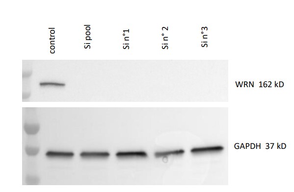
Western Blot: WRN Antibody [NB100-471]
Western Blot: WRN Antibody [NB100-471] - Human colon cancer cells with siRNA Control, siRNA pool of siWrn and 3 different sets of siRNA Wrn. GAPDH laoding control. Western blot image submitted by a verified customer review.![Immunocytochemistry/ Immunofluorescence: WRN Antibody [NB100-471]](https://resources.rndsystems.com/images/products/WRN-Antibody-Immunocytochemistry-Immunofluorescence-NB100-471-img0004.jpg "Immunocytochemistry/ Immunofluorescence: WRN Antibody [NB100-471]")
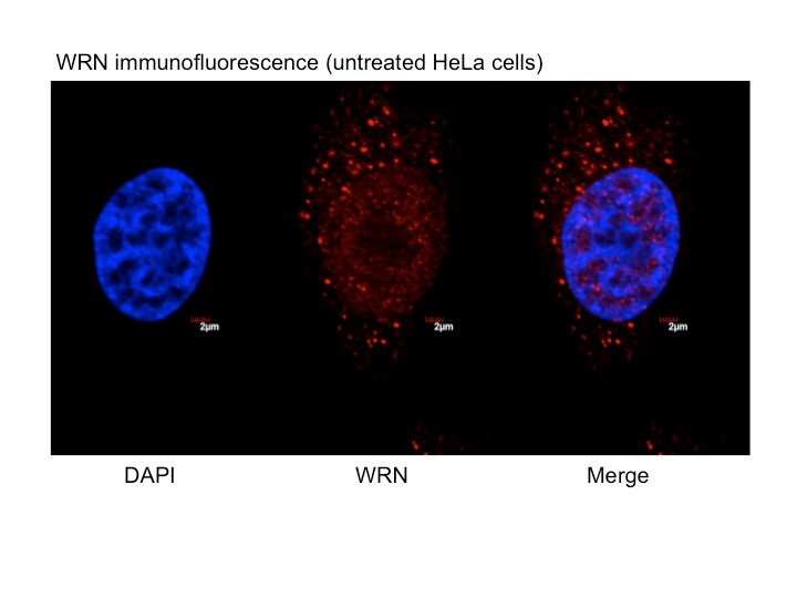
Immunocytochemistry/ Immunofluorescence: WRN Antibody [NB100-471]
Immunocytochemistry/Immunofluorescence: WRN Antibody [NB100-471] - Analysis of WRN in untreated, asynchronous HeLa cells using anti-WRN antibody. The primary antibody was used at a dilution of 1:100, incubated for 1 hour at room temperature in 1XPBS. Image from verified customer review.![Western Blot: WRN Antibody [NB100-471]](https://resources.rndsystems.com/images/products/WRN-Antibody-Western-Blot-NB100-471-img0005.jpg "Western Blot: WRN Antibody [NB100-471]")
Western Blot: WRN Antibody [NB100-471]
Western Blot: WRN Antibody [NB100-471] - Whole cell lysate (5, 15 and 50 ug) from HeLa cells.![Western Blot: WRN Antibody [NB100-471]](https://resources.rndsystems.com/images/products/WRN-Antibody-Western-Blot-NB100-471-img0006.jpg "Western Blot: WRN Antibody [NB100-471]")
Western Blot: WRN Antibody [NB100-471]
Western Blot: WRN Antibody [NB100-471] - Detection of WRN in 293T whole cell lysate using NB 100-471.![Western Blot: WRN Antibody [NB100-471]](https://resources.rndsystems.com/images/products/WRN-Antibody-Western-Blot-NB100-471-img0007.jpg "Western Blot: WRN Antibody [NB100-471]")
![Immunoprecipitation: WRN Antibody [NB100-471]](https://resources.rndsystems.com/images/products/WRN-Antibody-Immunoprecipitation-NB100-471-img0008.jpg "Immunoprecipitation: WRN Antibody [NB100-471]")
Immunoprecipitation: WRN Antibody [NB100-471]
Immunoprecipitation: WRN Antibody [NB100-471] - Detection of human WRN by western blot and immunoprecipitation. Samples: Whole cell lysate (5, 15 and 50 ug for WB; 1 mg for IP, 20% of IP loaded) from HeLa cells. Antibodies: Affinity purified rabbit anti-WRN antibody NB100-471 used for WB at 0.2 ug/ml (A) and 1 ug/ml (B) and used for IP at 3 ug/mg lysate. WRN was also immunoprecipitated by rabbit anti-WRN antibody NB100-472, which recognizes a downstream epitope. Detection: Chemiluminescence with exposure times of 30 seconds (A) and 3 seconds (B).Applications for WRN Antibody - BSA Free
Application
Recommended Usage
Immunocytochemistry/ Immunofluorescence
1:50 - 1:200
Immunoprecipitation
2 - 5 ug/mg lysate
Simple Western
1:25
Western Blot
1:5000 - 1:25000
Application Notes
Use in ICC/IF reported in scientific literature (PMID 25294835). Use in chromatin immunoprecipitation reported in scientific literature (PMID: 20065033). Use in Immunohistochemistry reported in scientific literature (PMID: 30146558). WRN antibody validated for WB from a verified customer review.
See Simple Western Antibody Database for Simple Western validation: Tested in Cell Lysate, separated by Size, antibody dilution of 1:25, apparent MW was 200 kDa
See Simple Western Antibody Database for Simple Western validation: Tested in Cell Lysate, separated by Size, antibody dilution of 1:25, apparent MW was 200 kDa
Reviewed Applications
Read 2 reviews rated 4 using NB100-471 in the following applications:
Formulation, Preparation, and Storage
Purification
Immunogen affinity purified
Formulation
Tris-Citrate/Phosphate (pH 7.0 - 8.0)
Format
BSA Free
Preservative
0.09% Sodium Azide
Concentration
1.0 mg/ml
Shipping
The product is shipped with polar packs. Upon receipt, store it immediately at the temperature recommended below.
Stability & Storage
Store at 4C. Do not freeze.
Background: WRN
Long Name
Werner syndrome ATP-dependent helicase
Alternate Names
EC 3.1.-.-, Exonuclease WRN, RecQ protein-like 2, RecQ3, RECQL2
Entrez Gene IDs
7486 (Human)
Gene Symbol
WRN
UniProt
Additional WRN Products
Product Documents for WRN Antibody - BSA Free
Certificate of Analysis
To download a Certificate of Analysis, please enter a lot or batch number in the search box below.
Product Specific Notices for WRN Antibody - BSA Free
This product is for research use only and is not approved for use in humans or in clinical diagnosis. Primary Antibodies are guaranteed for 1 year from date of receipt.
Citations for WRN Antibody - BSA Free
 Powered by Bioz
Customer Reviews for WRN Antibody - BSA Free (2)
4 out of 5
2 Customer Ratings
Have you used WRN Antibody - BSA Free?
Submit a review and receive an Amazon gift card!
$25/€18/£15/$25CAN/¥2500 Yen for a review with an image
$10/€7/£6/$10CAN/¥1110 Yen for a review without an image
Submit a review
Customer Images
Showing
1
-
2 of
2 reviews
Showing All
Filter By:
-
Application: Western BlotSample Tested: Colon cancer cell lineSpecies: HumanVerified Customer | Posted 12/09/2019Human colon cancer cells with siRNA CTRL/siRNA pool of siWrn/ 3 differents siRNA Wrn GAPDH control for loadingAb used at 1/1000_ON

-
Application: ImmunofluorescenceSample Tested: Asynchronous HeLa cellsSpecies: HumanVerified Customer | Posted 01/12/2015Localization of WRN in untreated, asynchronous HeLa cells (red = WRN; blue = DAPI).
There are no reviews that match your criteria.
Protocols
Find general support by application which include: protocols, troubleshooting, illustrated assays, videos and webinars.
- Antigen Retrieval Protocol (PIER)
- Antigen Retrieval for Frozen Sections Protocol
- Appropriate Fixation of IHC/ICC Samples
- Cellular Response to Hypoxia Protocols
- ChIP Protocol Video
- Chromatin Immunoprecipitation (ChIP) Protocol
- Chromatin Immunoprecipitation Protocol
- Chromogenic IHC Staining of Formalin-Fixed Paraffin-Embedded (FFPE) Tissue Protocol
- Chromogenic Immunohistochemistry Staining of Frozen Tissue
- ClariTSA™ Fluorophore Kits
- Detection & Visualization of Antibody Binding
- Fluorescent IHC Staining of Frozen Tissue Protocol
- Graphic Protocol for Heat-induced Epitope Retrieval
- Graphic Protocol for the Preparation and Fluorescent IHC Staining of Frozen Tissue Sections
- Graphic Protocol for the Preparation and Fluorescent IHC Staining of Paraffin-embedded Tissue Sections
- Graphic Protocol for the Preparation of Gelatin-coated Slides for Histological Tissue Sections
- ICC Cell Smear Protocol for Suspension Cells
- ICC Immunocytochemistry Protocol Videos
- ICC for Adherent Cells
- IHC Sample Preparation (Frozen sections vs Paraffin)
- Immunocytochemistry (ICC) Protocol
- Immunocytochemistry Troubleshooting
- Immunofluorescence of Organoids Embedded in Cultrex Basement Membrane Extract
- Immunofluorescent IHC Staining of Formalin-Fixed Paraffin-Embedded (FFPE) Tissue Protocol
- Immunohistochemistry (IHC) and Immunocytochemistry (ICC) Protocols
- Immunohistochemistry Frozen Troubleshooting
- Immunohistochemistry Paraffin Troubleshooting
- Immunoprecipitation Protocol
- Preparing Samples for IHC/ICC Experiments
- Preventing Non-Specific Staining (Non-Specific Binding)
- Primary Antibody Selection & Optimization
- Protocol for Heat-Induced Epitope Retrieval (HIER)
- Protocol for Making a 4% Formaldehyde Solution in PBS
- Protocol for VisUCyte™ HRP Polymer Detection Reagent
- Protocol for the Fluorescent ICC Staining of Cell Smears - Graphic
- Protocol for the Fluorescent ICC Staining of Cultured Cells on Coverslips - Graphic
- Protocol for the Preparation & Fixation of Cells on Coverslips
- Protocol for the Preparation and Chromogenic IHC Staining of Frozen Tissue Sections
- Protocol for the Preparation and Chromogenic IHC Staining of Frozen Tissue Sections - Graphic
- Protocol for the Preparation and Chromogenic IHC Staining of Paraffin-embedded Tissue Sections
- Protocol for the Preparation and Chromogenic IHC Staining of Paraffin-embedded Tissue Sections - Graphic
- Protocol for the Preparation and Fluorescent ICC Staining of Cells on Coverslips
- Protocol for the Preparation and Fluorescent ICC Staining of Non-adherent Cells
- Protocol for the Preparation and Fluorescent ICC Staining of Stem Cells on Coverslips
- Protocol for the Preparation and Fluorescent IHC Staining of Frozen Tissue Sections
- Protocol for the Preparation and Fluorescent IHC Staining of Paraffin-embedded Tissue Sections
- Protocol for the Preparation of Gelatin-coated Slides for Histological Tissue Sections
- Protocol for the Preparation of a Cell Smear for Non-adherent Cell ICC - Graphic
- R&D Systems Quality Control Western Blot Protocol
- TUNEL and Active Caspase-3 Detection by IHC/ICC Protocol
- The Importance of IHC/ICC Controls
- Troubleshooting Guide: Immunohistochemistry
- Troubleshooting Guide: Western Blot Figures
- Western Blot Conditions
- Western Blot Protocol
- Western Blot Protocol for Cell Lysates
- Western Blot Troubleshooting
- Western Blot Troubleshooting Guide
- View all Protocols, Troubleshooting, Illustrated assays and Webinars
Loading...