xCT Antibody - BSA Free
Novus Biologicals | Catalog # NB300-318
Key Product Details
Validated by
Orthogonal Validation, Biological Validation
Species Reactivity
Validated:
Human, Mouse, Rat
Cited:
Human, Mouse, Rat
Applications
Validated:
Immunohistochemistry, Immunohistochemistry-Paraffin, Western Blot, Flow Cytometry, Dual RNAscope ISH-IHC, Immunocytochemistry/ Immunofluorescence, Simple Western, Immunoprecipitation (Negative)
Cited:
Immunohistochemistry-Paraffin, Western Blot, Flow Cytometry, Immunocytochemistry/ Immunofluorescence, IF/IHC
Label
Unconjugated
Antibody Source
Polyclonal Rabbit IgG
Format
BSA Free
Loading...
Product Specifications
Immunogen
This xCT Antibody was prepared from a synthetic peptide made to an N-terminal region of the human xCT protein (between residues 1-50) [UniProt Q9UPY5].
Reactivity Notes
Rat reactivity reported in scientific literature (PMID: 21540084).
Localization
Membrane
Clonality
Polyclonal
Host
Rabbit
Isotype
IgG
Scientific Data Images for xCT Antibody - BSA Free
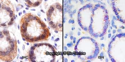
Dual RNAscope ISH-IHC: xCT Antibody [NB300-318] - Formalin-fixed paraffin-embedded tissue sections of human stomach were probed for xCT mRNA (ACD RNAScope Probe, catalog #422688; Fast Red chromogen, ACD catalog # 322750). Adjacent tissue section was processed for immunohistochemistry using Rabbit Polyclonal (Novus Biologicals catalog # NB300-318) at 0.25ug/mL with 1 hour incubation at room temperature followed by incubation with anti-rabbit IgG VisUCyte HRP Polymer Antibody (Catalog # VC003) and DAB chromogen (yellow-brown). Tissue was counterstained with hematoxylin (blue). Specific staining was localized to glandular cells.
![Simple Western: xCT AntibodyBSA Free [NB300-318]](https://resources.rndsystems.com/images/products/xCT-Antibody-Simple-Western-NB300-318-img0013.jpg "Simple Western: xCT AntibodyBSA Free [NB300-318]")
Simple Western: xCT AntibodyBSA Free [NB300-318]
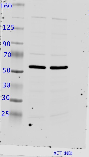
Simple Western: xCT Antibody [NB300-318] - Simple Western lane view shows a specific band for xCT using NB300-318 at 25 ug/ml in HeLa and HeLa + DEM cell lysates. This experiment was performed under reducing conditions using the 12-230 kDa separation system.![Western Blot: xCT AntibodyBSA Free [NB300-318]](https://resources.rndsystems.com/images/products/xCT-Antibody-Western-Blot-NB300-318-img0015.jpg "Western Blot: xCT AntibodyBSA Free [NB300-318]")
Western Blot: xCT AntibodyBSA Free [NB300-318]
xCT-Antibody-Western-Blot-NB300-318-img0015.jpg![Immunohistochemistry: xCT Antibody - BSA Free [NB300-318]](https://resources.rndsystems.com/images/products/xCT-Antibody-Immunohistochemistry-NB300-318-img0006.jpg "Immunohistochemistry: xCT Antibody - BSA Free [NB300-318]")
Immunohistochemistry: xCT Antibody - BSA Free [NB300-318]
Immunohistochemistry: xCT Antibody [NB300-318] - xCT staining in the absorptive epithelia of small intestinal villi detected using NB300-318.![Flow Cytometry: xCT Antibody - BSA Free [NB300-318]](https://resources.rndsystems.com/images/products/xCT-Antibody-Flow-Cytometry-NB300-318-img0009.jpg "Flow Cytometry: xCT Antibody - BSA Free [NB300-318]")
Flow Cytometry: xCT Antibody - BSA Free [NB300-318]
Flow Cytometry: xCT Antibody [NB300-318] - An intracellular stain was performed on HeLa cells with NB300-318AF647 (blue) and a matched isotype control (orange). Cells were fixed with 4% PFA and then permeabilized with 0.1% saponin. Cells were incubated in an antibody dilution of 2.5 ug/mL for 30 minutes at room temperature. Both antibodies were conjugated to Alexa Fluor 647.![Immunocytochemistry/ Immunofluorescence: xCT Antibody - BSA Free [NB300-318]](https://resources.rndsystems.com/images/products/xCT-Antibody-Immunocytochemistry-Immunofluorescence-NB300-318-img0005.jpg "Immunocytochemistry/ Immunofluorescence: xCT Antibody - BSA Free [NB300-318]")
Immunocytochemistry/ Immunofluorescence: xCT Antibody - BSA Free [NB300-318]
Immunocytochemistry/Immunofluorescence: xCT Antibody [NB300-318] - xCT antibody was tested in HepG2 cells with DyLight 488 (green). Nuclei and alpha-tubulin were counterstained with DAPI (blue) and DyLight 550 (red).![Flow Cytometry: xCT Antibody - BSA Free [NB300-318]](https://resources.rndsystems.com/images/products/xCT-Antibody-Flow-Cytometry-NB300-318-img0010.jpg "Flow Cytometry: xCT Antibody - BSA Free [NB300-318]")
Flow Cytometry: xCT Antibody - BSA Free [NB300-318]
Flow Cytometry: xCT Antibody [NB300-318] - An intracellular stain was performed on HeLa cells with xCT Antibody NB300-318AF488 (blue) and a matched isotype control (orange). Cells were fixed with 4% PFA and then permeabilized with 0.1% saponin. Cells were incubated in an antibody dilution of 5 ug/mL for 30 minutes at room temperature. Both antibodies were conjugated to Alexa Fluor 488.![Western Blot: xCT AntibodyBSA Free [NB300-318]](https://resources.rndsystems.com/images/products/xCT-Antibody-Western-Blot-NB300-318-img0007.jpg "Western Blot: xCT AntibodyBSA Free [NB300-318]")
Western Blot: xCT AntibodyBSA Free [NB300-318]
Western Blot: xCT Antibody [NB300-318] - Total protein from Human HeLa cells treated with and without 0.1 mM Diethyl Maleate for 24 hours was separated on a 12% gel by SDS-PAGE, transferred to PVDF membrane and blocked in 5% non-fat milk in TBST. The membrane was probed with 2.0 ug/ml anti-xCT in 1% non-fat milk in TBST and detected with an anti-rabbit HRP secondary antibody using chemiluminescence. Note the increase in xCT expression with treatment.![Flow Cytometry: xCT Antibody - BSA Free [NB300-318]](https://resources.rndsystems.com/images/products/xCT-Antibody-Flow-Cytometry-NB300-318-img0008.jpg "Flow Cytometry: xCT Antibody - BSA Free [NB300-318]")
Flow Cytometry: xCT Antibody - BSA Free [NB300-318]
Flow Cytometry: xCT Antibody [NB300-318] - An intracellular stain was performed on HeLa with xCT Antibody NB300-318 and a matched isotype control. Cells were fixed with 4% PFA and then permeablized with 0.1% saponin. Cells were incubated in an antibody dilution of 2.5 ug/mL for 30 minutes at room temperature, followed by Rabbit IgG APC-conjugated Secondary Antibody (F0111, R&D Systems).![Simple Western: xCT AntibodyBSA Free [NB300-318]](https://resources.rndsystems.com/images/products/xCT-Antibody-Simple-Western-NB300-318-img0014.jpg "Simple Western: xCT AntibodyBSA Free [NB300-318]")
Simple Western: xCT AntibodyBSA Free [NB300-318]
Simple Western: xCT Antibody [NB300-318] - Simple Western lane view shows a specific band for xCT using NB300-318 at 25 ug/mL in HepG2 cell lysates and antibody at 25 ug/mL. Electropherogram image of corresponding Simple Western lane view at WES molecular weight of 63 kDa. Image reported by internal validation.
Western Blot: xCT Antibody - BSA Free [NB300-318] -
Western Blot: xCT Antibody - BSA Free [NB300-318] - Subcutaneous injection into nude mice revealed that (A) MDA-MB-231 SH-4-54-resistant clone #2 proliferated at a slower rate than its wild-type (WT) counterpart in vivo. (B) qPCR demonstrated that xCT mRNA levels were lower in tumours isolated from animals injected with clone #2 relative to WT cells (2 animals per treatment group). (C) Western blot analysis of protein isolated from subcutaneous tumours derived from in vivo growth of the clones relative to WT-derived tumours revealed that xCT levels remained low, phospho-STAT5 (p-STAT5) levels remained high, & phospho-STAT3 (p-STAT3) levels remained unchanged in the absence of SH-4-54. Image collected & cropped by CiteAb from the following publication (https://dx.plos.org/10.1371/journal.pone.0161202), licensed under a CC-BY license. Not internally tested by Novus Biologicals.
Western Blot: xCT Antibody - BSA Free [NB300-318] -
Western Blot: xCT Antibody - BSA Free [NB300-318] - A link between KSHV latency & the MC1-R signaling axis in skin-derived cell lines. (a) Western blot analysis of phosphorylated NF-kappa B p65, MC1-R, & TRP-1 in total cell lysates extracted from MeWo (a) or Mel1700 (b) cells either uninfected (control) or acutely infected with KSHV for 0.3 h, 1 h, 3 h, or 6 h. GAPDH was used as loading control. (c) ImageJ quantitation of the p65 band intensities in (a) & (b) relative to GAPDH controls. (d) Mel1700 cells were infected in 6-well plates with increasing volumes (mL/well) of concentrated supernatant containing infectious KSHV, & total RNA from infected cells was subjected to RT-PCR using primer sets for host anti-inflammatory MC1R, POMC, & SLC7A11. (e) Equal aliquots from the same RNA used in (d) were subjected to RT-PCR analysis for select viral latency & cell growth control genes (i.e., GPCR, LANA, & v-FLIP). (f) Western blot analysis of the melanoma cell marker, Melan A, & anti-inflammatory genes MC1-R, TRP1, & SLC7A11 in total cell lysates of uninfected (−) or chronically infected (+) long-term cultures of MeWo-KSHV & Mel1700-KSHV cells. GAPDH was used as an internal control for both the RT-PCR (e) & western blot assays. Image collected & cropped by CiteAb from the following publication (https://pubmed.ncbi.nlm.nih.gov/24701351), licensed under a CC-BY license. Not internally tested by Novus Biologicals.
Western Blot: xCT Antibody - BSA Free [NB300-318] -
Western Blot: xCT Antibody - BSA Free [NB300-318] - A link between KSHV latency & the MC1-R signaling axis in skin-derived cell lines. (a) Western blot analysis of phosphorylated NF-kappa B p65, MC1-R, & TRP-1 in total cell lysates extracted from MeWo (a) or Mel1700 (b) cells either uninfected (control) or acutely infected with KSHV for 0.3 h, 1 h, 3 h, or 6 h. GAPDH was used as loading control. (c) ImageJ quantitation of the p65 band intensities in (a) & (b) relative to GAPDH controls. (d) Mel1700 cells were infected in 6-well plates with increasing volumes (mL/well) of concentrated supernatant containing infectious KSHV, & total RNA from infected cells was subjected to RT-PCR using primer sets for host anti-inflammatory MC1R, POMC, & SLC7A11. (e) Equal aliquots from the same RNA used in (d) were subjected to RT-PCR analysis for select viral latency & cell growth control genes (i.e., GPCR, LANA, & v-FLIP). (f) Western blot analysis of the melanoma cell marker, Melan A, & anti-inflammatory genes MC1-R, TRP1, & SLC7A11 in total cell lysates of uninfected (−) or chronically infected (+) long-term cultures of MeWo-KSHV & Mel1700-KSHV cells. GAPDH was used as an internal control for both the RT-PCR (e) & western blot assays. Image collected & cropped by CiteAb from the following publication (https://pubmed.ncbi.nlm.nih.gov/24701351), licensed under a CC-BY license. Not internally tested by Novus Biologicals.Applications for xCT Antibody - BSA Free
Application
Recommended Usage
Flow Cytometry
2-3ug/ml. Use reported in scientific literature (PMID 20028852)
Immunocytochemistry/ Immunofluorescence
1:100-1:1000
Immunohistochemistry
5 u/gml
Immunohistochemistry-Paraffin
5 ug/ml
Simple Western
10 ug/ml
Western Blot
1:1000
Application Notes
Immunoprecipitation is not recommended. In Western blot this antibody recognizes a band at approx. 35 kDa, and in ICC/IF membrane staining was observed in HeLa cells. Permeablization is recommended prior to performing Flow analysis.
In Simple Western only 10 - 15 uL of the recommended dilution is used per data point.
See Simple Western Antibody Database for Simple Western validation: Tested in HepG2 lysate, HeLa and HeLa + DEM cell lysates, separated by Size, antibody dilution of 25 ug/mL, apparent MW was 63 kDa. Separated by Size-Wes, Sally Sue/Peggy Sue.
Reviewed Applications
Read 3 reviews rated 4.3 using NB300-318 in the following applications:
Flow Cytometry Panel Builder
Bio-Techne Knows Flow Cytometry
Save time and reduce costly mistakes by quickly finding compatible reagents using the Panel Builder Tool.
Advanced Features
- Spectra Viewer - Custom analysis of spectra from multiple fluorochromes
- Spillover Popups - Visualize the spectra of individual fluorochromes
- Antigen Density Selector - Match fluorochrome brightness with antigen density

Formulation, Preparation, and Storage
Purification
Immunogen affinity purified
Formulation
PBS
Format
BSA Free
Preservative
0.02% Sodium Azide
Concentration
1.0 mg/ml
Shipping
The product is shipped with polar packs. Upon receipt, store it immediately at the temperature recommended below.
Stability & Storage
Store at 4C short term. Aliquot and store at -20C long term. Avoid freeze-thaw cycles.
Background: xCT/SLC7A11
References
1. Liu, J., Xia, X., & Huang, P. (2020). xCT: A Critical Molecule That Links Cancer Metabolism to Redox Signaling. Molecular therapy : the journal of the American Society of Gene Therapy. https://doi.org/10.1016/j.ymthe.2020.08.021
2. Koppula, P., Zhang, Y., Zhuang, L., & Gan, B. (2018). Amino acid transporter SLC7A11/xCT at the crossroads of regulating redox homeostasis and nutrient dependency of cancer. Cancer communications. https://doi.org/10.1186/s40880-018-0288-x
3. Lin, W., Wang, C., Liu, G., Bi, C., Wang, X., Zhou, Q., & Jin, H. (2020). SLC7A11/xCT in cancer: biological functions and therapeutic implications. American journal of cancer research.
4. xCT: Uniprot (Q9UPY5)
5. Koppula, P., Zhuang, L., & Gan, B. (2020). Cystine transporter SLC7A11/xCT in cancer: ferroptosis, nutrient dependency, and cancer therapy. Protein & cell. https://doi.org/10.1007/s13238-020-00789-5
6. Liu, L., Liu, R., Liu, Y., Li, G., Chen, Q., Liu, X., & Ma, S. (2020). Cystine-glutamate antiporter xCT as a therapeutic target for cancer. Cell biochemistry and function. https://doi.org/10.1002/cbf.3581
7. Cui, Q., Wang, J. Q., Assaraf, Y. G., Ren, L., Gupta, P., Wei, L., Ashby, C. R., Jr, Yang, D. H., & Chen, Z. S. (2018). Modulating ROS to overcome multidrug resistance in cancer. Drug resistance updates : reviews and commentaries in antimicrobial and anticancer chemotherapy. https://doi.org/10.1016/j.drup.2018.11.001
Long Name
Cationic 1/Solute Carrier Family 7 Member 11
Alternate Names
CCBR1, SLC7A11
Gene Symbol
SLC7A11
UniProt
Additional xCT/SLC7A11 Products
Product Documents for xCT Antibody - BSA Free
Certificate of Analysis
To download a Certificate of Analysis, please enter a lot or batch number in the search box below.
Product Specific Notices for xCT Antibody - BSA Free
This product is for research use only and is not approved for use in humans or in clinical diagnosis. Primary Antibodies are guaranteed for 1 year from date of receipt.
Related Research Areas
Citations for xCT Antibody - BSA Free
 Powered by Bioz
Customer Reviews for xCT Antibody - BSA Free (3)
4.3 out of 5
3 Customer Ratings
Have you used xCT Antibody - BSA Free?
Submit a review and receive an Amazon gift card!
$25/€18/£15/$25CAN/¥2500 Yen for a review with an image
$10/€7/£6/$10CAN/¥1110 Yen for a review without an image
Submit a review
Customer Images
Showing
1
-
3 of
3 reviews
Showing All
Filter By:
-
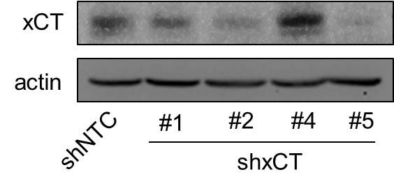
Application: Western BlotSample Tested: Human Cancer CellsSpecies: HumanVerified Customer | Posted 09/18/2018This antibody was used to detect xCT expression in control (NTC) and different xCT knockdown subclones in human breast cancer cell line by western blot.

-
Application: Western BlotSample Tested: pancreatic cancer cell line lysateSpecies: HumanVerified Customer | Posted 12/24/2015anti-xCT (1:1000 dilution)
-
Application: Western BlotSample Tested: Mouse Whole BrainSpecies: MouseVerified Customer | Posted 05/01/2013
There are no reviews that match your criteria.
Protocols
View specific protocols for xCT Antibody - BSA Free (NB300-318):
xCT Antibody:
Protocol for Flow Cytometry Intracellular Staining
Sample Preparation.
1. Grow cells to 60-85% confluency. Flow cytometry requires between 2 x 10⁵ and 1 x 10⁶ cells for optimal performance.
2. If cells are adherent, harvest gently by washing once with staining buffer and then scraping. Avoid using trypsin as this can disrupt certain epitopes of interest. If enzymatic harvest is required, use Accutase, Collagenase, or TrypLE Express for a less damaging option.
3. Reserve 100 uL for counting, then transfer cell volume into a 50 mL conical tube and centrifuge for 8 minutes at 400 RCF.
a. Count cells using a hemocytometer and a 1:1 trypan blue exclusion stain to determine cell viability before starting the flow protocol. If cells appear blue, do not proceed.
4. Re-suspend cells to a concentration of 1 x 10⁶ cells/mL in staining buffer (NBP2-26247).
5. Aliquot out 100 uL samples in accordance with your experimental samples.
Tip: When cell surface and intracellular staining are required in the same sample, it is advisable that the cell surface staining be performed first since the fixation and permeablization steps might reduce the availability of surface antigens.
Intracellular Staining.
Tip: When performing intracellular staining, it is important to use appropriate fixation and permeabilization reagents based upon the target and its subcellular location. Generally, our Intracellular Flow Assay Kit (NBP2-29450) is a good place to start as it contains an optimized combination of reagents for intracellular staining as well as an inhibitor of intracellular protein transport (necessary if staining secreted proteins). Certain targets may require more gentle or transient permeabilization protocols such as the commonly employed methanol or saponin-based methods.
Protocol for Cytoplasmic Targets:
1. Fix the cells by adding 100 uL fixation solution (such as 4% PFA) to each sample for 10-15 minutes.
2. Permeabilize cells by adding 100 uL of a permeabization buffer to every 1 x 10⁶ cells present in the sample. Mix well and incubate at room temperature for 15 minutes.
a. For cytoplasmic targets, use a gentle permeabilization solution such as 1X PBS + 0.5% Saponin or 1X PBS + 0.5% Tween-20.
b. To maintain the permeabilized state throughout your experiment, use staining buffer + 0.1% of the permeabilization reagent (i.e. 0.1% Tween-20 or 0.1% Saponin).
3. Following the 15 minute incubation, add 2 mL of the staining buffer + 0.1% permeabilizer to each sample.
4. Centrifuge for 1 minute at 400 RCF.
5. Discard supernatant and re-suspend in 100 uL of staining buffer + 0.1% permeabilizer.
6. Add appropriate amount of each antibody (eg. 1 test or 1 ug per sample, as experimentally determined).
7. Mix well and incubate at room temperature for 30 minutes- 1 hour. Gently mix samples every 10-15 minutes.
8. Following the primary/conjugate incubation, add 1-2 mL/sample of staining buffer +0.1% permeabilizer and centrifuge for 1 minute at 400 RCF.
9. Wash twice by re-suspending cells in staining buffer (2 mL for tubes or 200 uL for wells) and centrifuging at 400 RCF for 5 minutes. Discard supernatant.
10. Add appropriate amount of secondary antibody (as experimentally determined) to each sample.
11. Incubate at room temperature in dark for 20 minutes.
12. Add 1-2 mL of staining buffer and centrifuge at 400 RCF for 1 minute and discard supernatant.
13. Wash twice by re-suspending cells in staining buffer (2 mL for tubes or 200 uL for wells) and centrifuging at 400 RCF for 5 minutes. Discard supernatant.
14. Resuspend in an appropriate volume of staining buffer (usually 500 uL per sample) and proceed with analysis on your flow cytometer.
Immunocytochemistry Protocol
Culture cells to appropriate density in 35 mm culture dishes or 6-well plates.
1. Remove culture medium and wash the cells briefly in PBS. Add 10% formalin to the dish and fix at room temperature for 10 minutes.
2. Remove the formalin and wash the cells in PBS.
3. Permeablize the cells with 0.1% Triton X100 or other suitable detergent for 10 min.
4. Remove the permeablization buffer and wash three times for 10 minutes each in PBS. Be sure to not let the specimen dry out.
5. To block nonspecific antibody binding, incubate in 10% normal goat serum from 1 hour to overnight at room temperature.
6. Add primary antibody at appropriate dilution and incubate overnight at 4C.
7. Remove primary antibody and replace with PBS. Wash three times for 10 minutes each.
8. Add secondary antibody at appropriate dilution. Incubate for 1 hour at room temperature.
9. Remove secondary antibody and replace with PBS. Wash three times for 10 minutes each.
10. Counter stain DNA with DAPi if required.
*The above information is only intended as a guide. The researcher should determine what protocol best meets their needs. Please follow safe laboratory procedures.
xCT Antibody:
Immunohistochemistry-Paraffin Embedded Sections
Antigen Unmasking:
Bring slides to a boil in 10 mM sodium citrate buffer (pH 6.0) then maintain at a sub-boiling temperature for 10 minutes. Cool slides on bench-top for 30 minutes.
Staining:
1. Wash sections in deionized water three times for 5 minutes each.
2. Wash sections in wash buffer for 5 minutes.
3. Block each section with 100-400 ul blocking solution for 1 hour at room temperature.
4. Remove blocking solution and add 100-400 ul diluted primary antibody. Incubate overnight at 4 C.
5. Remove antibody solution and wash sections in wash buffer three times for 5 minutes each.
6. Add 100-400 ul biotinylated diluted secondary antibody. Incubate 30 minutes at room temperature.
7. Remove secondary antibody solution and wash sections three times with wash buffer for 5 minutes each.
8. Add 100-400 ul Streptavidin-HRP reagent to each section and incubate for 30 minutes at room temperature.
9. Wash sections three times in wash buffer for 5 minutes each.
10. Add 100-400 ul DAB substrate to each section and monitor staining closely.
11. As soon as the sections develop, immerse slides in deionized water.
12. Counterstain sections in hematoxylin.
13. Wash sections in deionized water two times for 5 minutes each.
14. Dehydrate sections.
15. Mount coverslips.
Western Blot Protocol
1. Perform SDS-PAGE on samples to be analyzed, loading 40 ug of total protein per lane.
2. Transfer proteins to membrane according to the instructions provided by the manufacturer of the membrane and transfer apparatus.
3. Stain according to standard Ponceau S procedure (or similar product) to assess transfer success, and mark molecular weight standards where appropriate.
4. Rinse the blot.
5. Block the membrane using standard blocking buffer for at least 1 hour.
6. Wash the membrane in wash buffer three times for 10 minutes each.
7. Dilute primary antibody in blocking buffer and incubate 1 hour at room temperature.
8. Wash the membrane in wash buffer three times for 10 minutes each.
9. Apply the diluted HRP conjugated secondary antibody in blocking buffer (as per manufacturers instructions) and incubate 1 hour at room temperature.
10. Wash the blot in wash buffer three times for 10 minutes each (this step can be repeated as required to reduce background).
11. Apply the detection reagent of choice in accordance with the manufacturers instructions.
Note: Tween-20 can be added to the blocking or antibody dilution buffer at a final concentration of 0.05-0.2%.
Find general support by application which include: protocols, troubleshooting, illustrated assays, videos and webinars.
- 7-Amino Actinomycin D (7-AAD) Cell Viability Flow Cytometry Protocol
- Antigen Retrieval Protocol (PIER)
- Antigen Retrieval for Frozen Sections Protocol
- Appropriate Fixation of IHC/ICC Samples
- Cellular Response to Hypoxia Protocols
- Chromogenic IHC Staining of Formalin-Fixed Paraffin-Embedded (FFPE) Tissue Protocol
- Chromogenic Immunohistochemistry Staining of Frozen Tissue
- ClariTSA™ Fluorophore Kits
- Detection & Visualization of Antibody Binding
- Extracellular Membrane Flow Cytometry Protocol
- Flow Cytometry Protocol for Cell Surface Markers
- Flow Cytometry Protocol for Staining Membrane Associated Proteins
- Flow Cytometry Staining Protocols
- Flow Cytometry Troubleshooting Guide
- Fluorescent IHC Staining of Frozen Tissue Protocol
- Graphic Protocol for Heat-induced Epitope Retrieval
- Graphic Protocol for the Preparation and Fluorescent IHC Staining of Frozen Tissue Sections
- Graphic Protocol for the Preparation and Fluorescent IHC Staining of Paraffin-embedded Tissue Sections
- Graphic Protocol for the Preparation of Gelatin-coated Slides for Histological Tissue Sections
- ICC Cell Smear Protocol for Suspension Cells
- ICC Immunocytochemistry Protocol Videos
- ICC for Adherent Cells
- IHC Sample Preparation (Frozen sections vs Paraffin)
- ISH-IHC Protocol for Chromogenic Detection on Formalin Fixed Paraffin Embedded (FFPE) Tissue
- Immunocytochemistry (ICC) Protocol
- Immunocytochemistry Troubleshooting
- Immunofluorescence of Organoids Embedded in Cultrex Basement Membrane Extract
- Immunofluorescent IHC Staining of Formalin-Fixed Paraffin-Embedded (FFPE) Tissue Protocol
- Immunohistochemistry (IHC) and Immunocytochemistry (ICC) Protocols
- Immunohistochemistry Frozen Troubleshooting
- Immunohistochemistry Paraffin Troubleshooting
- Intracellular Flow Cytometry Protocol Using Alcohol (Methanol)
- Intracellular Flow Cytometry Protocol Using Detergents
- Intracellular Nuclear Staining Flow Cytometry Protocol Using Detergents
- Intracellular Staining Flow Cytometry Protocol Using Alcohol Permeabilization
- Intracellular Staining Flow Cytometry Protocol Using Detergents to Permeabilize Cells
- Preparing Samples for IHC/ICC Experiments
- Preventing Non-Specific Staining (Non-Specific Binding)
- Primary Antibody Selection & Optimization
- Propidium Iodide Cell Viability Flow Cytometry Protocol
- Protocol for Heat-Induced Epitope Retrieval (HIER)
- Protocol for Liperfluo
- Protocol for Making a 4% Formaldehyde Solution in PBS
- Protocol for VisUCyte™ HRP Polymer Detection Reagent
- Protocol for the Characterization of Human Th22 Cells
- Protocol for the Characterization of Human Th9 Cells
- Protocol for the Fluorescent ICC Staining of Cell Smears - Graphic
- Protocol for the Fluorescent ICC Staining of Cultured Cells on Coverslips - Graphic
- Protocol for the Preparation & Fixation of Cells on Coverslips
- Protocol for the Preparation and Chromogenic IHC Staining of Frozen Tissue Sections
- Protocol for the Preparation and Chromogenic IHC Staining of Frozen Tissue Sections - Graphic
- Protocol for the Preparation and Chromogenic IHC Staining of Paraffin-embedded Tissue Sections
- Protocol for the Preparation and Chromogenic IHC Staining of Paraffin-embedded Tissue Sections - Graphic
- Protocol for the Preparation and Fluorescent ICC Staining of Cells on Coverslips
- Protocol for the Preparation and Fluorescent ICC Staining of Non-adherent Cells
- Protocol for the Preparation and Fluorescent ICC Staining of Stem Cells on Coverslips
- Protocol for the Preparation and Fluorescent IHC Staining of Frozen Tissue Sections
- Protocol for the Preparation and Fluorescent IHC Staining of Paraffin-embedded Tissue Sections
- Protocol for the Preparation of Gelatin-coated Slides for Histological Tissue Sections
- Protocol for the Preparation of a Cell Smear for Non-adherent Cell ICC - Graphic
- Protocol: Annexin V and PI Staining by Flow Cytometry
- Protocol: Annexin V and PI Staining for Apoptosis by Flow Cytometry
- R&D Systems Quality Control Western Blot Protocol
- TUNEL and Active Caspase-3 Detection by IHC/ICC Protocol
- The Importance of IHC/ICC Controls
- Troubleshooting Guide: Fluorokine Flow Cytometry Kits
- Troubleshooting Guide: Immunohistochemistry
- Troubleshooting Guide: Western Blot Figures
- Western Blot Conditions
- Western Blot Protocol
- Western Blot Protocol for Cell Lysates
- Western Blot Troubleshooting
- Western Blot Troubleshooting Guide
- View all Protocols, Troubleshooting, Illustrated assays and Webinars
FAQs for xCT Antibody - BSA Free
Showing
1
-
5 of
10 FAQs
Showing All
-
Q: Do you have a slc7a11 antibody that is conjugated, reacts to mouse, works with cell surface staining, does not need permeabilization?
A: SLC7A11 or xCT antibody NB300-318AF594 targets a cytoplasmic region of the protein, so it should work without permeabilization. However, the data does say it was permeabilized with saponin, so we cannot say for certain if it will work without permeabilization.
-
Q: For xCT antibody [NB300-318], why do you see a band at 35kDa when the predicted MW is ~55kDa and xCT is not known to undergo cleavage?
A: Although the theoretical molecular weight of xCT is ~55kDa, the protein’s amino acid structure, with multiple charged residues, results in its fast migration. The research paper by Liefferinge et al 2016 (The Journal of Comparative Neurology 524:1015-1032) shows the testing of several different xCT antibodies in knockout study and it was confirmed that the protein indeed runs at 35kDa (Fig.2. lane 1&2 lanes are xCT positive and lanes 3&4 are from xCT knockouts).
-
Q: Hello, I'm trying to run a western blot with NB300-318. What is the blocking buffer used?
A: For NB300-318 we recommend using 5% milk to block Blocking solution 1X TBST 5% non-fat dry milk
-
Q: I am using the xCT antibody NB300-318 for IHC. I was wondering if you could recommend a rabbit negative isotype control IGg/serum?
A: I would recommend NB810-56910 as an isotype control for you
-
Q: I am using the xCT antibody, NB300-318, for IHC. I was wondering if you could recommend a peptide to use for a negative control?
A: I regret to inform you that we do not have a blocking peptide available for this antibody at this time.
-
Q: I am using the xCT antibody, NB300-318, for IHC. I was wondering if you could recommend a rabbit negative isotype control IGg/serum?
A: I would recommend catalog number NB810-56910 as a rabbit isotype control for NB300-318.
-
Q: Is NB300-318 suitable for live cell flow cytometry? (is its epitope in the external side of the cells?) I would need it for FACS sorting application
A: The immunogen for this antibody is cytoplamic, so this antibody would not be suitable for live flow.
-
Q: Is there a blocking peptide available for use with NB300-318?
A: We unfortunately do not carry the blocking peptide to NB300-318.
-
Q: We are wondering if it is possible to know if there is -NH2 residues on this synthetic peptide. If yes, how many -NH2 or lysine are there?
A: The immunogen for NB300-318 contains 1 lysine residue.
-
Q: We ordered some xCT antibody (NB300-318). Is itpossible to know if there is -NH2 residues on this synthetic peptide. If so, how many -NH2 or lysine are there?
A: The immunogen for NB300-318 contains 1 lysine residue.
-
Q: Do you have a slc7a11 antibody that is conjugated, reacts to mouse, works with cell surface staining, does not need permeabilization?
A: SLC7A11 or xCT antibody NB300-318AF594 targets a cytoplasmic region of the protein, so it should work without permeabilization. However, the data does say it was permeabilized with saponin, so we cannot say for certain if it will work without permeabilization.
-
Q: For xCT antibody [NB300-318], why do you see a band at 35kDa when the predicted MW is ~55kDa and xCT is not known to undergo cleavage?
A: Although the theoretical molecular weight of xCT is ~55kDa, the protein’s amino acid structure, with multiple charged residues, results in its fast migration. The research paper by Liefferinge et al 2016 (The Journal of Comparative Neurology 524:1015-1032) shows the testing of several different xCT antibodies in knockout study and it was confirmed that the protein indeed runs at 35kDa (Fig.2. lane 1&2 lanes are xCT positive and lanes 3&4 are from xCT knockouts).
-
Q: Hello, I'm trying to run a western blot with NB300-318. What is the blocking buffer used?
A: For NB300-318 we recommend using 5% milk to block Blocking solution 1X TBST 5% non-fat dry milk
-
Q: I am using the xCT antibody NB300-318 for IHC. I was wondering if you could recommend a rabbit negative isotype control IGg/serum?
A: I would recommend NB810-56910 as an isotype control for you
-
Q: I am using the xCT antibody, NB300-318, for IHC. I was wondering if you could recommend a peptide to use for a negative control?
A: I regret to inform you that we do not have a blocking peptide available for this antibody at this time.
-
Q: I am using the xCT antibody, NB300-318, for IHC. I was wondering if you could recommend a rabbit negative isotype control IGg/serum?
A: I would recommend catalog number NB810-56910 as a rabbit isotype control for NB300-318.
-
Q: Is NB300-318 suitable for live cell flow cytometry? (is its epitope in the external side of the cells?) I would need it for FACS sorting application
A: The immunogen for this antibody is cytoplamic, so this antibody would not be suitable for live flow.
-
Q: Is there a blocking peptide available for use with NB300-318?
A: We unfortunately do not carry the blocking peptide to NB300-318.
-
Q: We are wondering if it is possible to know if there is -NH2 residues on this synthetic peptide. If yes, how many -NH2 or lysine are there?
A: The immunogen for NB300-318 contains 1 lysine residue.
-
Q: We ordered some xCT antibody (NB300-318). Is itpossible to know if there is -NH2 residues on this synthetic peptide. If so, how many -NH2 or lysine are there?
A: The immunogen for NB300-318 contains 1 lysine residue.
Loading...