XIAP Antibody - BSA Free
Novus Biologicals | Catalog # NB100-56183
![Western Blot: XIAP Antibody [NB100-56183]](https://resources.rndsystems.com/images/products/XIAP-Antibody-Western-Blot-NB100-56183-img0006.jpg "Western Blot: XIAP Antibody [NB100-56183]")
Loading...
Key Product Details
Species Reactivity
Validated:
Human, Mouse, Rat
Cited:
Human, Mouse
Applications
Validated:
Immunohistochemistry, Immunohistochemistry-Paraffin, Western Blot, Immunocytochemistry/ Immunofluorescence, Immunoprecipitation
Cited:
Western Blot, Immunocytochemistry/ Immunofluorescence
Label
Unconjugated
Antibody Source
Polyclonal Rabbit IgG
Format
BSA Free
Loading...
Product Specifications
Immunogen
Recombinant BIR2 domain protein fragment of human XIAP was used as immunogen. The BIR2 domain used for immunogen corresponds to amino acids 163-230 of human XIAP (Deveraux et al, 1999).
Specificity
The antibody recognizes epitopes in the BIR2 domain of XIAP. Therefore it can recognize full-length XIAP and XIAP cleavage fragments containing the BIR2 domain. However, XIAP cleavage fragments may be biologically unstable, and therefore cleavage fragments may be difficult to detect.
Clonality
Polyclonal
Host
Rabbit
Isotype
IgG
Scientific Data Images for XIAP Antibody - BSA Free
Western Blot: XIAP Antibody [NB100-56183]
Western Blot: XIAP Antibody - Unpurified [NB100-56183] - Analysis of XIAP in various tumor cell lines using this antibody at 1:2000.![Immunocytochemistry/ Immunofluorescence: XIAP Antibody [NB100-56183]](https://resources.rndsystems.com/images/products/XIAP-Antibody-Immunocytochemistry-Immunofluorescence-NB100-56183-img0007.jpg "Immunocytochemistry/ Immunofluorescence: XIAP Antibody [NB100-56183]")
Immunocytochemistry/ Immunofluorescence: XIAP Antibody [NB100-56183]
XIAP-Antibody-Immunocytochemistry-Immunofluorescence-NB100-56183-img0007.jpg![Immunohistochemistry-Paraffin: XIAP Antibody [NB100-56183]](https://resources.rndsystems.com/images/products/XIAP-Antibody-Immunohistochemistry-Paraffin-NB100-56183-img0005.jpg "Immunohistochemistry-Paraffin: XIAP Antibody [NB100-56183]")
Immunohistochemistry-Paraffin: XIAP Antibody [NB100-56183]
Immunohistochemistry-Paraffin: XIAP Antibody - Unpurified [NB100-56183] - Human breast carcinoma using this antibody at 1:2000. A-A3, successively higher magnifications of the breast carcinoma tissue section. Hematoxylin-eosin counterstain.![Western Blot: XIAP Antibody [NB100-56183]](https://resources.rndsystems.com/images/products/XIAP-Antibody-Western-Blot-NB100-56183-img0004.jpg "Western Blot: XIAP Antibody [NB100-56183]")
Western Blot: XIAP Antibody [NB100-56183]
Western Blot: XIAP Antibody - Unpurified [NB100-56183] - XIAP Antibody [NB100-56183] - Analysis of recombinant full-length IAP proteins using this antibody to XIAP at 1:2000. The data shows that the antibody recognizes only XIAP and not the other IAP proteins.
Immunocytochemistry/ Immunofluorescence: XIAP Antibody [NB100-56183] -
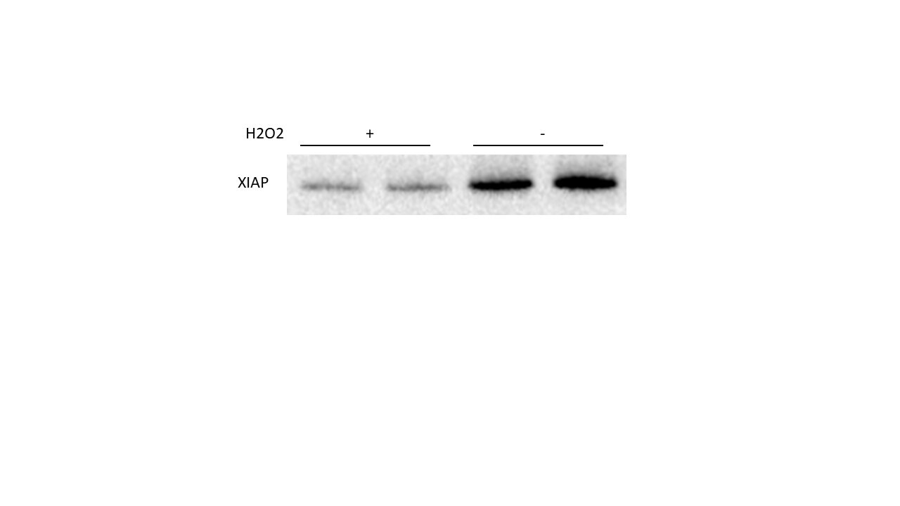
Immunocytochemistry/ Immunofluorescence: XIAP Antibody [NB100-56183] - Kaempferol inhibits the activity of caspase-3 enzyme. (a & b) Caspase-3 inhibition by kaempferol was measured using recombinant caspase-3 (enzyme) & Z-DEVD-aminoluciferin (substrate) with respect to time (a) & concentration (b). Ac-DEVD-CHO served as a positive control. (c) Dose response curve for kaempferol inhibition on caspase-3 enzyme activity. (d) Analysis of the inhibitory potential of estrogen receptor modulators on caspase-3 enzyme activity. Withanaloide A served as (−) control & Ac-DEVE-CHO as (+) control. Data represented as mean ± SEM of two independent experiments performed in duplicate. (e) Immunocytochemistry for XIAP expression levels in IMR32 cells after 24 hours of incubation in respective conditions (Scale bar: 67 µm). Image collected & cropped by CiteAb from the following publication (https://www.nature.com/articles/s41598-018-20499-7), licensed under a CC-BY license. Not internally tested by Novus Biologicals.Applications for XIAP Antibody - BSA Free
Application
Recommended Usage
Immunocytochemistry/ Immunofluorescence
reported in scientific literature (PMID 29391535)
Immunohistochemistry
1:10-1:500
Immunohistochemistry-Paraffin
1:1000-1:5000
Immunoprecipitation
1:50-1:200
Western Blot
1:1000-1:2000
Application Notes
Although not tested this antibody may be useful in Immunohistochemistry-Frozen.
Reviewed Applications
Read 3 reviews rated 4.3 using NB100-56183 in the following applications:
Formulation, Preparation, and Storage
Purification
Unpurified
Formulation
Whole antisera
Format
BSA Free
Preservative
0.05% Sodium Azide
Concentration
This product is unpurified. The exact concentration of antibody is not quantifiable.
Shipping
The product is shipped with polar packs. Upon receipt, store it immediately at the temperature recommended below.
Stability & Storage
Store at -20C. Avoid freeze-thaw cycles.
Background: XIAP
Long Name
X-linked Inhibitor of Apoptosis
Alternate Names
BIRC4
Gene Symbol
XIAP
Additional XIAP Products
Product Documents for XIAP Antibody - BSA Free
Certificate of Analysis
To download a Certificate of Analysis, please enter a lot or batch number in the search box below.
Product Specific Notices for XIAP Antibody - BSA Free
This product is for research use only and is not approved for use in humans or in clinical diagnosis. Primary Antibodies are guaranteed for 1 year from date of receipt.
Related Research Areas
Citations for XIAP Antibody - BSA Free
 Powered by Bioz
Customer Reviews for XIAP Antibody - BSA Free (3)
4.3 out of 5
3 Customer Ratings
Have you used XIAP Antibody - BSA Free?
Submit a review and receive an Amazon gift card!
$25/€18/£15/$25CAN/¥2500 Yen for a review with an image
$10/€7/£6/$10CAN/¥1110 Yen for a review without an image
Submit a review
Customer Images
Showing
1
-
3 of
3 reviews
Showing All
Filter By:
-
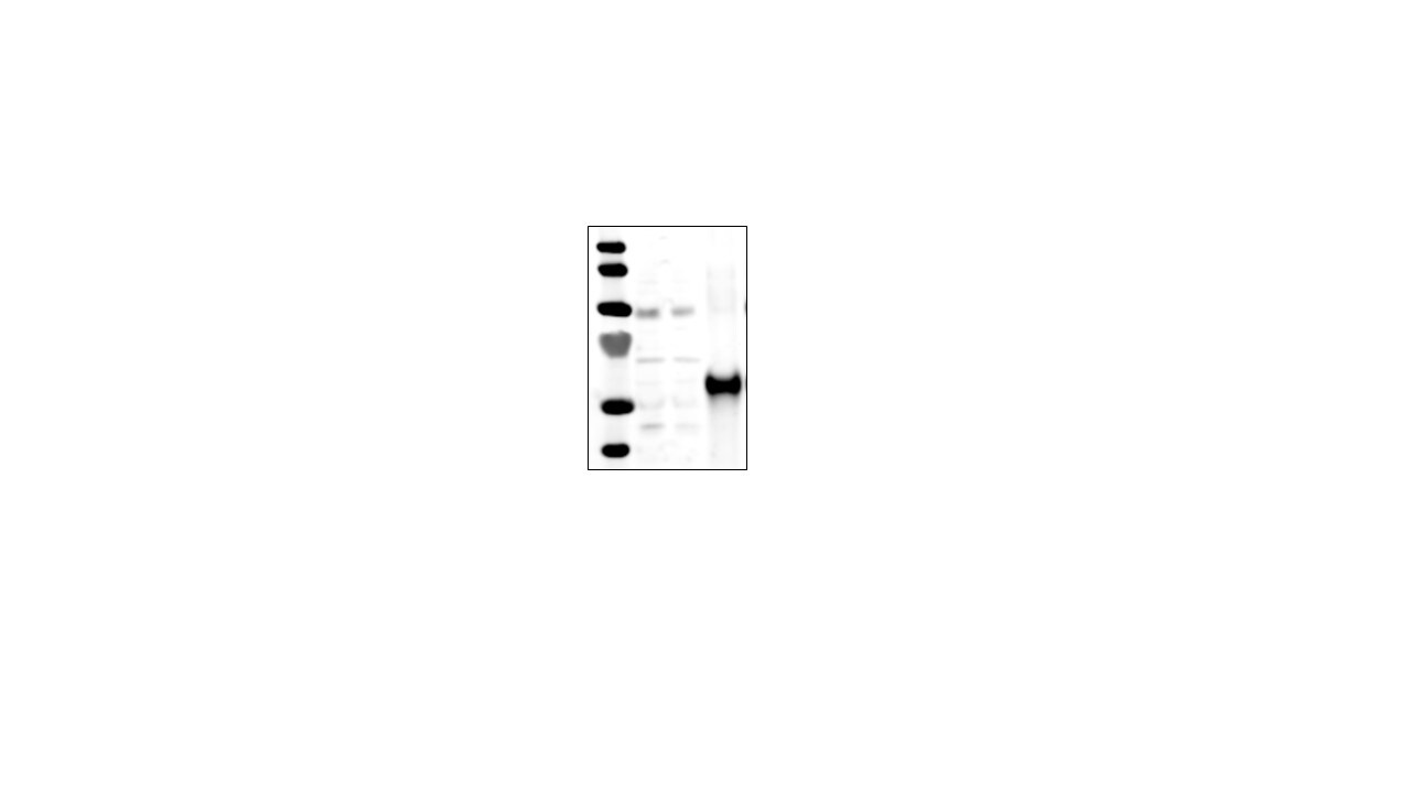
Application: Western BlotSample Tested: H9c2 whole cell lysateSpecies: RatVerified Customer | Posted 02/07/2022XIAP

-
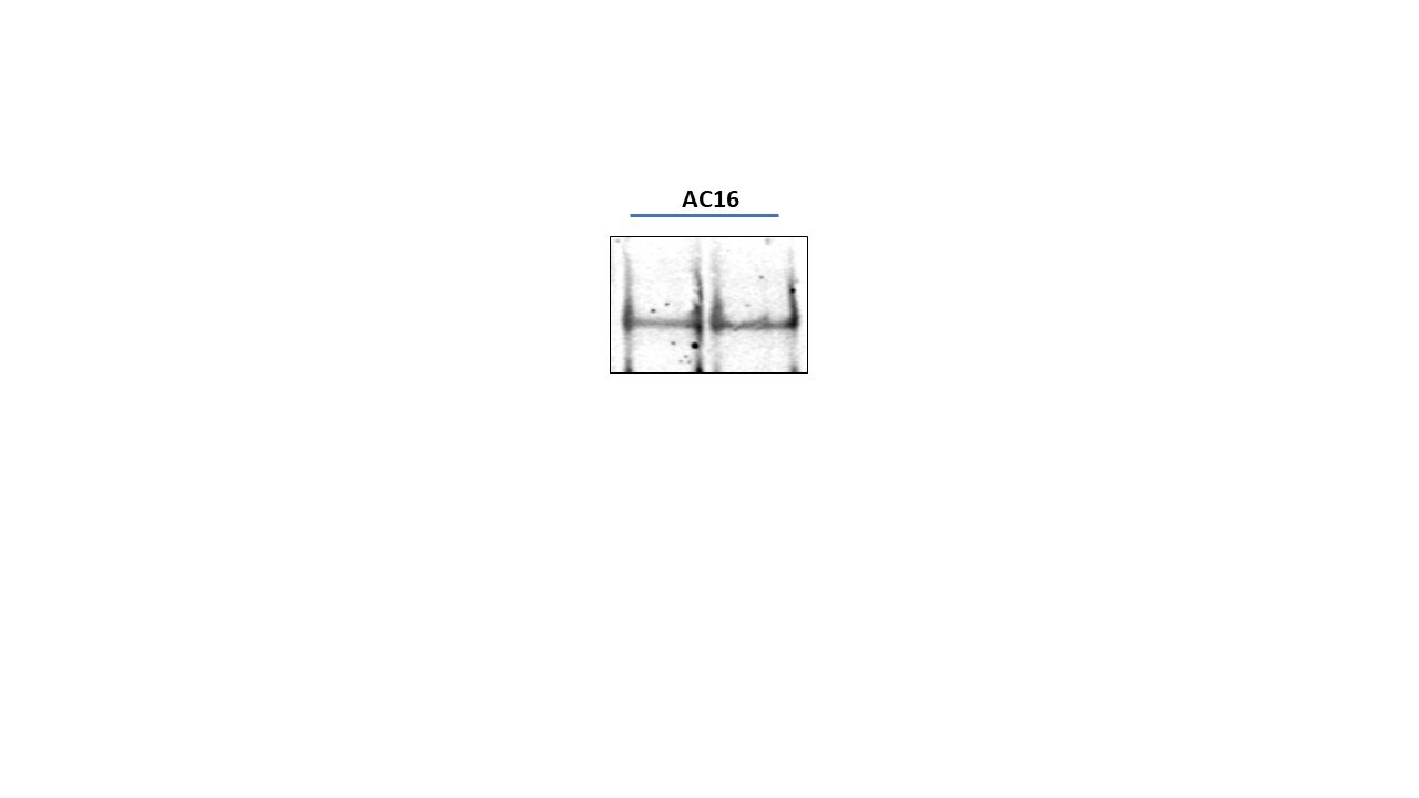
Application: ImmunoprecipitationSample Tested: AC16Species: HumanVerified Customer | Posted 04/20/2021XIAP
-
Application: ImmunoprecipitationSample Tested: AC16Species: HumanVerified Customer | Posted 05/17/2019Excellent Antibody
There are no reviews that match your criteria.
Protocols
Find general support by application which include: protocols, troubleshooting, illustrated assays, videos and webinars.
- Antigen Retrieval Protocol (PIER)
- Antigen Retrieval for Frozen Sections Protocol
- Appropriate Fixation of IHC/ICC Samples
- Cellular Response to Hypoxia Protocols
- Chromogenic IHC Staining of Formalin-Fixed Paraffin-Embedded (FFPE) Tissue Protocol
- Chromogenic Immunohistochemistry Staining of Frozen Tissue
- ClariTSA™ Fluorophore Kits
- Detection & Visualization of Antibody Binding
- Fluorescent IHC Staining of Frozen Tissue Protocol
- Graphic Protocol for Heat-induced Epitope Retrieval
- Graphic Protocol for the Preparation and Fluorescent IHC Staining of Frozen Tissue Sections
- Graphic Protocol for the Preparation and Fluorescent IHC Staining of Paraffin-embedded Tissue Sections
- Graphic Protocol for the Preparation of Gelatin-coated Slides for Histological Tissue Sections
- ICC Cell Smear Protocol for Suspension Cells
- ICC Immunocytochemistry Protocol Videos
- ICC for Adherent Cells
- IHC Sample Preparation (Frozen sections vs Paraffin)
- Immunocytochemistry (ICC) Protocol
- Immunocytochemistry Troubleshooting
- Immunofluorescence of Organoids Embedded in Cultrex Basement Membrane Extract
- Immunofluorescent IHC Staining of Formalin-Fixed Paraffin-Embedded (FFPE) Tissue Protocol
- Immunohistochemistry (IHC) and Immunocytochemistry (ICC) Protocols
- Immunohistochemistry Frozen Troubleshooting
- Immunohistochemistry Paraffin Troubleshooting
- Immunoprecipitation Protocol
- Preparing Samples for IHC/ICC Experiments
- Preventing Non-Specific Staining (Non-Specific Binding)
- Primary Antibody Selection & Optimization
- Protocol for Heat-Induced Epitope Retrieval (HIER)
- Protocol for Making a 4% Formaldehyde Solution in PBS
- Protocol for VisUCyte™ HRP Polymer Detection Reagent
- Protocol for the Fluorescent ICC Staining of Cell Smears - Graphic
- Protocol for the Fluorescent ICC Staining of Cultured Cells on Coverslips - Graphic
- Protocol for the Preparation & Fixation of Cells on Coverslips
- Protocol for the Preparation and Chromogenic IHC Staining of Frozen Tissue Sections
- Protocol for the Preparation and Chromogenic IHC Staining of Frozen Tissue Sections - Graphic
- Protocol for the Preparation and Chromogenic IHC Staining of Paraffin-embedded Tissue Sections
- Protocol for the Preparation and Chromogenic IHC Staining of Paraffin-embedded Tissue Sections - Graphic
- Protocol for the Preparation and Fluorescent ICC Staining of Cells on Coverslips
- Protocol for the Preparation and Fluorescent ICC Staining of Non-adherent Cells
- Protocol for the Preparation and Fluorescent ICC Staining of Stem Cells on Coverslips
- Protocol for the Preparation and Fluorescent IHC Staining of Frozen Tissue Sections
- Protocol for the Preparation and Fluorescent IHC Staining of Paraffin-embedded Tissue Sections
- Protocol for the Preparation of Gelatin-coated Slides for Histological Tissue Sections
- Protocol for the Preparation of a Cell Smear for Non-adherent Cell ICC - Graphic
- R&D Systems Quality Control Western Blot Protocol
- TUNEL and Active Caspase-3 Detection by IHC/ICC Protocol
- The Importance of IHC/ICC Controls
- Troubleshooting Guide: Immunohistochemistry
- Troubleshooting Guide: Western Blot Figures
- Western Blot Conditions
- Western Blot Protocol
- Western Blot Protocol for Cell Lysates
- Western Blot Troubleshooting
- Western Blot Troubleshooting Guide
- View all Protocols, Troubleshooting, Illustrated assays and Webinars
Loading...
Associated Pathways

