ZEB1 Antibody (2A8A6) - BSA Free
Novus Biologicals | Catalog # NBP2-23484
Key Product Details
Validated by
Species Reactivity
Validated:
Cited:
Applications
Validated:
Cited:
Label
Antibody Source
Format
Product Specifications
Immunogen
Reactivity Notes
Marker
Clonality
Host
Isotype
Scientific Data Images for ZEB1 Antibody (2A8A6) - BSA Free
![Western Blot: ZEB1 Antibody (2A8A6)BSA Free [NBP2-23484]](https://resources.rndsystems.com/images/products/ZEB1-Antibody-2A8A6-Western-Blot-NBP2-23484-img0014.jpg "Western Blot: ZEB1 Antibody (2A8A6)BSA Free [NBP2-23484]")
![Immunocytochemistry/ Immunofluorescence: ZEB1 Antibody (2A8A6) - BSA Free [NBP2-23484]](https://resources.rndsystems.com/images/products/ZEB1-Antibody-2A8A6-Immunocytochemistry-Immunofluorescence-NBP2-23484-img0009.jpg "Immunocytochemistry/ Immunofluorescence: ZEB1 Antibody (2A8A6) - BSA Free [NBP2-23484]")
Immunocytochemistry/ Immunofluorescence: ZEB1 Antibody (2A8A6) - BSA Free [NBP2-23484]
Immunocytochemistry/Immunofluorescence: ZEB1 Antibody (2A8A6) [NBP2-23484] - ZEB1 antibody was tested in HeLa cells with DyLight 488 (green). Nuclei and alpha-tubulin were counterstained with DAPI (blue) and DyLight 550 (red).![Immunohistochemistry: ZEB1 Antibody (2A8A6) - BSA Free [NBP2-23484]](https://resources.rndsystems.com/images/products/ZEB1-Antibody-2A8A6-Immunohistochemistry-NBP2-23484-img0006.jpg "Immunohistochemistry: ZEB1 Antibody (2A8A6) - BSA Free [NBP2-23484]")
Immunohistochemistry: ZEB1 Antibody (2A8A6) - BSA Free [NBP2-23484]
Immunohistochemistry: ZEB1 Antibody (2A8A6) [NBP2-23484] - IHC analysis of paraffin-embedded human cervical cancer tissues with DAB staining.![Flow Cytometry: ZEB1 Antibody (2A8A6) - BSA Free [NBP2-23484]](https://resources.rndsystems.com/images/products/ZEB1-Antibody-2A8A6-Flow-Cytometry-NBP2-23484-img0012.jpg "Flow Cytometry: ZEB1 Antibody (2A8A6) - BSA Free [NBP2-23484]")
Flow Cytometry: ZEB1 Antibody (2A8A6) - BSA Free [NBP2-23484]
Flow Cytometry: ZEB1 Antibody (2A8A6) [NBP2-23484] - An intracellular stain was performed on U-87 cells with ZEB1 [2A8A6] Antibody NBP2-23484AF700 (blue) and a matched isotype control (orange). Cells were fixed with 4% PFA and then permeabilized with 0.1% saponin. Cells were incubated in an antibody dilution of 5 ug/mL for 30 minutes at room temperature. Both antibodies were conjugated to Alexa Fluor 700.![Western Blot: ZEB1 Antibody (2A8A6)BSA Free [NBP2-23484]](https://resources.rndsystems.com/images/products/ZEB1-Antibody-2A8A6-Western-Blot-NBP2-23484-img0013.jpg "Western Blot: ZEB1 Antibody (2A8A6)BSA Free [NBP2-23484]")
Western Blot: ZEB1 Antibody (2A8A6)BSA Free [NBP2-23484]
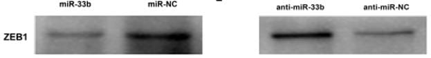
Western Blot: ZEB1 Antibody (2A8A6) [NBP2-23484] - ZEB1 expression was assayed in lung adenocarcinoma cells after transfectionwith miR-33b or miR-NC, anti-miR-33b or anti-miR-NC in H1299 and A549 cells. Image submitted by verified customer review.![Immunohistochemistry: ZEB1 Antibody (2A8A6) - BSA Free [NBP2-23484]](https://resources.rndsystems.com/images/products/ZEB1-Antibody-2A8A6-Immunohistochemistry-NBP2-23484-img0004.jpg "Immunohistochemistry: ZEB1 Antibody (2A8A6) - BSA Free [NBP2-23484]")
Immunohistochemistry: ZEB1 Antibody (2A8A6) - BSA Free [NBP2-23484]
Immunohistochemistry: ZEB1 Antibody (2A8A6) [NBP2-23484] - IHC analysis of paraffin-embedded human recturm cancer tissues with DAB staining.![Flow Cytometry: ZEB1 Antibody (2A8A6) - BSA Free [NBP2-23484]](https://resources.rndsystems.com/images/products/ZEB1-Antibody-2A8A6-Flow-Cytometry-NBP2-23484-img0007.jpg "Flow Cytometry: ZEB1 Antibody (2A8A6) - BSA Free [NBP2-23484]")
Flow Cytometry: ZEB1 Antibody (2A8A6) - BSA Free [NBP2-23484]
Flow Cytometry: ZEB1 Antibody (2A8A6) [NBP2-23484] - Flow cytometric analysis of Hela cells using ZEB1 mouse mAb (green) and negative control (red).![Flow Cytometry: ZEB1 Antibody (2A8A6) - BSA Free [NBP2-23484]](https://resources.rndsystems.com/images/products/ZEB1-Antibody-2A8A6-Flow-Cytometry-NBP2-23484-img0010.jpg "Flow Cytometry: ZEB1 Antibody (2A8A6) - BSA Free [NBP2-23484]")
Flow Cytometry: ZEB1 Antibody (2A8A6) - BSA Free [NBP2-23484]
Flow Cytometry: ZEB1 Antibody (2A8A6) [NBP2-23484] - An intracellular stain was performed on with A549 cells with ZEB1 Antibody NBP2-23484PE and a a matched isotype control (orange). Cells were fixed with 4% PFA and then permeabilized with 0.1% saponin. Cells were incubated in an antibody dilution of 5 ug/mL for 30 minutes at room temperature. Both antibodies were conjugated to phycoerythrin.![Flow Cytometry: ZEB1 Antibody (2A8A6) - BSA Free [NBP2-23484]](https://resources.rndsystems.com/images/products/ZEB1-Antibody-2A8A6-Flow-Cytometry-NBP2-23484-img0011.jpg "Flow Cytometry: ZEB1 Antibody (2A8A6) - BSA Free [NBP2-23484]")
Flow Cytometry: ZEB1 Antibody (2A8A6) - BSA Free [NBP2-23484]
Flow Cytometry: ZEB1 Antibody (2A8A6) [NBP2-23484] - An intracellular stain was performed on Jurkat with ZEB1 Antibody (2A8A6) NBP2-23484 and a matched isotype control. Cells were fixed with 4% PFA and then permeablized with 0.1% saponin. Cells were incubated in an antibody dilution of 1 ug/mL for 30 minutes at room temperature, followed by Mouse F(ab)2 IgG (H+L) PE-conjugated Antibody (R&D Systems, F0102B)Applications for ZEB1 Antibody (2A8A6) - BSA Free
Flow Cytometry
Immunocytochemistry/ Immunofluorescence
Immunohistochemistry
Immunohistochemistry-Paraffin
Western Blot
Reviewed Applications
Read 1 review rated 5 using NBP2-23484 in the following applications:
Flow Cytometry Panel Builder
Bio-Techne Knows Flow Cytometry
Save time and reduce costly mistakes by quickly finding compatible reagents using the Panel Builder Tool.
Advanced Features
- Spectra Viewer - Custom analysis of spectra from multiple fluorochromes
- Spillover Popups - Visualize the spectra of individual fluorochromes
- Antigen Density Selector - Match fluorochrome brightness with antigen density

Formulation, Preparation, and Storage
Purification
Formulation
Format
Preservative
Concentration
Shipping
Stability & Storage
Background: ZEB1
Long Name
Alternate Names
Entrez Gene IDs
Gene Symbol
UniProt
Additional ZEB1 Products
Product Documents for ZEB1 Antibody (2A8A6) - BSA Free
Certificate of Analysis
To download a Certificate of Analysis, please enter a lot or batch number in the search box below.
Product Specific Notices for ZEB1 Antibody (2A8A6) - BSA Free
This product is for research use only and is not approved for use in humans or in clinical diagnosis. Primary Antibodies are guaranteed for 1 year from date of receipt.
Citations for ZEB1 Antibody (2A8A6) - BSA Free
 Powered by Bioz
Customer Reviews for ZEB1 Antibody (2A8A6) - BSA Free (1)
Have you used ZEB1 Antibody (2A8A6) - BSA Free?
Submit a review and receive an Amazon gift card!
$25/€18/£15/$25CAN/¥2500 Yen for a review with an image
$10/€7/£6/$10CAN/¥1110 Yen for a review without an image
Submit a review
Customer Images
-
Application: Western BlotSample Tested: H1299 and A549 cells of lung adenocarcinoma cellsSpecies: HumanVerified Customer | Posted 09/14/2017ZEB1 expression was assayed in lung adenocarcinoma cells after transfection with miR-33b or miR-NC, anti-miR-33b or anti-miR-NC in H1299 and A549 cells.at a dilution of 1:800;secondary goat anti-mouse (1:5,000) immunoglobulin G.

There are no reviews that match your criteria.
Protocols
Find general support by application which include: protocols, troubleshooting, illustrated assays, videos and webinars.
- 7-Amino Actinomycin D (7-AAD) Cell Viability Flow Cytometry Protocol
- Antigen Retrieval Protocol (PIER)
- Antigen Retrieval for Frozen Sections Protocol
- Appropriate Fixation of IHC/ICC Samples
- Cellular Response to Hypoxia Protocols
- Chromogenic IHC Staining of Formalin-Fixed Paraffin-Embedded (FFPE) Tissue Protocol
- Chromogenic Immunohistochemistry Staining of Frozen Tissue
- ClariTSA™ Fluorophore Kits
- Detection & Visualization of Antibody Binding
- Extracellular Membrane Flow Cytometry Protocol
- Flow Cytometry Protocol for Cell Surface Markers
- Flow Cytometry Protocol for Staining Membrane Associated Proteins
- Flow Cytometry Staining Protocols
- Flow Cytometry Troubleshooting Guide
- Fluorescent IHC Staining of Frozen Tissue Protocol
- Graphic Protocol for Heat-induced Epitope Retrieval
- Graphic Protocol for the Preparation and Fluorescent IHC Staining of Frozen Tissue Sections
- Graphic Protocol for the Preparation and Fluorescent IHC Staining of Paraffin-embedded Tissue Sections
- Graphic Protocol for the Preparation of Gelatin-coated Slides for Histological Tissue Sections
- ICC Cell Smear Protocol for Suspension Cells
- ICC Immunocytochemistry Protocol Videos
- ICC for Adherent Cells
- IHC Sample Preparation (Frozen sections vs Paraffin)
- Immunocytochemistry (ICC) Protocol
- Immunocytochemistry Troubleshooting
- Immunofluorescence of Organoids Embedded in Cultrex Basement Membrane Extract
- Immunofluorescent IHC Staining of Formalin-Fixed Paraffin-Embedded (FFPE) Tissue Protocol
- Immunohistochemistry (IHC) and Immunocytochemistry (ICC) Protocols
- Immunohistochemistry Frozen Troubleshooting
- Immunohistochemistry Paraffin Troubleshooting
- Intracellular Flow Cytometry Protocol Using Alcohol (Methanol)
- Intracellular Flow Cytometry Protocol Using Detergents
- Intracellular Nuclear Staining Flow Cytometry Protocol Using Detergents
- Intracellular Staining Flow Cytometry Protocol Using Alcohol Permeabilization
- Intracellular Staining Flow Cytometry Protocol Using Detergents to Permeabilize Cells
- Preparing Samples for IHC/ICC Experiments
- Preventing Non-Specific Staining (Non-Specific Binding)
- Primary Antibody Selection & Optimization
- Propidium Iodide Cell Viability Flow Cytometry Protocol
- Protocol for Heat-Induced Epitope Retrieval (HIER)
- Protocol for Liperfluo
- Protocol for Making a 4% Formaldehyde Solution in PBS
- Protocol for VisUCyte™ HRP Polymer Detection Reagent
- Protocol for the Characterization of Human Th22 Cells
- Protocol for the Characterization of Human Th9 Cells
- Protocol for the Fluorescent ICC Staining of Cell Smears - Graphic
- Protocol for the Fluorescent ICC Staining of Cultured Cells on Coverslips - Graphic
- Protocol for the Preparation & Fixation of Cells on Coverslips
- Protocol for the Preparation and Chromogenic IHC Staining of Frozen Tissue Sections
- Protocol for the Preparation and Chromogenic IHC Staining of Frozen Tissue Sections - Graphic
- Protocol for the Preparation and Chromogenic IHC Staining of Paraffin-embedded Tissue Sections
- Protocol for the Preparation and Chromogenic IHC Staining of Paraffin-embedded Tissue Sections - Graphic
- Protocol for the Preparation and Fluorescent ICC Staining of Cells on Coverslips
- Protocol for the Preparation and Fluorescent ICC Staining of Non-adherent Cells
- Protocol for the Preparation and Fluorescent ICC Staining of Stem Cells on Coverslips
- Protocol for the Preparation and Fluorescent IHC Staining of Frozen Tissue Sections
- Protocol for the Preparation and Fluorescent IHC Staining of Paraffin-embedded Tissue Sections
- Protocol for the Preparation of Gelatin-coated Slides for Histological Tissue Sections
- Protocol for the Preparation of a Cell Smear for Non-adherent Cell ICC - Graphic
- Protocol: Annexin V and PI Staining by Flow Cytometry
- Protocol: Annexin V and PI Staining for Apoptosis by Flow Cytometry
- R&D Systems Quality Control Western Blot Protocol
- TUNEL and Active Caspase-3 Detection by IHC/ICC Protocol
- The Importance of IHC/ICC Controls
- Troubleshooting Guide: Fluorokine Flow Cytometry Kits
- Troubleshooting Guide: Immunohistochemistry
- Troubleshooting Guide: Western Blot Figures
- Western Blot Conditions
- Western Blot Protocol
- Western Blot Protocol for Cell Lysates
- Western Blot Troubleshooting
- Western Blot Troubleshooting Guide
- View all Protocols, Troubleshooting, Illustrated assays and Webinars
FAQs for ZEB1 Antibody (2A8A6) - BSA Free
-
Q: In looking at the Zeb1 antibodies on your website, the molecular weights of Zeb1 detected by several antibodies are different. Why is this?
A: ZEB1 is a rather large protein and undergoes a lot of post translational modifications such as phosphorylation and glycosylation (please see: UniProt P37275). The reason band patterns are different in the images may be due to sample type and how highly modified the ZEB1 protein is that the antibody is detecting. The more post translational modifications the higher you will detect the protein.