Zika Virus (H/PF/2013) Envelope Antibody (GT363) - Azide and BSA Free
Novus Biologicals | Catalog # NBP3-13587
![Immunocytochemistry/ Immunofluorescence: Zika Virus (H/PF/2013) Envelope Antibody (GT363) [NBP3-13587]](https://resources.rndsystems.com/images/products/Zika-Virus-H-PF-2013-Envelope-Antibody-GT363-Immunocytochemistry-Immunofluorescence-NBP3-13587-img0008.jpg "Immunocytochemistry/ Immunofluorescence: Zika Virus (H/PF/2013) Envelope Antibody (GT363) [NBP3-13587]")
Loading...
Key Product Details
Species Reactivity
Virus
Applications
Immunohistochemistry, Western Blot, ELISA, Sandwich ELISA, Immunocytochemistry/ Immunofluorescence
Label
Unconjugated
Antibody Source
Monoclonal Mouse IgG2B Clone # GT363
Format
Azide and BSA Free
Loading...
Product Specifications
Immunogen
Carrier-protein conjugated synthetic peptide encompassing a sequence within the center region of Zika Virus (H/PF/2013) Envelope (Zika virus (strain H/PF/2013)). The exact sequence is proprietary.
Reactivity Notes
Zika virus
Clonality
Monoclonal
Host
Mouse
Isotype
IgG2B
Theoretical MW
54 kDa.
Disclaimer note: The observed molecular weight of the protein may vary from the listed predicted molecular weight due to post translational modifications, post translation cleavages, relative charges, and other experimental factors.
Disclaimer note: The observed molecular weight of the protein may vary from the listed predicted molecular weight due to post translational modifications, post translation cleavages, relative charges, and other experimental factors.
Description
Centrifuge briefly prior to opening.
Scientific Data Images for Zika Virus (H/PF/2013) Envelope Antibody (GT363) - Azide and BSA Free
Immunocytochemistry/ Immunofluorescence: Zika Virus (H/PF/2013) Envelope Antibody (GT363) [NBP3-13587]
Immunocytochemistry/Immunofluorescence: Zika Virus (H/PF/2013) Envelope Antibody (GT363) [NBP3-13587] - Immunofluorescent analysis of arboviruses infected cells using Zika Virus (H/PF/2013) Envelope antibody [GT363] (NBP3-13587). Sample: Mock and zika virus-infected cells. Green: Zika Virus (H/PF/2013) Envelope antibody [GT363] (NBP3-13587) diluted at 1:100.![Immunocytochemistry/ Immunofluorescence: Zika Virus (H/PF/2013) Envelope Antibody (GT363) [NBP3-13587]](https://resources.rndsystems.com/images/products/Zika-Virus-H-PF-2013-Envelope-Antibody-GT363-Immunocytochemistry-Immunofluorescence-NBP3-13587-img0002.jpg "Immunocytochemistry/ Immunofluorescence: Zika Virus (H/PF/2013) Envelope Antibody (GT363) [NBP3-13587]")
Immunocytochemistry/ Immunofluorescence: Zika Virus (H/PF/2013) Envelope Antibody (GT363) [NBP3-13587]
Immunocytochemistry/Immunofluorescence: Zika Virus (H/PF/2013) Envelope Antibody (GT363) [NBP3-13587] - Immunofluorescent analysis of non-infected and infected vero cells using Zika Virus (H/PF/2013) Envelope antibody [GT363] (NBP3-13587). Green: Zika Virus (H/PF/2013) Envelope antibody [GT363] (NBP3-13587) diluted at 1:500.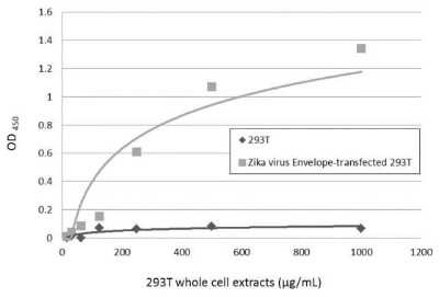
Sandwich ELISA: Zika Virus (H/PF/2013) Envelope Antibody (GT363) [NBP3-13587] - Sandwich ELISA detection of non-transfected and transfected 293T whole cell extracts using NBP3-13587 as capture antibody at concentration of 5 ug/mL and NBP3-13205 as detection antibody at concentration of 1 ug/mL. Rabbit IgG antibody (HRP) (NBP2-19301) was diluted at 1:10000 and used to detect the primary antibody.
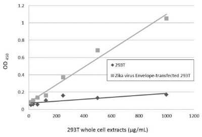
Sandwich ELISA: Zika Virus (H/PF/2013) Envelope Antibody (GT363) [NBP3-13587] - Sandwich ELISA detection of non-transfected and transfected 293T whole cell extracts using NBP3-13206 as capture antibody at concentration of 5 ug/mL and NBP3-13587 as detection antibody at concentration of 1 ug/mL. Mouse IgG antibody (HRP) (NBP2-19382) was diluted at 1:10000 and used to detect the primary antibody.
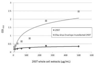
Sandwich ELISA: Zika Virus (H/PF/2013) Envelope Antibody (GT363) [NBP3-13587] - Sandwich ELISA detection of non-transfected and transfected 293T whole cell extracts using NBP3-13587 as capture antibody at concentration of 5 ug/mL and NBP3-13206 as detection antibody at concentration of 1 ug/mL. Rabbit IgG antibody (HRP) (NBP2-19301) was diluted at 1:10000 and used to detect the primary antibody.
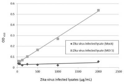
Sandwich ELISA: Zika Virus (H/PF/2013) Envelope Antibody (GT363) [NBP3-13587] - Sandwich ELISA detection of Zika virus-infected lysate using NBP3-13587 as capture antibody at concentration of 5 ug/mL and NBP3-13206 as detection antibody at concentration of 1 ug/mL. Rabbit IgG antibody (HRP) (NBP2-19301) was diluted at 1:10000 and used to detect the primary antibody.
 Envelope Antibody (GT363) [NBP3-13587] -")
Western Blot: Zika Virus (H/PF/2013) Envelope Antibody (GT363) [NBP3-13587] -
Zika Virus-PRVABC59 infected Vero cells were separated by 4-20% SDS-PAGE, and the membrane was blotted with Zika virus Envelope protein antibody [GT363] (HRP) (NBP3-13587-01) diluted at 1:2000. Envelope Antibody (GT363) [NBP3-13587] -")
Western Blot: Zika Virus (H/PF/2013) Envelope Antibody (GT363) [NBP3-13587] -
Zika virus Viral Lysate (0.5 ug) was separated by 10% SDS-PAGE, and the membrane was blotted with Zika virus Envelope protein antibody [GT363] (HRP) (NBP3-13587-01) diluted at 1:2000. Envelope Antibody (GT363) [NBP3-13587] -")
Western Blot: Zika Virus (H/PF/2013) Envelope Antibody (GT363) [NBP3-13587] -
Non-transfected (-) and transfected (+) 293T whole cell extracts (30 ug) were separated by 10% SDS-PAGE, and the membrane was blotted with Zika virus Envelope protein antibody [GT363] (NBP3-13587) diluted at 1:5000. The HRP-conjugated anti-mouse IgG antibody was used to detect the primary antibody.Applications for Zika Virus (H/PF/2013) Envelope Antibody (GT363) - Azide and BSA Free
Application
Recommended Usage
ELISA
1:1000-1:10000
Immunocytochemistry/ Immunofluorescence
1:100-1:1000
Immunohistochemistry
Assay dependent
Sandwich ELISA
Assay dependent
Western Blot
1:50-1:3000
Formulation, Preparation, and Storage
Purification
Protein A purified
Formulation
PBS
Format
Azide and BSA Free
Preservative
No Preservative
Concentration
Concentrations vary lot to lot. See vial label for concentration. If unlisted please contact technical services.
Shipping
The product is shipped with polar packs. Upon receipt, store it immediately at the temperature recommended below.
Stability & Storage
Store at 4C short term. Aliquot and store at -20C long term. Avoid freeze-thaw cycles.
Background: Zika Virus (H/PF/2013) Envelope
Long Name
Zika Virus Polyprotein
Alternate Names
ZIKV-E, ZIKV-Env, ZIKV_E
Additional Zika Virus (H/PF/2013) Envelope Products
Product Documents for Zika Virus (H/PF/2013) Envelope Antibody (GT363) - Azide and BSA Free
Certificate of Analysis
To download a Certificate of Analysis, please enter a lot or batch number in the search box below.
Product Specific Notices for Zika Virus (H/PF/2013) Envelope Antibody (GT363) - Azide and BSA Free
This product is for research use only and is not approved for use in humans or in clinical diagnosis. Primary Antibodies are guaranteed for 1 year from date of receipt.
Related Research Areas
Customer Reviews for Zika Virus (H/PF/2013) Envelope Antibody (GT363) - Azide and BSA Free
There are currently no reviews for this product. Be the first to review Zika Virus (H/PF/2013) Envelope Antibody (GT363) - Azide and BSA Free and earn rewards!
Have you used Zika Virus (H/PF/2013) Envelope Antibody (GT363) - Azide and BSA Free?
Submit a review and receive an Amazon gift card!
$25/€18/£15/$25CAN/¥2500 Yen for a review with an image
$10/€7/£6/$10CAN/¥1110 Yen for a review without an image
Submit a review
Protocols
Find general support by application which include: protocols, troubleshooting, illustrated assays, videos and webinars.
- Antigen Retrieval Protocol (PIER)
- Antigen Retrieval for Frozen Sections Protocol
- Appropriate Fixation of IHC/ICC Samples
- Cellular Response to Hypoxia Protocols
- Chromogenic IHC Staining of Formalin-Fixed Paraffin-Embedded (FFPE) Tissue Protocol
- Chromogenic Immunohistochemistry Staining of Frozen Tissue
- ClariTSA™ Fluorophore Kits
- Detection & Visualization of Antibody Binding
- ELISA Sample Preparation & Collection Guide
- ELISA Troubleshooting Guide
- Fluorescent IHC Staining of Frozen Tissue Protocol
- Graphic Protocol for Heat-induced Epitope Retrieval
- Graphic Protocol for the Preparation and Fluorescent IHC Staining of Frozen Tissue Sections
- Graphic Protocol for the Preparation and Fluorescent IHC Staining of Paraffin-embedded Tissue Sections
- Graphic Protocol for the Preparation of Gelatin-coated Slides for Histological Tissue Sections
- How to Run an R&D Systems DuoSet ELISA
- How to Run an R&D Systems Quantikine ELISA
- How to Run an R&D Systems Quantikine™ QuicKit™ ELISA
- ICC Cell Smear Protocol for Suspension Cells
- ICC Immunocytochemistry Protocol Videos
- ICC for Adherent Cells
- IHC Sample Preparation (Frozen sections vs Paraffin)
- Immunocytochemistry (ICC) Protocol
- Immunocytochemistry Troubleshooting
- Immunofluorescence of Organoids Embedded in Cultrex Basement Membrane Extract
- Immunofluorescent IHC Staining of Formalin-Fixed Paraffin-Embedded (FFPE) Tissue Protocol
- Immunohistochemistry (IHC) and Immunocytochemistry (ICC) Protocols
- Immunohistochemistry Frozen Troubleshooting
- Immunohistochemistry Paraffin Troubleshooting
- Preparing Samples for IHC/ICC Experiments
- Preventing Non-Specific Staining (Non-Specific Binding)
- Primary Antibody Selection & Optimization
- Protocol for Heat-Induced Epitope Retrieval (HIER)
- Protocol for Making a 4% Formaldehyde Solution in PBS
- Protocol for VisUCyte™ HRP Polymer Detection Reagent
- Protocol for the Fluorescent ICC Staining of Cell Smears - Graphic
- Protocol for the Fluorescent ICC Staining of Cultured Cells on Coverslips - Graphic
- Protocol for the Preparation & Fixation of Cells on Coverslips
- Protocol for the Preparation and Chromogenic IHC Staining of Frozen Tissue Sections
- Protocol for the Preparation and Chromogenic IHC Staining of Frozen Tissue Sections - Graphic
- Protocol for the Preparation and Chromogenic IHC Staining of Paraffin-embedded Tissue Sections
- Protocol for the Preparation and Chromogenic IHC Staining of Paraffin-embedded Tissue Sections - Graphic
- Protocol for the Preparation and Fluorescent ICC Staining of Cells on Coverslips
- Protocol for the Preparation and Fluorescent ICC Staining of Non-adherent Cells
- Protocol for the Preparation and Fluorescent ICC Staining of Stem Cells on Coverslips
- Protocol for the Preparation and Fluorescent IHC Staining of Frozen Tissue Sections
- Protocol for the Preparation and Fluorescent IHC Staining of Paraffin-embedded Tissue Sections
- Protocol for the Preparation of Gelatin-coated Slides for Histological Tissue Sections
- Protocol for the Preparation of a Cell Smear for Non-adherent Cell ICC - Graphic
- Quantikine HS ELISA Kit Assay Principle, Alkaline Phosphatase
- Quantikine HS ELISA Kit Principle, Streptavidin-HRP Polymer
- R&D Systems Quality Control Western Blot Protocol
- Sandwich ELISA (Colorimetric) – Biotin/Streptavidin Detection Protocol
- Sandwich ELISA (Colorimetric) – Direct Detection Protocol
- TUNEL and Active Caspase-3 Detection by IHC/ICC Protocol
- The Importance of IHC/ICC Controls
- Troubleshooting Guide: ELISA
- Troubleshooting Guide: Immunohistochemistry
- Troubleshooting Guide: Western Blot Figures
- Western Blot Conditions
- Western Blot Protocol
- Western Blot Protocol for Cell Lysates
- Western Blot Troubleshooting
- Western Blot Troubleshooting Guide
- View all Protocols, Troubleshooting, Illustrated assays and Webinars
Loading...