ZNF24 Antibody - BSA Free
Novus Biologicals | Catalog # NBP1-82866
![Western Blot: ZNF24 Antibody [NBP1-82866]](https://resources.rndsystems.com/images/products/ZNF24-Antibody-Western-Blot-NBP1-82866-img0009.jpg "Western Blot: ZNF24 Antibody [NBP1-82866]")
Loading...
Key Product Details
Validated by
Knockout/Knockdown
Species Reactivity
Validated:
Human
Cited:
Human
Predicted:
Rat (91%). Backed by our 100% Guarantee.
Applications
Validated:
Immunohistochemistry, Immunohistochemistry-Paraffin, Western Blot, Immunocytochemistry/ Immunofluorescence, Knockdown Validated
Cited:
Western Blot
Label
Unconjugated
Antibody Source
Polyclonal Rabbit IgG
Format
BSA Free
Loading...
Product Specifications
Immunogen
This antibody was developed against Recombinant Protein corresponding to amino acids: LESELDDPGQPVSLRRRKREVLVEDMVSQEEAQGLPSSELDAVENQLKWASWELHSLRHCDDDGRTENGALAPKQELPSALESHEVPGTLSMGVPQIFKYGETCFPKGRFERKRNPSRKKQHICDECGKHFS
Reactivity Notes
Immunogen displays the following percentage of sequence identity for non-tested species: Mouse (89%)
Clonality
Polyclonal
Host
Rabbit
Isotype
IgG
Scientific Data Images for ZNF24 Antibody - BSA Free
Western Blot: ZNF24 Antibody [NBP1-82866]
Western Blot: ZNF24 Antibody [NBP1-82866] - Analysis in human cell line SCLC-21H.![Immunohistochemistry-Paraffin: ZNF24 Antibody [NBP1-82866]](https://resources.rndsystems.com/images/products/ZNF24-Antibody-Immunohistochemistry-Paraffin-NBP1-82866-img0001.jpg "Immunohistochemistry-Paraffin: ZNF24 Antibody [NBP1-82866]")
Immunohistochemistry-Paraffin: ZNF24 Antibody [NBP1-82866]
Immunohistochemistry-Paraffin: ZNF24 Antibody [NBP1-82866] - Staining of human lymph node shows strong cytoplasmic and nuclear positivity in subsets of cells.![Western Blot: ZNF24 Antibody [NBP1-82866]](https://resources.rndsystems.com/images/products/ZNF24-Antibody-Western-Blot-NBP1-82866-img0005.jpg "Western Blot: ZNF24 Antibody [NBP1-82866]")

Western Blot: ZNF24 Antibody [NBP1-82866] -
Western Blot: ZNF24 Antibody [NBP1-82866] - ZNF24 promoted PD-L1 expression through binding to its promoter in FFA-treated LO2 cells. ZNF24 and PD-L1 expression in LO2 cells pretreated with siRNA against ZNF24, followed by FFA treatment detected by western blot. ∗∗P < 0.01. PD-L1 expression was suppressed following the ZNF24 knockdown. Image collected and cropped by CiteAb from the following publication (//pubmed.ncbi.nlm.nih.gov/35615575/) licensed under a CC-BY license.
Western Blot: ZNF24 Antibody [NBP1-82866] -
ZNF24 promoted PD-L1 expression through binding to its promoter in FFA-treated LO2 cells. (a) A schematic of the target sites (wild & mutant) of ZNF24 in the promoter of PD-L1. (b–d) Dual-luciferase reporter assays performed in LO2 cells transfected with WT or MT plasmid containing ZNF24-binding sites in the PD-L1 promoter using Lipofectamine 2000 after ZNF24 overexpression. (e–g) ZNF24 expression after FFA treatment determined by qRT-PCR & western blot. (h & i) ZNF24 expression in LO2 cells pretreated with siRNA against NOX4, MitoTEMPO (10 μM), or NAC (5 mM) followed by FFA treatment, measured by western blot. (j & k) ZNF24 & PD-L1 expression in LO2 cells pretreated with siRNA against ZNF24, followed by FFA treatment detected by western blot. ∗∗P < 0.01. Image collected & cropped by CiteAb from the following publication (https://pubmed.ncbi.nlm.nih.gov/35615575), licensed under a CC-BY license. Not internally tested by Novus Biologicals.
Western Blot: ZNF24 Antibody [NBP1-82866] -
Identification of ZNF24–UBE2I protein interactions. (a & b) Analysis of protein interactions between ZNF24 & UBE2I using the BioGRID database, further confirmed by Co-IP assays. (c–f) UBE2I expression in LO2 cells treated by FFA or pretreated with siRNA against NOX4, MitoTEMPO (10 μM), or NAC (5 mM) followed by FFA treatment measured by western blot. (g & h) After FFA treatment, Sumo-1: ZNF24 & ZNF24 expression levels detected by western blot. (i & j) After UBE2I overexpression, Sumo-1: ZNF24, ZNF24, PD-L1, & UBE2I expression levels were determined by western blot. (k–m) Dual-luciferase reporter assays performed in LO2 cells transfected with WT plasmid containing ZNF24-binding sites in the PD-L1 promoter using Lipofectamine 2000 after ZNF24 overexpression with or without Sumo-1 overexpression. ∗∗P < 0.01. Image collected & cropped by CiteAb from the following publication (https://pubmed.ncbi.nlm.nih.gov/35615575), licensed under a CC-BY license. Not internally tested by Novus Biologicals.
Western Blot: ZNF24 Antibody [NBP1-82866] -
ROS/ZNF24/PD-L1 pathway activation in NAFLD models. (a) Intracellular ROS in hepatocytes in NAFLD significantly increased. (b) JC-1 probes were used to detect mitochondrial membrane potential. (c & d) NOX4 & ZNF24 expressions were determined by immunohistochemistry. (e & f) ROS/ZNF24/PD-L1 pathway activation examined by western blot. Image collected & cropped by CiteAb from the following publication (https://pubmed.ncbi.nlm.nih.gov/35615575), licensed under a CC-BY license. Not internally tested by Novus Biologicals.
Western Blot: ZNF24 Antibody [NBP1-82866] -
Western Blot: ZNF24 Antibody [NBP1-82866] - ZNF24 promoted PD-L1 expression through binding to its promoter in FFA-treated LO2 cells. (a) A schematic of the target sites (wild & mutant) of ZNF24 in the promoter of PD-L1. (b–d) Dual-luciferase reporter assays performed in LO2 cells transfected with WT or MT plasmid containing ZNF24-binding sites in the PD-L1 promoter using Lipofectamine 2000 after ZNF24 overexpression. (e–g) ZNF24 expression after FFA treatment determined by qRT-PCR & western blot. (h & i) ZNF24 expression in LO2 cells pretreated with siRNA against NOX4, MitoTEMPO (10 μM), or NAC (5 mM) followed by FFA treatment, measured by western blot. (j & k) ZNF24 & PD-L1 expression in LO2 cells pretreated with siRNA against ZNF24, followed by FFA treatment detected by western blot. ∗∗P < 0.01. Image collected & cropped by CiteAb from the following publication (https://pubmed.ncbi.nlm.nih.gov/35615575), licensed under a CC-BY license. Not internally tested by Novus Biologicals.
Western Blot: ZNF24 Antibody [NBP1-82866] -
Western Blot: ZNF24 Antibody [NBP1-82866] - Identification of ZNF24–UBE2I protein interactions. (a & b) Analysis of protein interactions between ZNF24 & UBE2I using the BioGRID database, further confirmed by Co-IP assays. (c–f) UBE2I expression in LO2 cells treated by FFA or pretreated with siRNA against NOX4, MitoTEMPO (10 μM), or NAC (5 mM) followed by FFA treatment measured by western blot. (g & h) After FFA treatment, Sumo-1: ZNF24 & ZNF24 expression levels detected by western blot. (i & j) After UBE2I overexpression, Sumo-1: ZNF24, ZNF24, PD-L1, & UBE2I expression levels were determined by western blot. (k–m) Dual-luciferase reporter assays performed in LO2 cells transfected with WT plasmid containing ZNF24-binding sites in the PD-L1 promoter using Lipofectamine 2000 after ZNF24 overexpression with or without Sumo-1 overexpression. ∗∗P < 0.01. Image collected & cropped by CiteAb from the following publication (https://pubmed.ncbi.nlm.nih.gov/35615575), licensed under a CC-BY license. Not internally tested by Novus Biologicals.
Western Blot: ZNF24 Antibody [NBP1-82866] -
Western Blot: ZNF24 Antibody [NBP1-82866] - Identification of ZNF24–UBE2I protein interactions. (a & b) Analysis of protein interactions between ZNF24 & UBE2I using the BioGRID database, further confirmed by Co-IP assays. (c–f) UBE2I expression in LO2 cells treated by FFA or pretreated with siRNA against NOX4, MitoTEMPO (10 μM), or NAC (5 mM) followed by FFA treatment measured by western blot. (g & h) After FFA treatment, Sumo-1: ZNF24 & ZNF24 expression levels detected by western blot. (i & j) After UBE2I overexpression, Sumo-1: ZNF24, ZNF24, PD-L1, & UBE2I expression levels were determined by western blot. (k–m) Dual-luciferase reporter assays performed in LO2 cells transfected with WT plasmid containing ZNF24-binding sites in the PD-L1 promoter using Lipofectamine 2000 after ZNF24 overexpression with or without Sumo-1 overexpression. ∗∗P < 0.01. Image collected & cropped by CiteAb from the following publication (https://pubmed.ncbi.nlm.nih.gov/35615575), licensed under a CC-BY license. Not internally tested by Novus Biologicals.![ZNF24 Antibody - BSA Free Immunocytochemistry/ Immunofluorescence: ZNF24 Antibody [NBP1-82866]](https://resources.rndsystems.com/images/products/nbp1-82866_-immunocytochemistry-immunofluorescence-639174076444827290.jpg "Immunocytochemistry/ Immunofluorescence: ZNF24 Antibody [NBP1-82866]")
Immunocytochemistry/ Immunofluorescence: ZNF24 Antibody [NBP1-82866]
Staining of human cell line U-251 MG shows localization to nucleus.Applications for ZNF24 Antibody - BSA Free
Application
Recommended Usage
Immunocytochemistry/ Immunofluorescence
0.25-2 ug/ml
Immunohistochemistry
1:200 - 1:500
Immunohistochemistry-Paraffin
1:200-1:500
Western Blot
0.04 - 0.4 ug/ml
Application Notes
For IHC-Paraffin, HIER pH 6 retrieval is recommended. ICC/IF, Fixation Permeabilization: Use PFA/Triton X-100.
Reviewed Applications
Read 1 review rated 5 using NBP1-82866 in the following applications:
Formulation, Preparation, and Storage
Purification
Affinity purified
Formulation
PBS (pH 7.2) and 40% Glycerol
Format
BSA Free
Preservative
0.02% Sodium Azide
Concentration
Concentrations vary lot to lot. See vial label for concentration. If unlisted please contact technical services.
Shipping
The product is shipped with polar packs. Upon receipt, store it immediately at the temperature recommended below.
Stability & Storage
Store at 4C short term. Aliquot and store at -20C long term. Avoid freeze-thaw cycles.
Background: ZNF24
Long Name
Zinc Finger Protein 24
Alternate Names
KOX17, RSG-A, Zfp191, ZSCAN3
Gene Symbol
ZNF24
Additional ZNF24 Products
Product Documents for ZNF24 Antibody - BSA Free
Certificate of Analysis
To download a Certificate of Analysis, please enter a lot or batch number in the search box below.
Product Specific Notices for ZNF24 Antibody - BSA Free
This product is for research use only and is not approved for use in humans or in clinical diagnosis. Primary Antibodies are guaranteed for 1 year from date of receipt.
Citations for ZNF24 Antibody - BSA Free
 Powered by Bioz
Customer Reviews for ZNF24 Antibody - BSA Free (1)
5 out of 5
1 Customer Rating
Have you used ZNF24 Antibody - BSA Free?
Submit a review and receive an Amazon gift card!
$25/€18/£15/$25CAN/¥2500 Yen for a review with an image
$10/€7/£6/$10CAN/¥1110 Yen for a review without an image
Submit a review
Customer Images
Showing
1
-
1 of
1 review
Showing All
Filter By:
-
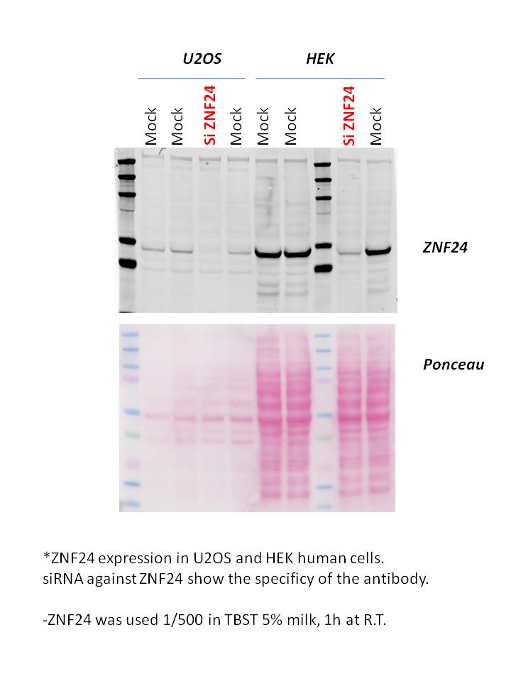
Application: Western BlotSample Tested: 293t HEK and U2OS cellsSpecies: HumanVerified Customer | Posted 06/03/2013

There are no reviews that match your criteria.
Protocols
Find general support by application which include: protocols, troubleshooting, illustrated assays, videos and webinars.
- Antigen Retrieval Protocol (PIER)
- Antigen Retrieval for Frozen Sections Protocol
- Appropriate Fixation of IHC/ICC Samples
- Cellular Response to Hypoxia Protocols
- Chromogenic IHC Staining of Formalin-Fixed Paraffin-Embedded (FFPE) Tissue Protocol
- Chromogenic Immunohistochemistry Staining of Frozen Tissue
- ClariTSA™ Fluorophore Kits
- Detection & Visualization of Antibody Binding
- Fluorescent IHC Staining of Frozen Tissue Protocol
- Graphic Protocol for Heat-induced Epitope Retrieval
- Graphic Protocol for the Preparation and Fluorescent IHC Staining of Frozen Tissue Sections
- Graphic Protocol for the Preparation and Fluorescent IHC Staining of Paraffin-embedded Tissue Sections
- Graphic Protocol for the Preparation of Gelatin-coated Slides for Histological Tissue Sections
- ICC Cell Smear Protocol for Suspension Cells
- ICC Immunocytochemistry Protocol Videos
- ICC for Adherent Cells
- IHC Sample Preparation (Frozen sections vs Paraffin)
- Immunocytochemistry (ICC) Protocol
- Immunocytochemistry Troubleshooting
- Immunofluorescence of Organoids Embedded in Cultrex Basement Membrane Extract
- Immunofluorescent IHC Staining of Formalin-Fixed Paraffin-Embedded (FFPE) Tissue Protocol
- Immunohistochemistry (IHC) and Immunocytochemistry (ICC) Protocols
- Immunohistochemistry Frozen Troubleshooting
- Immunohistochemistry Paraffin Troubleshooting
- Preparing Samples for IHC/ICC Experiments
- Preventing Non-Specific Staining (Non-Specific Binding)
- Primary Antibody Selection & Optimization
- Protocol for Heat-Induced Epitope Retrieval (HIER)
- Protocol for Making a 4% Formaldehyde Solution in PBS
- Protocol for VisUCyte™ HRP Polymer Detection Reagent
- Protocol for the Fluorescent ICC Staining of Cell Smears - Graphic
- Protocol for the Fluorescent ICC Staining of Cultured Cells on Coverslips - Graphic
- Protocol for the Preparation & Fixation of Cells on Coverslips
- Protocol for the Preparation and Chromogenic IHC Staining of Frozen Tissue Sections
- Protocol for the Preparation and Chromogenic IHC Staining of Frozen Tissue Sections - Graphic
- Protocol for the Preparation and Chromogenic IHC Staining of Paraffin-embedded Tissue Sections
- Protocol for the Preparation and Chromogenic IHC Staining of Paraffin-embedded Tissue Sections - Graphic
- Protocol for the Preparation and Fluorescent ICC Staining of Cells on Coverslips
- Protocol for the Preparation and Fluorescent ICC Staining of Non-adherent Cells
- Protocol for the Preparation and Fluorescent ICC Staining of Stem Cells on Coverslips
- Protocol for the Preparation and Fluorescent IHC Staining of Frozen Tissue Sections
- Protocol for the Preparation and Fluorescent IHC Staining of Paraffin-embedded Tissue Sections
- Protocol for the Preparation of Gelatin-coated Slides for Histological Tissue Sections
- Protocol for the Preparation of a Cell Smear for Non-adherent Cell ICC - Graphic
- R&D Systems Quality Control Western Blot Protocol
- TUNEL and Active Caspase-3 Detection by IHC/ICC Protocol
- The Importance of IHC/ICC Controls
- Troubleshooting Guide: Immunohistochemistry
- Troubleshooting Guide: Western Blot Figures
- Western Blot Conditions
- Western Blot Protocol
- Western Blot Protocol for Cell Lysates
- Western Blot Troubleshooting
- Western Blot Troubleshooting Guide
- View all Protocols, Troubleshooting, Illustrated assays and Webinars
Loading...