Alternative Biotinylation Protocol for the Phospho-Immunoreceptor Array
Introduction
Biotinylation is a commonly used method for labeling of cell-surface proteins. This method can be combined with antibody array analysis for profiling cell surface-expressed proteins. In this protocol the user prepares samples by labeling cell-surface proteins with Sulfo-NHS-LC-Biotin and lysing cells in a non-denaturing lysis buffer. For array analysis, cell lysates are diluted and incubated with the Human Phospho-Immunoreceptor Array (Catalog # ARY004B). After binding of immunoreceptors to their specific capture antibodies on the array, unbound material is washed away. Streptavidin-HRP and chemiluminescent detection is then used to visualize biotinylated immunoreceptors corresponding to those expressed on the cell surface.
Sample Biotinylation Data
Required Reagents
- PBS (adjusted to pH 8.0)
- Sulfo-NHS-LC-Biotin (Pierce, Catalog # 21335)
- Glycine
- Streptavidin-HRP (Catalog # DY998)
- Chemiluminescence Detection Reagents
Cell-surface biotinylation and Sample Preparation
The cell-surface biotinylation procedure is a modification of the procedure recommended by the manufacturer of the biotinylation reagent. For suspended cells, wash two times with ice-cold PBS (pH 8.0) to remove sources of primary amines in the culture media. After the final spin, resuspend the cells at a concentration of 2.5 x 10⁷ cells/mL in ice-cold PBS (pH 8.0) containing 0.25 mg of Sulfo-NHS-LC-Biotin reagent per mL and incubate at 4 °C for 30 minutes. Wash the cells twice with PBS containing 100 mM glycine to quench and remove excess biotin reagent. Solubilize the cells at 1 x 10⁷ cells/mL in Lysis Buffer 15. Pipette up and down to resuspend and rock the lysates gently at 2 to 8 °C for 30 minutes. Microcentrifuge at 14,000 x g for 5 minutes, and transfer the supernatant into a clean microcentrifuge tube. Determine the sample protein concentration using a total protein assay. For incubation with the Human Phospho-Immunoreceptor Array, use a smaller quantity of lysate than that used for immunoprecipitation or Western blot (5 to 50 µg ). Lysates should be used immediately or aliquoted and stored at ≤ 70 °C. Thawed lysates should be kept on ice prior to use.
Array Protocol
Bring all reagents to room temperature before use. Keep samples on ice.
- Add 1.5 mL of Array Buffer 1 into individual wells of the 4-Well Multi-dish that will be used for each array.
- Using flat-tip tweezers, remove each array to be used from between the protective sheets.
- Place one array into each well of the 4-Well Multi-dish. The array number should be facing upward.
Note: The blue dye will disappear from the spots. The capture antibodies are retained in their specific locations. - Incubate for 1 hour on a rocking platform shaker. Orient the tray so that each array rocks end to end in its well.
- Dilute the lysate to 1.5 mL with Array Buffer 1 and add dilute lysate.
- Remove Array Buffer 1 from the 4-Well Multi-dish.
- Incubate overnight at 2 to 8 º C (or 2 hours at room temperature) on a rocking platform shaker. Orient the tray so that each array rocks end to end in its well.
- Carefully remove each array and place into individual plastic containers with a minimum of 20 mL of 1X Wash Buffer. Rinse the 4-Well Multi-dish with deionized or distilled water and dry thoroughly.
- Wash each array with 1X Wash Buffer for 10 minutes on a rocking platform shaker. Repeat two times for a total of three washes.
- Dilute Streptavidin-HRP 1:4000 by adding 2.5 µL of Streptavidin-HRP to 10 mL of Array Buffer 2.
- Add 1.5 mL of the freshly diluted Streptavidin-HRP to each well of the 4-Well Multi-dish.
- Carefully remove each array from its wash container, return it to the 4-Well Multi-dish, and cover with the lid.
- Incubate for 1 hour at room temperature on a rocking platform shaker. Orient the tray so that each array rocks end to end in its well.
- Wash each array as described in steps 8 and 9.
- Remove each array from the wash container and place it on a plastic sheet protector. Expose each array to chemiluminescent substrate according to instructions provided in the kit insert.
- Cover with plastic wrap and expose to X-ray film for 1 to 10 minutes.
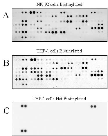
Lysates from biotinylated NK-92 cells (A; 25 µg), biotinylated THP-1 cells (B ; 20 µg), and unbiotinylated THP-1 cells (C;25 µg) were tested with the Human Phospho-Immunoreceptor Array using the above protocol. The results reveal the immunoreceptor expression profile of each cell line and only positive control spots in unbiotinylated cells.