
Arginase 1/ARG1/liver Arginase Antibody - BSA Free
Novus Biologicals | Catalog # NBP1-32731

![Knockdown Validated: Arginase 1/ARG1/liver Arginase Antibody - BSA Free [NBP1-32731]](https://resources.rndsystems.com/images/products/Arginase-1-ARG1-liver-Arginase-Antibody-Western-Blot-NBP1-32731-img0022.jpg "Western Blot: Arginase 1/ARG1/liver Arginase Antibody - BSA Free [NBP1-32731]")
Scientific images may be resized, cropped, or adjusted for brightness or contrast for presentation purposes. These updates are limited to presentation and do not affect the underlying scientific interpretation of the data. Where available, additional source data may be provided upon request.
Key Product Details
Validated by
Knockout/Knockdown
Species Reactivity
Validated:
Human, Mouse, Rat
Cited:
Human, Mouse, Rat
Predicted:
Bovine (90%), Rabbit (91%). Backed by our 100% Guarantee.
Applications
Validated:
Immunohistochemistry, Immunohistochemistry-Paraffin, Immunohistochemistry-Frozen, Western Blot, Flow Cytometry, Immunocytochemistry/ Immunofluorescence, Simple Western, Immunoprecipitation, Knockdown Validated
Cited:
Immunohistochemistry-Paraffin, Immunohistochemistry-Frozen, Western Blot, Flow Cytometry, Immunocytochemistry/ Immunofluorescence, IF/IHC
Label
Unconjugated
Antibody Source
Polyclonal Rabbit IgG
Format
BSA Free
Loading...
Product Specifications
Immunogen
Arginase 1/ARG1/liver Arginase Antibody made from full length human Arginase 1 Recombinant protein.
Reactivity Notes
Rat reactivity reported in scientific literature (PMID:32701588). Immunogen displays the following percentage of sequence identity for non-tested species: Porcine (89%).
Localization
Cytoplasm/Nucleus
Clonality
Polyclonal
Host
Rabbit
Isotype
IgG
Scientific Data Images for Arginase 1/ARG1/liver Arginase Antibody - BSA Free
Western Blot: Arginase 1/ARG1/liver Arginase Antibody - BSA Free [NBP1-32731]
Western Blot: Arginase 1/ARG1/liver Arginase Antibody [NBP1-32731] - Non-transfected (-) and transfected (+) HepG2 whole cell extracts (30 ug) were separated by 10% SDS-PAGE, and the membrane was blotted with Arginase 1 antibody.![Immunohistochemistry: Arginase 1/ARG1/liver Arginase Antibody - BSA Free [NBP1-32731]](https://resources.rndsystems.com/images/products/Arginase-1-ARG1-liver-Arginase-Antibody-Immunohistochemistry-NBP1-32731-img0033.jpg "Immunohistochemistry: Arginase 1/ARG1/liver Arginase Antibody - BSA Free [NBP1-32731]")
Immunohistochemistry: Arginase 1/ARG1/liver Arginase Antibody - BSA Free [NBP1-32731]
Arginase-1-ARG1-liver-Arginase-Antibody-Immunohistochemistry-NBP1-32731-img0033.jpg![Simple Western: Arginase 1/ARG1/liver Arginase AntibodyBSA Free [NBP1-32731]](https://resources.rndsystems.com/images/products/Arginase-1-ARG1-liver-Arginase-Antibody-Simple-Western-NBP1-32731-img0029.jpg "Simple Western: Arginase 1/ARG1/liver Arginase AntibodyBSA Free [NBP1-32731]")
Simple Western: Arginase 1/ARG1/liver Arginase AntibodyBSA Free [NBP1-32731]
Simple Western: Arginase 1/ARG1/liver Arginase Antibody [NBP1-32731] - Simple Western lane view shows a specific band for ARG1 in human and mouse Liver lysate using ARG1 antibody (NBP1-32731) at 25 ug/ml. This experiment was performed under reducing conditions using the 12-230 kDa separation system.![Flow Cytometry: Arginase 1/ARG1/liver Arginase Antibody - BSA Free [NBP1-32731]](https://resources.rndsystems.com/images/products/Arginase-1-ARG1-liver-Arginase-Antibody-Flow-Cytometry-NBP1-32731-img0032.jpg "Flow Cytometry: Arginase 1/ARG1/liver Arginase Antibody - BSA Free [NBP1-32731]")
Flow Cytometry: Arginase 1/ARG1/liver Arginase Antibody - BSA Free [NBP1-32731]
Arginase-1-ARG1-liver-Arginase-Antibody-Flow-Cytometry-NBP1-32731-img0032.jpg![Western Blot: Arginase 1/ARG1/liver Arginase AntibodyBSA Free [NBP1-32731]](https://resources.rndsystems.com/images/products/Arginase-1-ARG1-liver-Arginase-Antibody-Western-Blot-NBP1-32731-img0014.jpg "Western Blot: Arginase 1/ARG1/liver Arginase AntibodyBSA Free [NBP1-32731]")
Western Blot: Arginase 1/ARG1/liver Arginase AntibodyBSA Free [NBP1-32731]
Western Blot: Arginase 1/ARG1/liver Arginase Antibody [NBP1-32731] - Arginase 1 antibody detects Arginase 1 protein. Various whole cell extracts (30ug) were separated by 12% SDS-PAGE, and the membrane was blotted with Arginase 1 antibody diluted at 1:1000.![Immunoprecipitation: Arginase 1/ARG1/liver Arginase Antibody - BSA Free [NBP1-32731]](https://resources.rndsystems.com/images/products/Arginase-1-ARG1-liver-Arginase-Antibody-Immunoprecipitation-NBP1-32731-img0015.jpg "Immunoprecipitation: Arginase 1/ARG1/liver Arginase Antibody - BSA Free [NBP1-32731]")
Immunoprecipitation: Arginase 1/ARG1/liver Arginase Antibody - BSA Free [NBP1-32731]
Immunoprecipitation: Arginase 1/ARG1/liver Arginase Antibody [NBP1-32731] - Analysis was performed using Arginase 1 antibody EasyBlot anti-Rabbit IgG was used as a secondary reagent. Arginase 1 protein from HepG2 whole cell extracts using 5 ug of Arginase 1 antibody.![Western Blot: Arginase 1/ARG1/liver Arginase AntibodyBSA Free [NBP1-32731]](https://resources.rndsystems.com/images/products/Arginase-1-ARG1-liver-Arginase-Antibody-Western-Blot-NBP1-32731-img0020.jpg "Western Blot: Arginase 1/ARG1/liver Arginase AntibodyBSA Free [NBP1-32731]")
Western Blot: Arginase 1/ARG1/liver Arginase AntibodyBSA Free [NBP1-32731]
Western Blot: Arginase 1/ARG1/liver Arginase Antibody [NBP1-32731] - Rat tissue extract (50 ug) was separated by 12% SDS-PAGE, and the membrane was blotted with Arginase 1 antibody diluted at 1:10000.![Immunocytochemistry/ Immunofluorescence: Arginase 1/ARG1/liver Arginase Antibody - BSA Free [NBP1-32731]](https://resources.rndsystems.com/images/products/Arginase-1-ARG1-liver-Arginase-Antibody-Immunocytochemistry-Immunofluorescence-NBP1-32731-img0034.jpg "Immunocytochemistry/ Immunofluorescence: Arginase 1/ARG1/liver Arginase Antibody - BSA Free [NBP1-32731]")
Immunocytochemistry/ Immunofluorescence: Arginase 1/ARG1/liver Arginase Antibody - BSA Free [NBP1-32731]
Immunocytochemistry/Immunofluorescence: Arginase 1/ARG1/liver Arginase Antibody [NBP1-32731] - An intracellular stain was performed on HepG2 cells with Arginase 1/ARG1/liver Arginase Antibody NBP1-32731AF647 (blue) and a matched isotype control (orange). Cells were fixed with 4% PFA and then permeabilized with 0.1% saponin. Cells were incubated in an antibody dilution of 2.5 ug/mL for 30 minutes at room temperature. Both antibodies were conjugated to Alexa Fluor 647.![Western Blot: Arginase 1/ARG1/liver Arginase AntibodyBSA Free [NBP1-32731]](https://resources.rndsystems.com/images/products/Arginase-1-ARG1-liver-Arginase-Antibody-Western-Blot-NBP1-32731-img0018.jpg "Western Blot: Arginase 1/ARG1/liver Arginase AntibodyBSA Free [NBP1-32731]")
Western Blot: Arginase 1/ARG1/liver Arginase AntibodyBSA Free [NBP1-32731]
Western Blot: Arginase 1/ARG1/liver Arginase Antibody [NBP1-32731] - Mouse tissue extract (50 ug) was separated by 12% SDS-PAGE, and the membrane was blotted with Arginase 1 antibody diluted at 1:10000.![Immunocytochemistry/ Immunofluorescence: Arginase 1/ARG1/liver Arginase Antibody - BSA Free [NBP1-32731]](https://resources.rndsystems.com/images/products/Arginase-1-ARG1-liver-Arginase-Antibody-Immunocytochemistry-Immunofluorescence-NBP1-32731-img0009.jpg "Immunocytochemistry/ Immunofluorescence: Arginase 1/ARG1/liver Arginase Antibody - BSA Free [NBP1-32731]")
Immunocytochemistry/ Immunofluorescence: Arginase 1/ARG1/liver Arginase Antibody - BSA Free [NBP1-32731]
Immunocytochemistry/Immunofluorescence: Arginase 1/ARG1/liver Arginase Antibody [NBP1-32731] - Paraformaldehyde-fixed HeLa.![Immunocytochemistry/ Immunofluorescence: Arginase 1/ARG1/liver Arginase Antibody - BSA Free [NBP1-32731]](https://resources.rndsystems.com/images/products/Arginase-1-ARG1-liver-Arginase-Antibody-Immunocytochemistry-Immunofluorescence-NBP1-32731-img0027.jpg "Immunocytochemistry/ Immunofluorescence: Arginase 1/ARG1/liver Arginase Antibody - BSA Free [NBP1-32731]")
Immunocytochemistry/ Immunofluorescence: Arginase 1/ARG1/liver Arginase Antibody - BSA Free [NBP1-32731]
Immunocytochemistry/Immunofluorescence: Arginase 1/ARG1/liver Arginase Antibody [NBP1-32731] - HepG2 cells were fixed for 10 minutes using 10% formalin and then permeabilized for 5 minutes using 1X PBS + 0.05% Triton X-100. The cells were incubated with anti-ARG1 at 10 ug/ml overnight at 4C and detected with an anti-rabbit DyLight 488 (Green) at a 1:500 dilution. Nuclei were counterstained with DAPI (Blue). Cells were imaged using a 40X objective.![Immunohistochemistry-Paraffin: Arginase 1/ARG1/liver Arginase Antibody - BSA Free [NBP1-32731]](https://resources.rndsystems.com/images/products/Arginase-1-ARG1-liver-Arginase-Antibody-Immunohistochemistry-Paraffin-NBP1-32731-img0010.jpg "Immunohistochemistry-Paraffin: Arginase 1/ARG1/liver Arginase Antibody - BSA Free [NBP1-32731]")
Immunohistochemistry-Paraffin: Arginase 1/ARG1/liver Arginase Antibody - BSA Free [NBP1-32731]
Immunohistochemistry-Paraffin: Arginase 1/ARG1/liver Arginase Antibody [NBP1-32731] - Cal27 xenograft, using arginase I antibody at 1:500 dilution.![Immunohistochemistry-Paraffin: Arginase 1/ARG1/liver Arginase Antibody - BSA Free [NBP1-32731]](https://resources.rndsystems.com/images/products/Arginase-1-ARG1-liver-Arginase-Antibody-Immunohistochemistry-Paraffin-NBP1-32731-img0026.jpg "Immunohistochemistry-Paraffin: Arginase 1/ARG1/liver Arginase Antibody - BSA Free [NBP1-32731]")
Immunohistochemistry-Paraffin: Arginase 1/ARG1/liver Arginase Antibody - BSA Free [NBP1-32731]
Immunohistochemistry-Paraffin: Arginase 1/ARG1/liver Arginase Antibody [NBP1-32731] - Tissue section of the mouse liver using 1:200 dilution of ARG1 antibody (NBP1-32731). The signal was developed using HRP-DAB method which followed counterstaining of the cells with hematoxylin.![Immunohistochemistry-Paraffin: Arginase 1/ARG1/liver Arginase Antibody - BSA Free [NBP1-32731]](https://resources.rndsystems.com/images/products/Arginase-1-ARG1-liver-Arginase-Antibody-Immunohistochemistry-Paraffin-NBP1-32731-img0028.jpg "Immunohistochemistry-Paraffin: Arginase 1/ARG1/liver Arginase Antibody - BSA Free [NBP1-32731]")
Immunohistochemistry-Paraffin: Arginase 1/ARG1/liver Arginase Antibody - BSA Free [NBP1-32731]
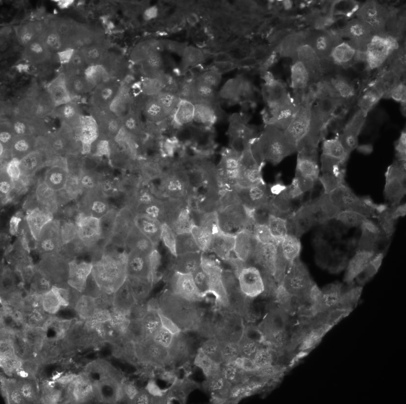
Immunohistochemistry-Paraffin: Arginase 1/ARG1/liver Arginase Antibody [NBP1-32731] - Hepatocellular carcinoma stained with Arginase1 1:100, pH9 antigen retrieval. Image from verified customer review.![Flow Cytometry: Arginase 1/ARG1/liver Arginase Antibody - BSA Free [NBP1-32731]](https://resources.rndsystems.com/images/products/Arginase-1-ARG1-liver-Arginase-Antibody-Flow-Cytometry-NBP1-32731-img0025.jpg "Flow Cytometry: Arginase 1/ARG1/liver Arginase Antibody - BSA Free [NBP1-32731]")
Flow Cytometry: Arginase 1/ARG1/liver Arginase Antibody - BSA Free [NBP1-32731]
Flow Cytometry: Arginase 1/ARG1/liver Arginase Antibody [NBP1-32731] - An intracellular stain was performed on HepG2 with NBP1-32731 and a matched isotype control. Cells were fixed with 4% PFA and then permeablized with 0.1% saponin. Cells were incubated in an antibody dilution of 5 ug/mL for 30 minutes at room temperature, followed by Rabbit IgG (H+L) Cross-Adsorbed Secondary Antibody.![Flow Cytometry: Arginase 1/ARG1/liver Arginase Antibody - BSA Free [NBP1-32731]](https://resources.rndsystems.com/images/products/Arginase-1-ARG1-liver-Arginase-Antibody-Flow-Cytometry-NBP1-32731-img0030.jpg "Flow Cytometry: Arginase 1/ARG1/liver Arginase Antibody - BSA Free [NBP1-32731]")
Flow Cytometry: Arginase 1/ARG1/liver Arginase Antibody - BSA Free [NBP1-32731]
Flow Cytometry: Arginase 1/ARG1/liver Arginase Antibody [NBP1-32731] - An intracellular stain was performed on RH30 cells with Arginase 1 Antibody NBP1-32731 (blue) and a matched isotype control (orange). Cells were fixed with 4% PFA and then permeabilized with 0.1% saponin. Cells were incubated in an antibody dilution of 5.0 ug/mL for 30 minutes at room temperature, followed by Rabbit IgG (H+L) Cross-Adsorbed Secondary Antibody.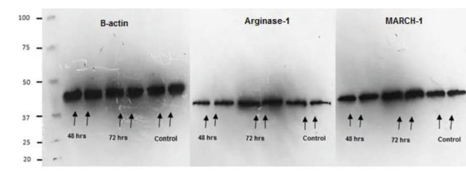
Western Blot: Arginase 1/ARG1/liver Arginase Antibody [NBP1-32731] -
nbp1-32731_rabbit-polyclonal-arginase-1-arg1-liver-arginase-antibody-862023835350.jpg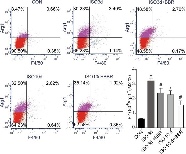
Flow Cytometry: Arginase 1/ARG1/liver Arginase Antibody - BSA Free [NBP1-32731] -
Effects of berberine on the M2 population of macrophages. Representative dot plot of M2 subpopulations in control group and the rats injected with ISO 3 and 7 days later. M2 cells were labelled with F4/80 and Arg1. *P < 0.05 as compared with the control group. #P < 0.05 vs. the corresponding ISO group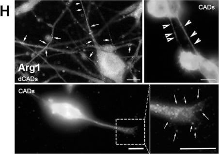
Immunocytochemistry/ Immunofluorescence: Arginase 1/ARG1/liver Arginase Antibody - BSA Free [NBP1-32731] -
Immunocytochemistry/ Immunofluorescence: Arginase 1/ARG1/liver Arginase Antibody - BSA Free [NBP1-32731] - Validation of the LCM/MS data from cellular protrusions using microscopy. Immunofluorescence (IF) of proteins identified by LCM/MS (A–D) “expected” or (E–H) “not expected” to be within protrusions based on their known functions/localizations. (A) Cd47 & (B) Anxa2 are found in both hCAD/dCAD protrusions. (C) Tenm2 is found within hCAD protrusions & (D) Cobl in GCs. Interestingly, (E) Grk5 was observed in hCAD protrusions; (F) Hist1h3b protein in hCAD protrusions & GCs (G) Hspa1b in dCAD protrusions & TNTs & (H) Arg1 in dCAD protrusions, GCs, & TNTs. White arrows/arrowheads show punctates within dCAD/hCAD protrusions & GCs or within TNTs, respectively. Scale bars = 10 μm. IF of all 8 proteins corroborate the LCM/MS protein identification for each subtypes of protrusions. (I) 1198 unique proteins were identified by LCM/MS to be in protrusions & 169 were found to be exclusive to protrusions. 904 of the 1198 proteins found in protrusions & 87 out of 169 exclusive proteins had images in the HPA Subcell database. IF images for the proteins identified by LCM/MS in protrusions, present in the HPA Subcell database, were observed & catalogued as being observed or not in protrusions (Figure S2). On average 4.7% of proteins are found in protrusions in the HPA database, 13.6% are found in our LCM isolated protrusions & 23% were found in the “exclusive” LCM protrusions. The fold enrichment increases by 2.9% or 4.8% when we looked at proteins identified by LCM/MS in protrusions or exclusive to protrusions, respectively. Image collected & cropped by CiteAb from the following publication (https://pubmed.ncbi.nlm.nih.gov/30866487), licensed under a CC-BY license. Not internally tested by Novus Biologicals.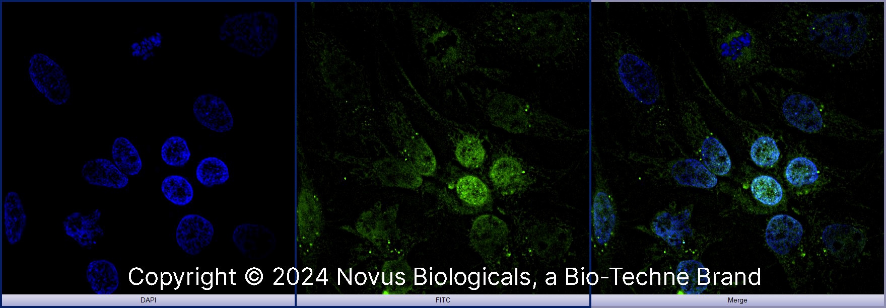
Arginase 1/ARG1/liver Arginase in HepG2 Human Cell Line.
Arginase 1/ARG1/liver Arginase was detected in immersion fixed HepG2 human hepatocellular carcinoma cell line using Rabbit anti-Arginase 1/ARG1/liver Arginase Affinity Purified Polyclonal Antibody conjugated to FITC (Catalog # NBP1-32731F) (green) at 10 µg/mL overnight at 4C. Cells were counterstained with DAPI (dark blue). Cells were imaged using a 100X objective and digitally deconvolved.Applications for Arginase 1/ARG1/liver Arginase Antibody - BSA Free
Application
Recommended Usage
Flow Cytometry
5 - 10 ug/ml
Immunocytochemistry/ Immunofluorescence
1:10 - 1:500
Immunohistochemistry
1:100-1:1000
Immunohistochemistry-Frozen
1:10 - 1:500
Immunohistochemistry-Paraffin
1:100-1:1000
Immunoprecipitation
1:10 - 1:500
Western Blot
1:1000-1:50000
Reviewed Applications
Read 1 review rated 4 using NBP1-32731 in the following applications:
Flow Cytometry Panel Builder
Bio-Techne Knows Flow Cytometry
Save time and reduce costly mistakes by quickly finding compatible reagents using the Panel Builder Tool.
Advanced Features
- Spectra Viewer - Custom analysis of spectra from multiple fluorochromes
- Spillover Popups - Visualize the spectra of individual fluorochromes
- Antigen Density Selector - Match fluorochrome brightness with antigen density

Formulation, Preparation, and Storage
Purification
Immunogen affinity purified
Formulation
PBS
Format
BSA Free
Preservative
0.02% Sodium Azide
Concentration
1.0 mg/ml
Shipping
The product is shipped with polar packs. Upon receipt, store it immediately at the temperature recommended below.
Stability & Storage
Aliquot and store at -20C or -80C. Avoid freeze-thaw cycles.
Background: Arginase 1/ARG1
Arginase and nitric oxidase synthase (NOS) compete for the same L-arginine substrate, creating a delicate balance between pathways (1). Furthermore, bioavailability of L-arginine and ARG1 expression has been implicated in several pathologies including vascular disease, neuronal disease, cardiovascular disease, immune dysfunction, inflammation, and cancer (1,3-5). For instance, ARG1 functions as a macrophage marker, defining the M2 population, while inducible nitric oxide synthase (iNOS) characterizes the M1 population; impaired M1/M2 polarization and changes in ARG1 expression is observed in diseases such as arteriogenesis, asthma, pulmonary fibrosis, and inflammatory bowel disease (1,3). In humans, arginase deficiency, known as argininemia, is an autosomal recessive metabolic disorder characterized by elevated ammonia (hyperammonemia) levels and arginine accumulation (6). Given that many arginase-associated diseases are characterized by upregulation in expression of ARG1, ARG2, or both, arginase inhibitors are currently being studied as a potential therapeutic approach (1,4).
References
1. S Clemente, G., van Waarde, A., F Antunes, I., Domling, A., & H Elsinga, P. (2020). Arginase as a Potential Biomarker of Disease Progression: A Molecular Imaging Perspective. International Journal of Molecular Sciences. https://doi.org/10.3390/ijms21155291
2. Uniprot (P05089)
3. Kieler, M., Hofmann, M., & Schabbauer, G. (2021). More than just protein building blocks: How amino acids and related metabolic pathways fuel macrophage polarization. The FEBS Journal. Advance online publication. https://doi.org/10.1111/febs.15715
4. Shosha, E., Fouda, A. Y., Narayanan, S. P., Caldwell, R. W., & Caldwell, R. B. (2020). Is the Arginase Pathway a Novel Therapeutic Avenue for Diabetic Retinopathy?. Journal of Clinical Medicine. https://doi.org/10.3390/jcm9020425
5. Correale J. (2021). Immunosuppressive Amino-Acid Catabolizing Enzymes in Multiple Sclerosis. Frontiers in Immunology. https://doi.org/10.3389/fimmu.2020.600428
6. Morales, J. A., & Sticco, K. L. (2020). Arginase Deficiency. In StatPearls. StatPearls Publishing.
Long Name
Liver-Type Arginase
Alternate Names
AI, ARG1, Arginase-1, Liver Arginase, PGIF, Type I Arginase
Entrez Gene IDs
383 (Human)
Gene Symbol
ARG1
UniProt
Additional Arginase 1/ARG1 Products
Product Documents for Arginase 1/ARG1/liver Arginase Antibody - BSA Free
Certificate of Analysis
To download a Certificate of Analysis, please enter a lot or batch number in the search box below.
Product Specific Notices for Arginase 1/ARG1/liver Arginase Antibody - BSA Free
This product is for research use only and is not approved for use in humans or in clinical diagnosis. Primary Antibodies are guaranteed for 1 year from date of receipt.
Related Research Areas
Citations for Arginase 1/ARG1/liver Arginase Antibody - BSA Free
 Powered by Bioz
Customer Reviews for Arginase 1/ARG1/liver Arginase Antibody - BSA Free (1)
4 out of 5
1 Customer Rating
Have you used Arginase 1/ARG1/liver Arginase Antibody - BSA Free?
Submit a review and receive an Amazon gift card!
$25/€18/£15/$25CAN/¥2500 for a review with an image
$10/€7/£6/$10CAN/¥1110 for a review without an image
Submit a review
Customer Images
Showing
1
-
1 的
1 review
Showing All
Filter By:
-
Application: Immunofluorescence - paraffinSample Tested: Hepatocellular carcinomaSpecies: HumanVerified Customer | Posted 03/09/2019Hepatocellular carcinoma stained with Arginase1 1:100pH9 antigen retrieval

There are no reviews that match your criteria.
Protocols
View specific protocols for Arginase 1/ARG1/liver Arginase Antibody - BSA Free (NBP1-32731):
Protocol for Flow Cytometry Intracellular Staining
Sample Preparation.
1. Grow cells to 60-85% confluency. Flow cytometry requires between 2 x 10⁵ and 1 x 10⁶ cells for optimal performance.
2. If cells are adherent, harvest gently by washing once with staining buffer and then scraping. Avoid using trypsin as this can disrupt certain epitopes of interest. If enzymatic harvest is required, use Accutase, Collagenase, or TrypLE Express for a less damaging option.
3. Reserve 100 uL for counting, then transfer cell volume into a 50 mL conical tube and centrifuge for 8 minutes at 400 RCF.
a. Count cells using a hemocytometer and a 1:1 trypan blue exclusion stain to determine cell viability before starting the flow protocol. If cells appear blue, do not proceed.
4. Re-suspend cells to a concentration of 1 x 10⁶ cells/mL in staining buffer (NBP2-26247).
5. Aliquot out 1 mL samples in accordance with your experimental samples.
Tip: When cell surface and intracellular staining are required in the same sample, it is advisable that the cell surface staining be performed first since the fixation and permeablization steps might reduce the availability of surface antigens.
Intracellular Staining.
Tip: When performing intracellular staining, it is important to use appropriate fixation and permeabilization reagents based upon the target and its subcellular location. Generally, our Intracellular Flow Assay Kit (NBP2-29450) is a good place to start as it contains an optimized combination of reagents for intracellular staining as well as an inhibitor of intracellular protein transport (necessary if staining secreted proteins). Certain targets may require more gentle or transient permeabilization protocols such as the commonly employed methanol or saponin-based methods.
Protocol for Cytoplasmic Targets:
Optional: Perform cell surface staining as described in the previous section.
1. Fix the cells by adding 100 uL fixation solution (such as 4% PFA) to each sample for 10-15 minutes.
2. Permeabilize cells by adding 100 uL of a permeabization buffer to every 1 x 10⁶ cells present in the sample. Mix well and incubate at room temperature for 15 minutes.
a. For cytoplasmic targets, use a gentle permeabilization solution such as 1X PBS + 0.5% Saponin or 1X PBS + 0.5% Tween-20.
b. To maintain the permeabilized state throughout your experiment, use staining buffer + 0.1% of the permeabilization reagent (i.e. 0.1% Tween-20 or 0.1% Saponin).
3. Following the 15 minute incubation, add 2 mL of the staining buffer + 0.1% permeabilizer to each sample.
4. Centrifuge for 5 minutes at 400 RCF.
5. Discard supernatant and re-suspend in 1 mL of staining buffer + 0.1% permeabilizer.
6. Stain each sample at 1 uL/ 1 x 10⁶ cells of primary antibody or 1-3 uL/ 1 x 10⁶ cells for directly conjugated antibodies. Mix well and incubate at room temperature for 30 minutes- 1 hour. Gently mix samples every 10-15 minutes.
7. Following the primary/conjugate incubation, add 2 mL/sample of staining buffer +0.1% permeabilizer and centrifuge for 5 minutes at 400 RCF.
8. Remove supernatant and re-suspend each sample in 2 mL staining buffer + 0.1% permeabilizer, repeat wash for 5 minutes at 400 RCF.
9. If using a directly conjugated antibody, after the second wash, re-suspend cell pellet to a final volume of 500 uL per sample and proceed with flow analysis.
Immunocytochemistry Protocol
Culture cells to appropriate density in 35 mm culture dishes or 6-well plates.
1. Remove culture medium and wash the cells briefly in PBS. Add 10% formalin to the dish and fix at room temperature for 10 minutes.
2. Remove the formalin and wash the cells in PBS.
3. Permeablize the cells with 0.1% Triton X100 or other suitable detergent for 10 min.
4. Remove the permeablization buffer and wash three times for 10 minutes each in PBS. Be sure to not let the specimen dry out.
5. To block nonspecific antibody binding, incubate in 10% normal goat serum from 1 hour to overnight at room temperature.
6. Add primary antibody at appropriate dilution and incubate overnight at 4C.
7. Remove primary antibody and replace with PBS. Wash three times for 10 minutes each.
8. Add secondary antibody at appropriate dilution. Incubate for 1 hour at room temperature.
9. Remove secondary antibody and replace with PBS. Wash three times for 10 minutes each.
10. Counter stain DNA with DAPi if required.
*The above information is only intended as a guide. The researcher should determine what protocol best meets their needs. Please follow safe laboratory procedures.
Immunohistochemistry-Paraffin Embedded Sections
Antigen Unmasking:
Bring slides to a boil in 10 mM sodium citrate buffer (pH 6.0) then maintain at a sub-boiling temperature for 10 minutes. Cool slides on bench-top for 30 minutes.
Staining:
1. Wash sections in deionized water three times for 5 minutes each.
2. Wash sections in wash buffer for 5 minutes.
3. Block each section with 100-400 ul blocking solution for 1 hour at room temperature.
4. Remove blocking solution and add 100-400 ul diluted primary antibody. Incubate overnight at 4 C.
5. Remove antibody solution and wash sections in wash buffer three times for 5 minutes each.
6. Add 100-400 ul biotinylated diluted secondary antibody. Incubate 30 minutes at room temperature.
7. Remove secondary antibody solution and wash sections three times with wash buffer for 5 minutes each.
8. Add 100-400 ul Streptavidin-HRP reagent to each section and incubate for 30 minutes at room temperature.
9. Wash sections three times in wash buffer for 5 minutes each.
10. Add 100-400 ul DAB substrate to each section and monitor staining closely.
11. As soon as the sections develop, immerse slides in deionized water.
12. Counterstain sections in hematoxylin.
13. Wash sections in deionized water two times for 5 minutes each.
14. Dehydrate sections.
15. Mount coverslips.
Western Blot Protocol
1. Perform SDS-PAGE on samples to be analyzed, loading 10-25 ug of total protein per lane.
2. Transfer proteins to PVDF membrane according to the instructions provided by the manufacturer of the membrane and transfer apparatus.
3. Stain the membrane with Ponceau S (or similar product) to assess transfer success, and mark molecular weight standards where appropriate.
4. Rinse the blot TBS -0.05% Tween 20 (TBST).
5. Block the membrane in 5% Non-fat milk in TBST (blocking buffer) for at least 1 hour.
6. Wash the membrane in TBST three times for 10 minutes each.
7. Dilute anti-ARG1 primary antibody in blocking buffer and incubate overnight at 4C with gentle rocking.
8. Wash the membrane in TBST three times for 10 minutes each.
9. Incubate the membrane in diluted HRP conjugated secondary antibody in blocking buffer (as per manufacturer's instructions) for 1 hour at room temperature.
10. Wash the blot in TBST three times for 10 minutes each (this step can be repeated as required to reduce background).
11. Apply the detection reagent of choice in accordance with the manufacturers instructions.
Find general support by application which include: protocols, troubleshooting, illustrated assays, videos and webinars.
- 7-Amino Actinomycin D (7-AAD) Cell Viability Flow Cytometry Protocol
- Antigen Retrieval Protocol (PIER)
- Antigen Retrieval for Frozen Sections Protocol
- Appropriate Fixation of IHC/ICC Samples
- Cellular Response to Hypoxia Protocols
- Chromogenic IHC Staining of Formalin-Fixed Paraffin-Embedded (FFPE) Tissue Protocol
- Chromogenic Immunohistochemistry Staining of Frozen Tissue
- ClariTSA™ Fluorophore Kits
- Detection & Visualization of Antibody Binding
- Extracellular Membrane Flow Cytometry Protocol
- Flow Cytometry Protocol for Cell Surface Markers
- Flow Cytometry Protocol for Staining Membrane Associated Proteins
- Flow Cytometry Staining Protocols
- Flow Cytometry Troubleshooting Guide
- Fluorescent IHC Staining of Frozen Tissue Protocol
- Graphic Protocol for Heat-induced Epitope Retrieval
- Graphic Protocol for the Preparation and Fluorescent IHC Staining of Frozen Tissue Sections
- Graphic Protocol for the Preparation and Fluorescent IHC Staining of Paraffin-embedded Tissue Sections
- Graphic Protocol for the Preparation of Gelatin-coated Slides for Histological Tissue Sections
- ICC Cell Smear Protocol for Suspension Cells
- ICC Immunocytochemistry Protocol Videos
- ICC for Adherent Cells
- IHC Sample Preparation (Frozen sections vs Paraffin)
- Immunocytochemistry (ICC) Protocol
- Immunocytochemistry Troubleshooting
- Immunofluorescence of Organoids Embedded in Cultrex Basement Membrane Extract
- Immunofluorescent IHC Staining of Formalin-Fixed Paraffin-Embedded (FFPE) Tissue Protocol
- Immunohistochemistry (IHC) and Immunocytochemistry (ICC) Protocols
- Immunohistochemistry Frozen Troubleshooting
- Immunohistochemistry Paraffin Troubleshooting
- Immunoprecipitation Protocol
- Intracellular Flow Cytometry Protocol Using Alcohol (Methanol)
- Intracellular Flow Cytometry Protocol Using Detergents
- Intracellular Nuclear Staining Flow Cytometry Protocol Using Detergents
- Intracellular Staining Flow Cytometry Protocol Using Alcohol Permeabilization
- Intracellular Staining Flow Cytometry Protocol Using Detergents to Permeabilize Cells
- Preparing Samples for IHC/ICC Experiments
- Preventing Non-Specific Staining (Non-Specific Binding)
- Primary Antibody Selection & Optimization
- Propidium Iodide Cell Viability Flow Cytometry Protocol
- Protocol for Heat-Induced Epitope Retrieval (HIER)
- Protocol for Liperfluo
- Protocol for Making a 4% Formaldehyde Solution in PBS
- Protocol for VisUCyte™ HRP Polymer Detection Reagent
- Protocol for the Characterization of Human Th22 Cells
- Protocol for the Characterization of Human Th9 Cells
- Protocol for the Fluorescent ICC Staining of Cell Smears - Graphic
- Protocol for the Fluorescent ICC Staining of Cultured Cells on Coverslips - Graphic
- Protocol for the Preparation & Fixation of Cells on Coverslips
- Protocol for the Preparation and Chromogenic IHC Staining of Frozen Tissue Sections
- Protocol for the Preparation and Chromogenic IHC Staining of Frozen Tissue Sections - Graphic
- Protocol for the Preparation and Chromogenic IHC Staining of Paraffin-embedded Tissue Sections
- Protocol for the Preparation and Chromogenic IHC Staining of Paraffin-embedded Tissue Sections - Graphic
- Protocol for the Preparation and Fluorescent ICC Staining of Cells on Coverslips
- Protocol for the Preparation and Fluorescent ICC Staining of Non-adherent Cells
- Protocol for the Preparation and Fluorescent ICC Staining of Stem Cells on Coverslips
- Protocol for the Preparation and Fluorescent IHC Staining of Frozen Tissue Sections
- Protocol for the Preparation and Fluorescent IHC Staining of Paraffin-embedded Tissue Sections
- Protocol for the Preparation of Gelatin-coated Slides for Histological Tissue Sections
- Protocol for the Preparation of a Cell Smear for Non-adherent Cell ICC - Graphic
- Protocol: Annexin V and PI Staining by Flow Cytometry
- Protocol: Annexin V and PI Staining for Apoptosis by Flow Cytometry
- R&D Systems Quality Control Western Blot Protocol
- TUNEL and Active Caspase-3 Detection by IHC/ICC Protocol
- The Importance of IHC/ICC Controls
- Troubleshooting Guide: Fluorokine Flow Cytometry Kits
- Troubleshooting Guide: Immunohistochemistry
- Troubleshooting Guide: Western Blot Figures
- Western Blot Conditions
- Western Blot Protocol
- Western Blot Protocol for Cell Lysates
- Western Blot Troubleshooting
- Western Blot Troubleshooting Guide
- View all Protocols, Troubleshooting, Illustrated assays and Webinars
FAQs for Arginase 1/ARG1/liver Arginase Antibody - BSA Free
Showing
1
-
1 的
1 FAQ
Showing All
-
Q: Does the anti-arginase 1 antibody have any specificity towards Arginase II?
A:
NBP1-32731 has not been epitope mapped, and cross-reactivity with Arginase II has not been explicitly tested.
As you are probably aware, ARG1 and ARG2 share about 60% homology with each other. Since this antibody has not been epitope mapped its hard to say whether this antibody will also detect ARG2. There are some stretches of conservation that are 15 AA long, so it is possible that ARG2 could be recognized with this antibody, especially considering it is a polyclonal antibody. We have 20 primary antibodies that target ARG1, so perhaps you could find one that has the immunogen listed here
Loading...