Bromodeoxyuridine/BrdU Antibody (BU-1)
R&D Systems | Catalog # MAB7225

Key Product Details
Validated by
Biological Validation
Species Reactivity
Validated:
Multi-Species
Cited:
Human, Mouse, Transgenic Mouse
Applications
Validated:
Immunohistochemistry, Intracellular Staining by Flow Cytometry, Immunocytochemistry, CyTOF-ready
Cited:
Neutralization, Immunofluorescence, Immunocytochemistry, In vivo assay, CyTOF-reported
Label
Unconjugated
Antibody Source
Monoclonal Mouse IgG2A Clone # BU-1
Loading...
Product Specifications
Immunogen
5-iodourindine (5-IO) coupled to ovalbumin
Specificity
Detects Bromodeoxyuridine/BrdU.
Clonality
Monoclonal
Host
Mouse
Isotype
IgG2A
Scientific Data Images for Bromodeoxyuridine/BrdU Antibody (BU-1)
Bromodeoxyuridine/BrdU in MCF‑7 Human Cell Line.
Bromodeoxyuridine/BrdU was detected in immersion fixed MCF-7 human breast cancer cell line stimulated with BrdU using Mouse Anti-Bromodeoxyuridine/ BrdU Antigen Affinity-purified Monoclonal Antibody (Catalog # MAB7225) at 10 µg/mL for 3 hours at room temperature. Cells were stained using the NorthernLights™ 557-conjugated Anti-Mouse IgG Secondary Antibody (red; Catalog # NL007) and counterstained with DAPI (blue). Specific staining was localized to nuclei. View our protocol for Fluorescent ICC Staining of Cells on Coverslips.
Bromodeoxyuridine/BrdU in Human Breast Cancer Tissue.
Bromodeoxyuridine/BrdU was detected in immersion fixed paraffin-embedded sections of human breast cancer tissue using Mouse Anti-Bromodeoxyuridine/BrdU Monoclonal Antibody (Catalog # MAB7225) at 1 µg/mL for 1 hour at room temperature followed by incubation with the Anti-Mouse IgG VisUCyte™ HRP Polymer Antibody (VC001). Before incubation with the primary antibody, tissue was subjected to heat-induced epitope retrieval using Antigen Retrieval Reagent-Basic (CTS013). Tissue was stained using DAB (brown) and counterstained with hematoxylin (blue). Specific staining was localized to cell nuclei. Staining was performed using our protocol for IHC Staining with VisUCyte HRP Polymer Detection Reagents.
Bromodeoxyuridine/BrdU in Human Testis.
Bromodeoxyuridine/BrdU was detected in immersion fixed paraffin-embedded sections of human testis using Mouse Anti-Bromodeoxyuridine/BrdU Monoclonal Antibody (Catalog # MAB7225) at 1 µg/mL for 1 hour at room temperature followed by incubation with the Anti-Mouse IgG VisUCyte™ HRP Polymer Antibody (VC001). Before incubation with the primary antibody, tissue was subjected to heat-induced epitope retrieval using Antigen Retrieval Reagent-Basic (CTS013). Tissue was stained using DAB (brown) and counterstained with hematoxylin (blue). Specific staining was localized to cell nuclei. Staining was performed using our protocol for IHC Staining with VisUCyte HRP Polymer Detection Reagents.
Detection of Bromodeoxyuridine/BrdU in Human PBMCs by Flow Cytometry
Human peripheral blood mononuclear cells (PBMCs) were treated overnight with 50 ng/mL PMA, 500 ng/mL Ionomycin, and 30 µg/mL BrdU, then stained with Mouse Anti-Bromodeoxyuridine/BrdU Monoclonal Antibody (Catalog # MAB7225, filled histogram) or isotype control antibody (Catalog # MAB003, open histo-gram), followed by Allophycocyanin-conjugated Anti-Mouse IgG Secondary Antibody (Catalog # F0101B). To facilitate intracellular staining, cells were fixed with cold, 70% ethanol for 5 minutes, DNA was denatured with 1.5M HCl for 30 minutes, and then cells were permeabilized with Flow Cytometry Permeabilization/Wash Buffer I (Catalog # FC005).
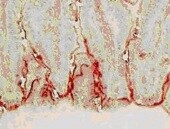
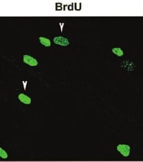
Detection of Bromodeoxyuridine/BrdU by Immunohistochemistry
SIGIRR delta E8 interacts with a mitochondrial protein ATP5A1. (A) Engineered HT-29 cells inducibly expressing SIGIRR delta E8 were incubated with 10 μM of BrdU for 6 h followed by fixation with PFA and immunofluoresence staining with anti-BrdU antibody. (B) HA pull-down experiment was performed from cells expressing a HA-tagged SIGIRR. These proteins were fractionated on an SDS-Page gel, the fractionated proteins were digested with trypsin, and the digests were analyzed by LC-MS/MS analysis. The protein, ATP5A1, was identified in the SIGIRR pull-down experiments by a total of 6 peptides (top). One of these peptides corresponds to the (134)TGAIVDVPVGEELLGR(149) peptide, which was identified as a doubly charged peptide with a mass of 812.950 Da. The CID spectra for this peptide are given above and are dominated by singly charged C-terminal y ions and N-terminal b ions (bottom). (C) HeLa cells were cotransfected with myc-tagged ATP5A1 and M2 flag-tagged SIGIRRFL and SIGIRR delta E8. Transfected cells were lysed, and the lysates were immunoprecipitated with anti-FLAG antibody followed by Western blot analysis. (D) SIGIRR protein was immunoprecipitated from indicated cell lines. Coimmunoprecipitated proteins were resolved on SDS-PAGE followed by Western blot for ATP5A1 and SIGIRR. The experiments (except MS) were repeated thrice and yielded consistent results; the representative results are shown. Data are shown as mean ± SEM. ****P < 0.0001 by 2-tailed t-test. Scale bar, 50 μm. CID, collision-induced dissociation; LC-MS/MS, liquid chromatography–mass spectrometry/mass spectrometry; PFA, paraformaldehyde; SDS-PAGE, sodium dodecyl sulfate–polyacrylamide gel electrophoresis. Image collected and cropped by CiteAb from the following open publication (https://pubmed.ncbi.nlm.nih.gov/35900274), licensed under a CC-BY license. Not internally tested by R&D Systems.
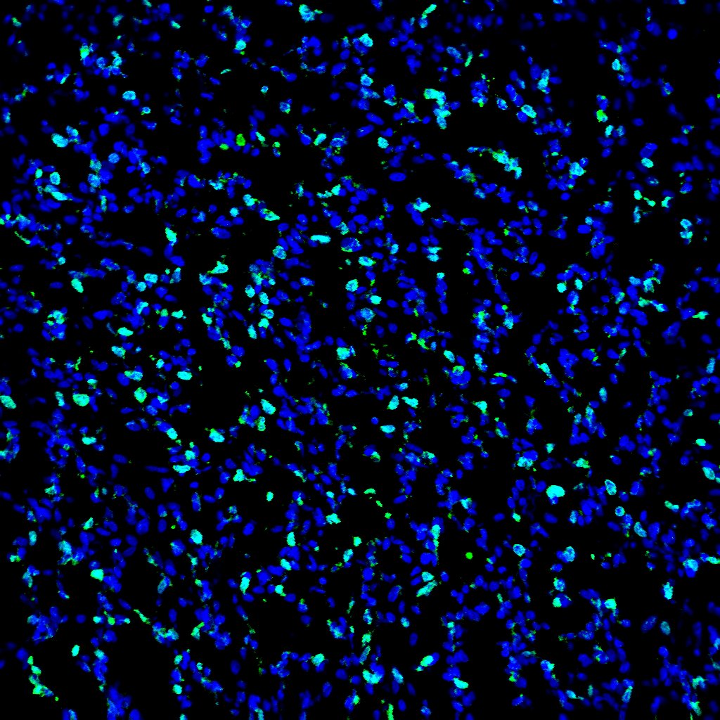
Detection of Bromodeoxyuridine/BrdU by Immunocytochemistry/ Immunofluorescence
Disruption of the TAGLN2-YBX1-AKT interaction reduces cytosolic ssDNA accumulation, ISGs expression, and drug resistance.A~C Multiplex immunofluorescence of TMA was performed using the Opal 7-color Manual IHC Kit and VECTASHIELD® HardSet Antifade Mounting Medium. The multiplex antibody panel was optimized as follows: TAGLN2, Opal 520 (yellow); CK, Opal 570 (green); YBX1, Opal 620 (red). The TMA was counterstained with DAPI (blue) and incubated with an antifluorescence quencher. Expression and spatial distribution of TAGLN2 or YBX1 in tissues and the correlation with patient clinical data. The DAPI channel was used to identify individual cells. A tissue segmentation algorithm combined with CK staining was applied to define tumoral and stromal areas. The scale bar is 200 μm. D Fisetin or MK2206 inhibited the accumulation of cytosolic ssDNA induced by overexpression of TAGLN2 by BrdU-gamma H2AX double labeling. HGC-27 cells stably transfected with TAGLN2 were prelabeled with BrdU and subsequently treated with 1 μg/ml Cisplatin with or without 10 μM Fisetin or 200 nM MK2206 in the medium. Cells were stained for DNA (DAPI, blue), the primary BrdU antibody (red) and phospho-histone H2AX (green). E Relative mRNA levels of the panel of IFN-related genes with or without 10 μM Fisetin or 200 nM MK2206 in the medium after 6 Gy X-ray treatment were evaluated by quantitative RT‒PCR analysis. The cytotoxicity induced by MK2206 from 16.25 nM to 13 μM (F) or MK2206 (200 nM) and Cisplatin (0.4 μg/ml) combination on tumor cells was detected (G). *P < 0.05, **P < 0.01, ***P < 0.001. Image collected and cropped by CiteAb from the following open publication (https://pubmed.ncbi.nlm.nih.gov/39168971), licensed under a CC-BY license. Not internally tested by R&D Systems.Applications for Bromodeoxyuridine/BrdU Antibody (BU-1)
Application
Recommended Usage
CyTOF-ready
Ready to be labeled using established conjugation methods. No BSA or other carrier proteins that could interfere with conjugation.
Immunocytochemistry
8-25 µg/mL
Sample: Immersion fixed MCF‑7 human breast cancer cell line stimulated with BrdU
Sample: Immersion fixed MCF‑7 human breast cancer cell line stimulated with BrdU
Immunohistochemistry
1-25 µg/mL
Sample: Immersion fixed paraffin-embedded sections of human breast cancer tissue and immersion fixed paraffin-embedded sections of human testis
Sample: Immersion fixed paraffin-embedded sections of human breast cancer tissue and immersion fixed paraffin-embedded sections of human testis
Intracellular Staining by Flow Cytometry
0.25 µg/106 cells
Sample: Human peripheral blood mononuclear cells (PBMCs), treated with PMA, Ionomycin, and BrdU, were fixed with ethanol, DNA was denatured with HCl, and then cells were permeabilized with Flow Cytometry Permeabilization/Wash Buffer I (Catalog # FC005)
Sample: Human peripheral blood mononuclear cells (PBMCs), treated with PMA, Ionomycin, and BrdU, were fixed with ethanol, DNA was denatured with HCl, and then cells were permeabilized with Flow Cytometry Permeabilization/Wash Buffer I (Catalog # FC005)
Reviewed Applications
Read 3 reviews rated 5 using MAB7225 in the following applications:
Flow Cytometry Panel Builder
Bio-Techne Knows Flow Cytometry
Save time and reduce costly mistakes by quickly finding compatible reagents using the Panel Builder Tool.
Advanced Features
- Spectra Viewer - Custom analysis of spectra from multiple fluorochromes
- Spillover Popups - Visualize the spectra of individual fluorochromes
- Antigen Density Selector - Match fluorochrome brightness with antigen density

Formulation, Preparation, and Storage
Purification
Protein A or G purified from hybridoma culture supernatant
Reconstitution
Sterile PBS to a final concentration of 0.5 mg/mL. For liquid material, refer to CoA for concentration.
Loading...
Formulation
Lyophilized from a 0.2 μm filtered solution in PBS with Trehalose. *Small pack size (SP) is supplied either lyophilized or as a 0.2 µm filtered solution in PBS.
Shipping
Lyophilized product is shipped at ambient temperature. Liquid small pack size (-SP) is shipped with polar packs. Upon receipt, store immediately at the temperature recommended below.
Stability & Storage
Use a manual defrost freezer and avoid repeated freeze-thaw cycles.
- 12 months from date of receipt, -20 to -70 °C as supplied.
- 1 month, 2 to 8 °C under sterile conditions after reconstitution.
- 6 months, -20 to -70 °C under sterile conditions after reconstitution.
Calculators
Background: Bromodeoxyuridine/BrdU
Alternate Names
5-bromo-2-deoxyuridine, BrdU
Additional Bromodeoxyuridine/BrdU Products
Product Documents for Bromodeoxyuridine/BrdU Antibody (BU-1)
Certificate of Analysis
To download a Certificate of Analysis, please enter a lot or batch number in the search box below.
Note: Certificate of Analysis not available for kit components.
Product Specific Notices for Bromodeoxyuridine/BrdU Antibody (BU-1)
For research use only
Related Research Areas
Citations for Bromodeoxyuridine/BrdU Antibody (BU-1)
 Powered by Bioz
Customer Reviews for Bromodeoxyuridine/BrdU Antibody (BU-1) (3)
5 out of 5
3 Customer Ratings
Have you used Bromodeoxyuridine/BrdU Antibody (BU-1)?
Submit a review and receive an Amazon gift card!
$25/€18/£15/$25CAN/¥2500 Yen for a review with an image
$10/€7/£6/$10CAN/¥1110 Yen for a review without an image
Submit a review
Customer Images
Showing
1
-
3 的
3 reviews
Showing All
Filter By:
-
Application: Immunocytochemistry/ImmunofluorescenceSample Tested: A375 human melanoma cell lineSpecies: HumanVerified Customer | Posted 09/27/2023

-
Application: ImmunohistochemistrySample Tested: Testis tissueSpecies: RatVerified Customer | Posted 10/19/2021
-
Application: Immunocytochemistry/ImmunofluorescenceSample Tested: Nasal Mucosa FibroblastsSpecies: HumanVerified Customer | Posted 07/16/2021
There are no reviews that match your criteria.
Protocols
Find general support by application which include: protocols, troubleshooting, illustrated assays, videos and webinars.
- 7-Amino Actinomycin D (7-AAD) Cell Viability Flow Cytometry Protocol
- Antigen Retrieval Protocol (PIER)
- Antigen Retrieval for Frozen Sections Protocol
- Appropriate Fixation of IHC/ICC Samples
- Cellular Response to Hypoxia Protocols
- Chromogenic IHC Staining of Formalin-Fixed Paraffin-Embedded (FFPE) Tissue Protocol
- Chromogenic Immunohistochemistry Staining of Frozen Tissue
- ClariTSA™ Fluorophore Kits
- Detection & Visualization of Antibody Binding
- Extracellular Membrane Flow Cytometry Protocol
- Flow Cytometry Protocol for Cell Surface Markers
- Flow Cytometry Protocol for Staining Membrane Associated Proteins
- Flow Cytometry Staining Protocols
- Flow Cytometry Troubleshooting Guide
- Fluorescent IHC Staining of Frozen Tissue Protocol
- Graphic Protocol for Heat-induced Epitope Retrieval
- Graphic Protocol for the Preparation and Fluorescent IHC Staining of Frozen Tissue Sections
- Graphic Protocol for the Preparation and Fluorescent IHC Staining of Paraffin-embedded Tissue Sections
- Graphic Protocol for the Preparation of Gelatin-coated Slides for Histological Tissue Sections
- ICC Cell Smear Protocol for Suspension Cells
- ICC Immunocytochemistry Protocol Videos
- ICC for Adherent Cells
- IHC Sample Preparation (Frozen sections vs Paraffin)
- Immunocytochemistry (ICC) Protocol
- Immunocytochemistry Troubleshooting
- Immunofluorescence of Organoids Embedded in Cultrex Basement Membrane Extract
- Immunofluorescent IHC Staining of Formalin-Fixed Paraffin-Embedded (FFPE) Tissue Protocol
- Immunohistochemistry (IHC) and Immunocytochemistry (ICC) Protocols
- Immunohistochemistry Frozen Troubleshooting
- Immunohistochemistry Paraffin Troubleshooting
- Intracellular Flow Cytometry Protocol Using Alcohol (Methanol)
- Intracellular Flow Cytometry Protocol Using Detergents
- Intracellular Nuclear Staining Flow Cytometry Protocol Using Detergents
- Intracellular Staining Flow Cytometry Protocol Using Alcohol Permeabilization
- Intracellular Staining Flow Cytometry Protocol Using Detergents to Permeabilize Cells
- Preparing Samples for IHC/ICC Experiments
- Preventing Non-Specific Staining (Non-Specific Binding)
- Primary Antibody Selection & Optimization
- Propidium Iodide Cell Viability Flow Cytometry Protocol
- Protocol for Heat-Induced Epitope Retrieval (HIER)
- Protocol for Liperfluo
- Protocol for Making a 4% Formaldehyde Solution in PBS
- Protocol for VisUCyte™ HRP Polymer Detection Reagent
- Protocol for the Characterization of Human Th22 Cells
- Protocol for the Characterization of Human Th9 Cells
- Protocol for the Fluorescent ICC Staining of Cell Smears - Graphic
- Protocol for the Fluorescent ICC Staining of Cultured Cells on Coverslips - Graphic
- Protocol for the Preparation & Fixation of Cells on Coverslips
- Protocol for the Preparation and Chromogenic IHC Staining of Frozen Tissue Sections
- Protocol for the Preparation and Chromogenic IHC Staining of Frozen Tissue Sections - Graphic
- Protocol for the Preparation and Chromogenic IHC Staining of Paraffin-embedded Tissue Sections
- Protocol for the Preparation and Chromogenic IHC Staining of Paraffin-embedded Tissue Sections - Graphic
- Protocol for the Preparation and Fluorescent ICC Staining of Cells on Coverslips
- Protocol for the Preparation and Fluorescent ICC Staining of Non-adherent Cells
- Protocol for the Preparation and Fluorescent ICC Staining of Stem Cells on Coverslips
- Protocol for the Preparation and Fluorescent IHC Staining of Frozen Tissue Sections
- Protocol for the Preparation and Fluorescent IHC Staining of Paraffin-embedded Tissue Sections
- Protocol for the Preparation of Gelatin-coated Slides for Histological Tissue Sections
- Protocol for the Preparation of a Cell Smear for Non-adherent Cell ICC - Graphic
- Protocol: Annexin V and PI Staining by Flow Cytometry
- Protocol: Annexin V and PI Staining for Apoptosis by Flow Cytometry
- TUNEL and Active Caspase-3 Detection by IHC/ICC Protocol
- The Importance of IHC/ICC Controls
- Troubleshooting Guide: Fluorokine Flow Cytometry Kits
- Troubleshooting Guide: Immunohistochemistry
- View all Protocols, Troubleshooting, Illustrated assays and Webinars
Loading...