CD14 Antibody (M5E2) - BSA Free
Novus Biologicals | Catalog # NB100-77758
Key Product Details
Species Reactivity
Validated:
Cited:
Applications
Validated:
Cited:
Label
Antibody Source
Format
Product Specifications
Immunogen
Reactivity Notes
Clonality
Host
Isotype
Scientific Data Images for CD14 Antibody (M5E2) - BSA Free
![Immunocytochemistry/ Immunofluorescence: CD14 Antibody (M5E2) - BSA Free [NB100-77758]](https://resources.rndsystems.com/images/products/CD14-Antibody-M5E2-Immunocytochemistry-Immunofluorescence-NB100-77758-img0009.jpg "Immunocytochemistry/ Immunofluorescence: CD14 Antibody (M5E2) - BSA Free [NB100-77758]")
Immunocytochemistry/ Immunofluorescence: CD14 Antibody (M5E2) - BSA Free [NB100-77758]
Immunocytochemistry/Immunofluorescence: CD14 Antibody (M5E2) [NB100-77758] - Representative immunostaining of CD14 (red) in adult human lung tissue sample with nuclei counterstained blue. This image was submitted by customer review. Image from the Alexa Fluor 647 version of this antibody.![Flow Cytometry: CD14 Antibody (M5E2) - BSA Free [NB100-77758]](https://resources.rndsystems.com/images/products/CD14-Antibody-M5E2-Flow-Cytometry-NB100-77758-img0007.jpg "Flow Cytometry: CD14 Antibody (M5E2) - BSA Free [NB100-77758]")
Flow Cytometry: CD14 Antibody (M5E2) - BSA Free [NB100-77758]
Flow Cytometry: CD14 Antibody (M5E2) [NB100-77758] - A surface stain was performed on human peripheral blood monocytes with CD163 (EDHu-1) antibody NB110-40686APC and a matched isotype control NBP2-27287APC. Cells were incubated in an antibody dilution of 1 ug/mL for 20 minutes at room temperature. A co-stain was performed with NB100-77758AF488.![Immunocytochemistry/ Immunofluorescence: CD14 Antibody (M5E2) - BSA Free [NB100-77758]](https://resources.rndsystems.com/images/products/CD14-Antibody-M5E2-Immunocytochemistry-Immunofluorescence-NB100-77758-img0008.jpg "Immunocytochemistry/ Immunofluorescence: CD14 Antibody (M5E2) - BSA Free [NB100-77758]")
Immunocytochemistry/ Immunofluorescence: CD14 Antibody (M5E2) - BSA Free [NB100-77758]
Immunocytochemistry/Immunofluorescence: CD14 Antibody (M5E2) [NB100-77758] - Human lung tissue was stained with FITC-conjugated CD14 (Green) for overnight. Image from the Alexa Fluor 488 version of this antibody.![Flow Cytometry: CD14 Antibody (M5E2) - BSA Free [NB100-77758]](https://resources.rndsystems.com/images/products/CD14-Antibody-M5E2-Flow-Cytometry-NB100-77758-img0001.jpg "Flow Cytometry: CD14 Antibody (M5E2) - BSA Free [NB100-77758]")
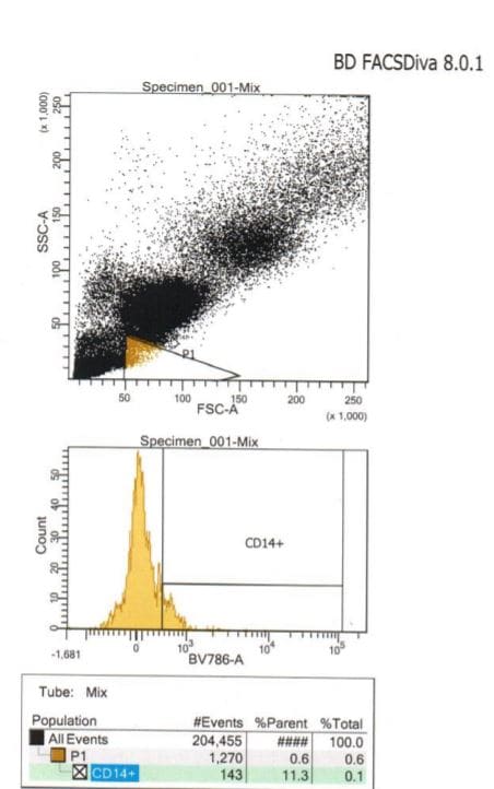
Flow Cytometry: CD14 Antibody (M5E2) - BSA Free [NB100-77758]
Flow Cytometry: CD14 Antibody (M5E2) [NB100-77758] - Human peripheral blood monocytes stained with M5E2 APC![Flow Cytometry: CD14 Antibody (M5E2) - BSA Free [NB100-77758]](https://resources.rndsystems.com/images/products/CD14-Antibody-M5E2-Flow-Cytometry-NB100-77758-img0004.jpg "Flow Cytometry: CD14 Antibody (M5E2) - BSA Free [NB100-77758]")
Flow Cytometry: CD14 Antibody (M5E2) - BSA Free [NB100-77758]
Flow Cytometry: CD14 Antibody (M5E2) [NB100-77758] - Analysis using the FITC conjugate of NB100-77758. Staining of Human peripheral blood monocytes with M5E2 FITC.![Flow Cytometry: CD14 Antibody (M5E2) - BSA Free [NB100-77758]](https://resources.rndsystems.com/images/products/CD14-Antibody-M5E2-Flow-Cytometry-NB100-77758-img0005.jpg "Flow Cytometry: CD14 Antibody (M5E2) - BSA Free [NB100-77758]")
Flow Cytometry: CD14 Antibody (M5E2) - BSA Free [NB100-77758]
Flow Cytometry: CD14 Antibody (M5E2) [NB100-77758] - Analysis using the PE conjugate of NB100-77758. Staining of CD14 in human PBMC using anti-CD14 antibody conjugated with PE. Image from verified customer review.![Flow Cytometry: CD14 Antibody (M5E2) - BSA Free [NB100-77758]](https://resources.rndsystems.com/images/products/CD14-Antibody-M5E2-Flow-Cytometry-NB100-77758-img0006.jpg "Flow Cytometry: CD14 Antibody (M5E2) - BSA Free [NB100-77758]")
Flow Cytometry: CD14 Antibody (M5E2) - BSA Free [NB100-77758]
Flow Cytometry: CD14 Antibody (M5E2) [NB100-77758] - Analysis using PE conjugate of NB100-77758. A cell surface stain was performed on hPBMCs with ILT5 antibody (MM0413-9S32) NBP2-11729 (top image) and a matched isotype control NBP2-27287 (bottom image). Cells were incubated in an antibody dilution of 1:200 for 20 minutes at room temperature. Both antibodies were conjugated to APC. A co-stain was performed using CD14 antibody (M5E2) NB100-77758PE.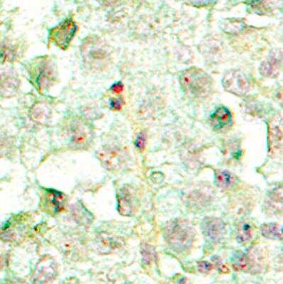
Applications for CD14 Antibody (M5E2) - BSA Free
Block/Neutralize
Flow Cytometry
Immunocytochemistry/ Immunofluorescence
Immunohistochemistry
Immunohistochemistry-Frozen
Simple Western
Reviewed Applications
Read 2 reviews rated 3 using NB100-77758 in the following applications:
Flow Cytometry Panel Builder
Bio-Techne Knows Flow Cytometry
Save time and reduce costly mistakes by quickly finding compatible reagents using the Panel Builder Tool.
Advanced Features
- Spectra Viewer - Custom analysis of spectra from multiple fluorochromes
- Spillover Popups - Visualize the spectra of individual fluorochromes
- Antigen Density Selector - Match fluorochrome brightness with antigen density

Formulation, Preparation, and Storage
Purification
Formulation
Format
Preservative
Concentration
Shipping
Stability & Storage
Background: CD14
Additional CD14 Products
Product Documents for CD14 Antibody (M5E2) - BSA Free
Certificate of Analysis
To download a Certificate of Analysis, please enter a lot or batch number in the search box below.
Product Specific Notices for CD14 Antibody (M5E2) - BSA Free
This product is for research use only and is not approved for use in humans or in clinical diagnosis. Primary Antibodies are guaranteed for 1 year from date of receipt.
Citations for CD14 Antibody (M5E2) - BSA Free
 Powered by Bioz
Customer Reviews for CD14 Antibody (M5E2) - BSA Free (2)
Have you used CD14 Antibody (M5E2) - BSA Free?
Submit a review and receive an Amazon gift card!
$25/€18/£15/$25CAN/¥2500 Yen for a review with an image
$10/€7/£6/$10CAN/¥1110 Yen for a review without an image
Submit a review
Customer Images
-
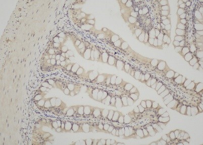
Application: Immunohistochemistry-ParaffinSample Tested: After bowel and After intestinal bowelSpecies: GrouperVerified Customer | Posted 10/16/2020This is the graph of the results of the experiment, repeated several times, the results are negativeDewaxing to water: 1, paraffin sections in turn sliced into 15 min - dimethylbenzene xylene Ⅰ Ⅱ 15 min - xylene III Ⅰ 5 min - 15 min - anhydrous ethanol anhydrous ethanol Ⅱ 5 min 5 min - 75% - 85% alcohol alcohol 5 min - the distilled water to wash. 2, antigen repair: biopsy under full antigen repair citrate buffer (PH6.0) repair box in a have a certain amount of water pressure cooker, induction cooker heated to stomatal began to jet, stop heating, release the pressure, the slice on the repair box, induction cooker heated to stomatal began to jet, 5 min after close the induction cooker, the process should prevent excessive evaporation buffer, do not dry.After natural cooling, the slides were placed in PBS (PH7.4) and shaken on the decolorizing shaper for 3 times, 5min each. 3. Break endogenous peroxidase: the slides were placed in 3% hydrogen peroxide solution and incubated at room temperature and away from light for 25 min. The slides were placed in PBS (PH7.4) and shaken on a decolorizing shaper for 3 times for 5min each. Serum sealing: 3%BSA was added to cover the tissue evenly in the chemochemical ring, and the tissue was sealed at room temperature for 30min.(Primary antibody is sealed with rabbit serum from goat and BSA from other sources.) 4. Primary antibody was added: the blocking solution was gently removed, the primary antibody prepared with PBS in a certain proportion was added to the sections, and the sections were placed flat in a wet box and incubated overnight at 4°C.(Add a small amount of water in the wet box to prevent evaporation of antibodies) 5. Secondary antibody was added: the slides were placed in PBS (PH7.4) and washed by shaking on the decolorizing shaker for 3 times, 5min each.After the sections were slightly shaken and dried, the tissues were dripped with secondary antibody (HRP-labeled) of the species corresponding to the primary antibody and incubated at room temperature for 50min. 7. DAB color development: The slides were placed in PBS (PH7.4) and shaken on the decoloring shaker for 3 times, 5min each.DAB color developing solution newly prepared was added in the circle after the sections were slightly dried. The color developing time was controlled under the microscope. The positive color was brownish yellow, and the color developing was stopped after the sections were washed under tap water. 8. Restaining nuclei: restaining with hematoxylin for about 3min; washing with tap water; differentiation with hematoxylin differentiation solution for several seconds; washing with tap water; flushing with hematoxylin returning blue solution; washing with running water., dehydration and 9: the slice in turn into 5 min 5 min 75% alcohol - 85% alcohol, anhydrous ethanol Ⅰ Ⅱ 5 min - the anhydrous ethanol dehydration in 5 min - xylene Ⅰ 5 min is transparent, the slice out of xylene is a bit dry, neutral rubber sealing piece. Microscope examination, image acquisition and analysis.
 Bio-Techne ResponseThis review was submitted through the legacy Novus Innovators Program, reflecting a new species or application tested on a primary antibody.
 -
Application: Flow CytometrySample Tested: Whole blood cellsSpecies: CanineVerified Customer | Posted 05/18/2018Monocyte detetction in dog cells using CD14The whole blood cells were lysed to get rid of the RBC. The washed cell were then incubated with the Antibody. They were washed and then incubated with a conjugated secondary. Allowed to sit for 20mins and then washed again and read on the BD Fortessa.
There are no reviews that match your criteria.
Protocols
Find general support by application which include: protocols, troubleshooting, illustrated assays, videos and webinars.
- 7-Amino Actinomycin D (7-AAD) Cell Viability Flow Cytometry Protocol
- Antigen Retrieval Protocol (PIER)
- Antigen Retrieval for Frozen Sections Protocol
- Appropriate Fixation of IHC/ICC Samples
- Cellular Response to Hypoxia Protocols
- Chromogenic IHC Staining of Formalin-Fixed Paraffin-Embedded (FFPE) Tissue Protocol
- Chromogenic Immunohistochemistry Staining of Frozen Tissue
- ClariTSA™ Fluorophore Kits
- Detection & Visualization of Antibody Binding
- Extracellular Membrane Flow Cytometry Protocol
- Flow Cytometry Protocol for Cell Surface Markers
- Flow Cytometry Protocol for Staining Membrane Associated Proteins
- Flow Cytometry Staining Protocols
- Flow Cytometry Troubleshooting Guide
- Fluorescent IHC Staining of Frozen Tissue Protocol
- Graphic Protocol for Heat-induced Epitope Retrieval
- Graphic Protocol for the Preparation and Fluorescent IHC Staining of Frozen Tissue Sections
- Graphic Protocol for the Preparation and Fluorescent IHC Staining of Paraffin-embedded Tissue Sections
- Graphic Protocol for the Preparation of Gelatin-coated Slides for Histological Tissue Sections
- ICC Cell Smear Protocol for Suspension Cells
- ICC Immunocytochemistry Protocol Videos
- ICC for Adherent Cells
- IHC Sample Preparation (Frozen sections vs Paraffin)
- ISH-IHC Protocol for Chromogenic Detection on Formalin Fixed Paraffin Embedded (FFPE) Tissue
- Immunocytochemistry (ICC) Protocol
- Immunocytochemistry Troubleshooting
- Immunofluorescence of Organoids Embedded in Cultrex Basement Membrane Extract
- Immunofluorescent IHC Staining of Formalin-Fixed Paraffin-Embedded (FFPE) Tissue Protocol
- Immunohistochemistry (IHC) and Immunocytochemistry (ICC) Protocols
- Immunohistochemistry Frozen Troubleshooting
- Immunohistochemistry Paraffin Troubleshooting
- Intracellular Flow Cytometry Protocol Using Alcohol (Methanol)
- Intracellular Flow Cytometry Protocol Using Detergents
- Intracellular Nuclear Staining Flow Cytometry Protocol Using Detergents
- Intracellular Staining Flow Cytometry Protocol Using Alcohol Permeabilization
- Intracellular Staining Flow Cytometry Protocol Using Detergents to Permeabilize Cells
- Preparing Samples for IHC/ICC Experiments
- Preventing Non-Specific Staining (Non-Specific Binding)
- Primary Antibody Selection & Optimization
- Propidium Iodide Cell Viability Flow Cytometry Protocol
- Protocol for Heat-Induced Epitope Retrieval (HIER)
- Protocol for Liperfluo
- Protocol for Making a 4% Formaldehyde Solution in PBS
- Protocol for VisUCyte™ HRP Polymer Detection Reagent
- Protocol for the Characterization of Human Th22 Cells
- Protocol for the Characterization of Human Th9 Cells
- Protocol for the Fluorescent ICC Staining of Cell Smears - Graphic
- Protocol for the Fluorescent ICC Staining of Cultured Cells on Coverslips - Graphic
- Protocol for the Preparation & Fixation of Cells on Coverslips
- Protocol for the Preparation and Chromogenic IHC Staining of Frozen Tissue Sections
- Protocol for the Preparation and Chromogenic IHC Staining of Frozen Tissue Sections - Graphic
- Protocol for the Preparation and Chromogenic IHC Staining of Paraffin-embedded Tissue Sections
- Protocol for the Preparation and Chromogenic IHC Staining of Paraffin-embedded Tissue Sections - Graphic
- Protocol for the Preparation and Fluorescent ICC Staining of Cells on Coverslips
- Protocol for the Preparation and Fluorescent ICC Staining of Non-adherent Cells
- Protocol for the Preparation and Fluorescent ICC Staining of Stem Cells on Coverslips
- Protocol for the Preparation and Fluorescent IHC Staining of Frozen Tissue Sections
- Protocol for the Preparation and Fluorescent IHC Staining of Paraffin-embedded Tissue Sections
- Protocol for the Preparation of Gelatin-coated Slides for Histological Tissue Sections
- Protocol for the Preparation of a Cell Smear for Non-adherent Cell ICC - Graphic
- Protocol: Annexin V and PI Staining by Flow Cytometry
- Protocol: Annexin V and PI Staining for Apoptosis by Flow Cytometry
- TUNEL and Active Caspase-3 Detection by IHC/ICC Protocol
- The Importance of IHC/ICC Controls
- Troubleshooting Guide: Fluorokine Flow Cytometry Kits
- Troubleshooting Guide: Immunohistochemistry
- View all Protocols, Troubleshooting, Illustrated assays and Webinars
FAQs for CD14 Antibody (M5E2) - BSA Free
-
Q: Can this antibody be used for testing frozen and not fixed human (CNS, brain) tissue?
A:
The CD14 Antibody (M5E2) antibody with catalog number NB100-77758 is covered by the Novus Guarantee to detect human protein in frozen samples by IHC. The recommended dilution range for IHC-Frozen is 1:10-1:2000. According to UniProt, CD14 is expressed strongly on the surface of monocytes, weakly on the surface of granulocytes, and is also expressed by most tissue macrophages. The following PubMed reference discusses the expression and release of CD14 in astrocytic brain tumors.
-
Q: We have an inquiry of your product Cat. ID # NB100-2735, NB600-1185, NB100-77758, NB100-78016, NB100-77855. We are interested in this product and would like to make sure that there is no any other protein added in the buffer. I saw the storage buffer in datasheet doesn't has any other protein but still would like to confirm with you again. Would you please help us to check and provide the purification method and buffer of the antibody?
A: Thank you for your inquiry. Catalog numbers NB100-77758, NB100-78016 and NB100-77855 are in a buffer containing PBS and sodium azide. NB600-1185 is in a buffer containing PBS, 150mM NaCl and sodium azide. NB100-2735 is whole tissue culture supernatant and may therefore contain other proteins.
-
Q: Can this antibody be used for testing frozen and not fixed human (CNS, brain) tissue?
A:
The CD14 Antibody (M5E2) antibody with catalog number NB100-77758 is covered by the Novus Guarantee to detect human protein in frozen samples by IHC. The recommended dilution range for IHC-Frozen is 1:10-1:2000. According to UniProt, CD14 is expressed strongly on the surface of monocytes, weakly on the surface of granulocytes, and is also expressed by most tissue macrophages. The following PubMed reference discusses the expression and release of CD14 in astrocytic brain tumors.
-
Q: We have an inquiry of your product Cat. ID # NB100-2735, NB600-1185, NB100-77758, NB100-78016, NB100-77855. We are interested in this product and would like to make sure that there is no any other protein added in the buffer. I saw the storage buffer in datasheet doesn't has any other protein but still would like to confirm with you again. Would you please help us to check and provide the purification method and buffer of the antibody?
A: Thank you for your inquiry. Catalog numbers NB100-77758, NB100-78016 and NB100-77855 are in a buffer containing PBS and sodium azide. NB600-1185 is in a buffer containing PBS, 150mM NaCl and sodium azide. NB100-2735 is whole tissue culture supernatant and may therefore contain other proteins.
Associated Pathways


