
CD163 Antibody (ED2) - BSA Free
Novus Biologicals | Catalog # NBP2-39099

![Immunocytochemistry/ Immunofluorescence: CD163 Antibody (ED2) - BSA Free [NBP2-39099]](https://resources.rndsystems.com/images/products/CD163-Antibody-ED2-Immunocytochemistry-Immunofluorescence-NBP2-39099-img0013.jpg "Immunocytochemistry/ Immunofluorescence: CD163 Antibody (ED2) - BSA Free [NBP2-39099]")
Scientific images may be resized, cropped, or adjusted for brightness or contrast for presentation purposes. These updates are limited to presentation and do not affect the underlying scientific interpretation of the data. Where available, additional source data may be provided upon request.
Key Product Details
Species Reactivity
Validated:
Rat
Cited:
Human, Rat
Applications
Validated:
Immunohistochemistry, Immunohistochemistry-Paraffin, Immunohistochemistry-Frozen, Western Blot, Flow Cytometry, Immunocytochemistry/ Immunofluorescence, Immunoprecipitation
Cited:
Flow Cytometry, Immunocytochemistry/ Immunofluorescence
Label
Unconjugated
Antibody Source
Monoclonal Mouse IgG1 Clone # ED2
Format
BSA Free
Loading...
Product Specifications
Immunogen
Rat spleen cell homogenate
Specificity
This antibody recognizes the rat ED2 cell surface glycoprotein.
Clonality
Monoclonal
Host
Mouse
Isotype
IgG1
Scientific Data Images for CD163 Antibody (ED2) - BSA Free
Immunocytochemistry/ Immunofluorescence: CD163 Antibody (ED2) - BSA Free [NBP2-39099]
Immunocytochemistry/Immunofluorescence: CD163 Antibody (ED2) [NBP2-39099] - Staining of rat lymph node cryosection. Image A in Red (NBP2-39099), and Image B in green using Mouse anti-Rat CD8. Image C is the merged image with nuclei counter-stained blue using DAPI. High power.![Immunohistochemistry-Paraffin: CD163 Antibody (ED2) - BSA Free [NBP2-39099]](https://resources.rndsystems.com/images/products/CD163-Antibody-ED2-BSA-Free-Immunohistochemistry-Paraffin-NBP2-39099-img0017.jpg "Immunohistochemistry-Paraffin: CD163 Antibody (ED2) - BSA Free [NBP2-39099]")
Immunohistochemistry-Paraffin: CD163 Antibody (ED2) - BSA Free [NBP2-39099]
Immunohistochemistry-Paraffin: CD163 Antibody (ED2) - BSA Free [NBP2-39099] - Alcohol-fixed paraffin embedded spleen tissue from healthy rat spleen. Primary antibody dilution: 1:100. Image from verified customer review.![Immunocytochemistry/ Immunofluorescence: CD163 Antibody (ED2) - BSA Free [NBP2-39099]](https://resources.rndsystems.com/images/products/CD163-Antibody-ED2-Immunocytochemistry-Immunofluorescence-NBP2-39099-img0012.jpg "Immunocytochemistry/ Immunofluorescence: CD163 Antibody (ED2) - BSA Free [NBP2-39099]")
Immunocytochemistry/ Immunofluorescence: CD163 Antibody (ED2) - BSA Free [NBP2-39099]
Immunocytochemistry/Immunofluorescence: CD163 Antibody (ED2) [NBP2-39099] - Staining of rat lymph node cryosection. Image A in Red (NBP2-39099), and Image B in green using Mouse anti-Rat CD8. Image C is the merged image with nuclei counter-stained blue using DAPI. Low power.![Immunohistochemistry-Frozen: CD163 Antibody (ED2) - BSA Free [NBP2-39099]](https://resources.rndsystems.com/images/products/CD163-Antibody-ED2-Immunohistochemistry-Frozen-NBP2-39099-img0014.jpg "Immunohistochemistry-Frozen: CD163 Antibody (ED2) - BSA Free [NBP2-39099]")
Immunohistochemistry-Frozen: CD163 Antibody (ED2) - BSA Free [NBP2-39099]
Immunohistochemistry-Frozen: CD163 Antibody (ED2) [NBP2-39099] - Staining of acetone fixed, cryostat sectioned rat spleen.![Immunohistochemistry-Frozen: CD163 Antibody (ED2) - BSA Free [NBP2-39099]](https://resources.rndsystems.com/images/products/CD163-Antibody-ED2-Immunohistochemistry-Frozen-NBP2-39099-img0015.jpg "Immunohistochemistry-Frozen: CD163 Antibody (ED2) - BSA Free [NBP2-39099]")
Immunohistochemistry-Frozen: CD163 Antibody (ED2) - BSA Free [NBP2-39099]
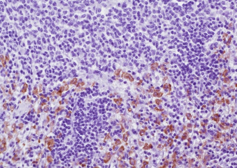
Immunohistochemistry-Frozen: CD163 Antibody (ED2) [NBP2-39099] - Immunoperoxidase staining of rat lymph node cryosection (NBP2-39099) followed by horseradish peroxidase conjugated Goat anti Mouse IgG1 as a detection reagent. Low power.![Immunohistochemistry-Frozen: CD163 Antibody (ED2) - BSA Free [NBP2-39099]](https://resources.rndsystems.com/images/products/CD163-Antibody-ED2-Immunohistochemistry-Frozen-NBP2-39099-img0016.jpg "Immunohistochemistry-Frozen: CD163 Antibody (ED2) - BSA Free [NBP2-39099]")
Immunohistochemistry-Frozen: CD163 Antibody (ED2) - BSA Free [NBP2-39099]
Immunohistochemistry-Frozen: CD163 Antibody (ED2) [NBP2-39099] - Immunoperoxidase staining of rat lymph node cryosection (NBP2-39099) followed by horseradish peroxidase conjugated Goat anti Mouse IgG1 as a detection reagent. High power.Applications for CD163 Antibody (ED2) - BSA Free
Application
Recommended Usage
Flow Cytometry
1:10 - 1:100
Immunocytochemistry/ Immunofluorescence
1:10-1:500
Immunohistochemistry
1:50 - 1:100
Immunohistochemistry-Frozen
1:50 - 1:100
Immunohistochemistry-Paraffin
1:50 - 1:100
Immunoprecipitation
1:10 - 1:500
Western Blot
1:100 - 1:2000
Application Notes
This product requires protein digestion pre-treatment of paraffin sections e.g. trypsin or pronase.
This antibody may be used in immunohistology using antigen retrieval, and has also been described reacting with paraffin-embedded material following PLP fixation (Periodate-lysine-paraformaldehyde). See Whiteland et al.
This antibody may be used in immunohistology using antigen retrieval, and has also been described reacting with paraffin-embedded material following PLP fixation (Periodate-lysine-paraformaldehyde). See Whiteland et al.
Reviewed Applications
Read 1 review rated 5 using NBP2-39099 in the following applications:
Flow Cytometry Panel Builder
Bio-Techne Knows Flow Cytometry
Save time and reduce costly mistakes by quickly finding compatible reagents using the Panel Builder Tool.
Advanced Features
- Spectra Viewer - Custom analysis of spectra from multiple fluorochromes
- Spillover Popups - Visualize the spectra of individual fluorochromes
- Antigen Density Selector - Match fluorochrome brightness with antigen density

Formulation, Preparation, and Storage
Purification
Protein A purified
Formulation
PBS
Format
BSA Free
Preservative
0.09% Sodium Azide
Concentration
0.5 mg/ml
Shipping
The product is shipped with polar packs. Upon receipt, store it immediately at the temperature recommended below.
Stability & Storage
Store at 4C short term. Aliquot and store at -20C long term. Avoid freeze-thaw cycles.
Background: CD163
One of the primary functions of CD163 is uptake of haptoglobin-hemoglobin (Hp-Hb) complexes from the liver, spleen, and bone marrow, ultimately triggering an anti-inflammatory response (3, 5, 7). CD163 also functions as an erythroblast adhesion receptor and promotes cell maturation and survival (3, 5, 7). Furthermore, CD163 functions in immune sensing of bacteria and as a receptor for tumor necrosis factor (TNF)-like weak inducer of apoptosis (TWEAK) (3, 5, 7). As mentioned above, CD163 is expressed on cells in the monocyte/macrophage lineage and, in general, anti-inflammatory signals including glucocorticoids, interleukin (IL)-6, and IL-10 stimulate CD163 synthesis and expression while, conversely, pro-inflammatory signals such as interferon-gamma (INF-gamma), TNF-alpha, and lipopolysaccharide (LPS) downregulate CD163 (3, 5). In addition to membrane-bound form of CD163, the protein can be cleaved by metalloproteinases (MMP) and induced by LPS or phorbol myristate acetate (PMA) to release a soluble form (sCD163) into the plasma (7). Increased levels of sCD163 in the plasma and an increased number of CD163-expressing macrophages at the site of inflammation are associated with a variety of pathologies (3, 5-7). CD163/sCD163 is often increased and a suitable clinical marker for inflammatory diseases including rheumatoid arthritis (RA), Gaucher disease, chronic kidney disease, diabetes, and Crohn's disease (3, 5-7).
Alternative names for CD163 includes GHI/61, HbSR, Hemoglobin scavenger receptor, M130, macrophage-associated antigen, MM130, RM3/1, SCARI1, scavenger receptor cysteine-rich type 1 protein M130, sCD163, and soluble CD163.
References
1. Law, S. K., Micklem, K. J., Shaw, J. M., Zhang, X. P., Dong, Y., Willis, A. C., & Mason, D. Y. (1993). A new macrophage differentiation antigen which is a member of the scavenger receptor superfamily. European journal of immunology. https://doi.org/10.1002/eji.1830230940
2. Onofre, G., Kolackova, M., Jankovicova, K., & Krejsek, J. (2009). Scavenger receptor CD163 and its biological functions. Acta medica (Hradec Kralove).
3. Van Gorp, H., Delputte, P. L., & Nauwynck, H. J. (2010). Scavenger receptor CD163, a Jack-of-all-trades and potential target for cell-directed therapy. Molecular immunology. https://doi.org/10.1016/j.molimm.2010.02.008
4. Sulahian, T. H., Hogger, P., Wahner, A. E., Wardwell, K., Goulding, N. J., Sorg, C., Droste, A., Stehling, M., Wallace, P. K., Morganelli, P. M., & Guyre, P. M. (2000). Human monocytes express CD163, which is upregulated by IL-10 and identical to p155. Cytokine. https://doi.org/10.1006/cyto.2000.0720
5. Etzerodt, A., & Moestrup, S. K. (2013). CD163 and inflammation: biological, diagnostic, and therapeutic aspects. Antioxidants & redox signaling. https://doi.org/10.1089/ars.2012.4834
6. Skytthe, M. K., Graversen, J. H., & Moestrup, S. K. (2020). Targeting of CD163+ Macrophages in Inflammatory and Malignant Diseases. International journal of molecular sciences, 21(15), 5497. https://doi.org/10.3390/ijms21155497
7. Moller H. J. (2012). Soluble CD163. Scandinavian journal of clinical and laboratory investigation. https://doi.org/10.3109/00365513.2011.626868
Additional CD163 Products
Product Documents for CD163 Antibody (ED2) - BSA Free
Certificate of Analysis
To download a Certificate of Analysis, please enter a lot or batch number in the search box below.
Product Specific Notices for CD163 Antibody (ED2) - BSA Free
This product is for research use only and is not approved for use in humans or in clinical diagnosis. Primary Antibodies are guaranteed for 1 year from date of receipt.
Citations for CD163 Antibody (ED2) - BSA Free
 Powered by Bioz
Customer Reviews for CD163 Antibody (ED2) - BSA Free (1)
5 out of 5
1 Customer Rating
Have you used CD163 Antibody (ED2) - BSA Free?
Submit a review and receive an Amazon gift card!
$25/€18/£15/$25CAN/¥2500 for a review with an image
$10/€7/£6/$10CAN/¥1110 for a review without an image
Submit a review
Customer Images
Showing
1
-
1 的
1 review
Showing All
Filter By:
-
Application: Immunohistochemistry-ParaffinSample Tested: Spleen tissueSpecies: RatVerified Customer | Posted 11/30/2022Alcohol-fixed paraffin embedded spleen tissue from healthy rat.Dilution: 1:100

There are no reviews that match your criteria.
Protocols
Find general support by application which include: protocols, troubleshooting, illustrated assays, videos and webinars.
- 7-Amino Actinomycin D (7-AAD) Cell Viability Flow Cytometry Protocol
- Antigen Retrieval Protocol (PIER)
- Antigen Retrieval for Frozen Sections Protocol
- Appropriate Fixation of IHC/ICC Samples
- Cellular Response to Hypoxia Protocols
- Chromogenic IHC Staining of Formalin-Fixed Paraffin-Embedded (FFPE) Tissue Protocol
- Chromogenic Immunohistochemistry Staining of Frozen Tissue
- ClariTSA™ Fluorophore Kits
- Detection & Visualization of Antibody Binding
- Extracellular Membrane Flow Cytometry Protocol
- Flow Cytometry Protocol for Cell Surface Markers
- Flow Cytometry Protocol for Staining Membrane Associated Proteins
- Flow Cytometry Staining Protocols
- Flow Cytometry Troubleshooting Guide
- Fluorescent IHC Staining of Frozen Tissue Protocol
- Graphic Protocol for Heat-induced Epitope Retrieval
- Graphic Protocol for the Preparation and Fluorescent IHC Staining of Frozen Tissue Sections
- Graphic Protocol for the Preparation and Fluorescent IHC Staining of Paraffin-embedded Tissue Sections
- Graphic Protocol for the Preparation of Gelatin-coated Slides for Histological Tissue Sections
- ICC Cell Smear Protocol for Suspension Cells
- ICC Immunocytochemistry Protocol Videos
- ICC for Adherent Cells
- IHC Sample Preparation (Frozen sections vs Paraffin)
- Immunocytochemistry (ICC) Protocol
- Immunocytochemistry Troubleshooting
- Immunofluorescence of Organoids Embedded in Cultrex Basement Membrane Extract
- Immunofluorescent IHC Staining of Formalin-Fixed Paraffin-Embedded (FFPE) Tissue Protocol
- Immunohistochemistry (IHC) and Immunocytochemistry (ICC) Protocols
- Immunohistochemistry Frozen Troubleshooting
- Immunohistochemistry Paraffin Troubleshooting
- Immunoprecipitation Protocol
- Intracellular Flow Cytometry Protocol Using Alcohol (Methanol)
- Intracellular Flow Cytometry Protocol Using Detergents
- Intracellular Nuclear Staining Flow Cytometry Protocol Using Detergents
- Intracellular Staining Flow Cytometry Protocol Using Alcohol Permeabilization
- Intracellular Staining Flow Cytometry Protocol Using Detergents to Permeabilize Cells
- Preparing Samples for IHC/ICC Experiments
- Preventing Non-Specific Staining (Non-Specific Binding)
- Primary Antibody Selection & Optimization
- Propidium Iodide Cell Viability Flow Cytometry Protocol
- Protocol for Heat-Induced Epitope Retrieval (HIER)
- Protocol for Liperfluo
- Protocol for Making a 4% Formaldehyde Solution in PBS
- Protocol for VisUCyte™ HRP Polymer Detection Reagent
- Protocol for the Characterization of Human Th22 Cells
- Protocol for the Characterization of Human Th9 Cells
- Protocol for the Fluorescent ICC Staining of Cell Smears - Graphic
- Protocol for the Fluorescent ICC Staining of Cultured Cells on Coverslips - Graphic
- Protocol for the Preparation & Fixation of Cells on Coverslips
- Protocol for the Preparation and Chromogenic IHC Staining of Frozen Tissue Sections
- Protocol for the Preparation and Chromogenic IHC Staining of Frozen Tissue Sections - Graphic
- Protocol for the Preparation and Chromogenic IHC Staining of Paraffin-embedded Tissue Sections
- Protocol for the Preparation and Chromogenic IHC Staining of Paraffin-embedded Tissue Sections - Graphic
- Protocol for the Preparation and Fluorescent ICC Staining of Cells on Coverslips
- Protocol for the Preparation and Fluorescent ICC Staining of Non-adherent Cells
- Protocol for the Preparation and Fluorescent ICC Staining of Stem Cells on Coverslips
- Protocol for the Preparation and Fluorescent IHC Staining of Frozen Tissue Sections
- Protocol for the Preparation and Fluorescent IHC Staining of Paraffin-embedded Tissue Sections
- Protocol for the Preparation of Gelatin-coated Slides for Histological Tissue Sections
- Protocol for the Preparation of a Cell Smear for Non-adherent Cell ICC - Graphic
- Protocol: Annexin V and PI Staining by Flow Cytometry
- Protocol: Annexin V and PI Staining for Apoptosis by Flow Cytometry
- R&D Systems Quality Control Western Blot Protocol
- TUNEL and Active Caspase-3 Detection by IHC/ICC Protocol
- The Importance of IHC/ICC Controls
- Troubleshooting Guide: Fluorokine Flow Cytometry Kits
- Troubleshooting Guide: Immunohistochemistry
- Troubleshooting Guide: Western Blot Figures
- Western Blot Conditions
- Western Blot Protocol
- Western Blot Protocol for Cell Lysates
- Western Blot Troubleshooting
- Western Blot Troubleshooting Guide
- View all Protocols, Troubleshooting, Illustrated assays and Webinars
Loading...
Associated Pathways

