CD45RA Antibody (CA4.1D3) - BSA Free
Novus Biologicals | Catalog # NB100-64897
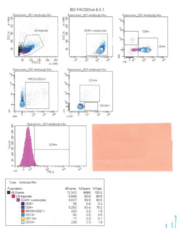
![Flow Cytometry: CD45RA Antibody (CA4.1D3) [NB100-64897]](https://resources.rndsystems.com/images/products/CD45RA-Antibody-CA4-1D3-Flow-Cytometry-NB100-64897-img0001.jpg "Flow Cytometry: CD45RA Antibody (CA4.1D3) [NB100-64897]")
Loading...
Key Product Details
Species Reactivity
Canine
Applications
Immunohistochemistry, Immunohistochemistry-Frozen, Flow Cytometry
Label
Unconjugated
Antibody Source
Monoclonal Mouse IgG1 Clone # CA4.1D3
Format
BSA Free
Loading...
Product Specifications
Immunogen
Made to Dog CD45RA
Specificity
Recognizes the canine homologue of CD45RA, a cell surface glycoprotein expressed by B lymphocytes and a subset of T-lymphocytes.
Clonality
Monoclonal
Host
Mouse
Isotype
IgG1
Scientific Data Images for CD45RA Antibody (CA4.1D3) - BSA Free
Flow Cytometry: CD45RA Antibody (CA4.1D3) [NB100-64897]
Flow Cytometry: CD45RA Antibody (CA4.1D3) [NB100-64897] - Pan leukocyte detection in Canine IP- whole blood cells. Image courtesy of customer.Applications for CD45RA Antibody (CA4.1D3) - BSA Free
Application
Recommended Usage
Flow Cytometry
Neat
Immunohistochemistry
1:10-1:500
Immunohistochemistry-Frozen
1:10-1:500
Reviewed Applications
Read 1 review rated 5 using NB100-64897 in the following applications:
Flow Cytometry Panel Builder
Bio-Techne Knows Flow Cytometry
Save time and reduce costly mistakes by quickly finding compatible reagents using the Panel Builder Tool.
Advanced Features
- Spectra Viewer - Custom analysis of spectra from multiple fluorochromes
- Spillover Popups - Visualize the spectra of individual fluorochromes
- Antigen Density Selector - Match fluorochrome brightness with antigen density

Formulation, Preparation, and Storage
Purification
Tissue culture supernatant
Formulation
Tissue culture supernatant
Format
BSA Free
Preservative
0.01% Sodium Azide
Concentration
This product is unpurified. The exact concentration of antibody is not quantifiable.
Shipping
The product is shipped with polar packs. Upon receipt, store it immediately at the temperature recommended below.
Stability & Storage
Store at 4C short term. Aliquot and store at -20C long term. Avoid freeze-thaw cycles.
Background: CD45RA
Long Name
CD45R, A isoform
Alternate Names
B220, CD45 antigen, CD45R, EC 3.1.3.48, L-CA, LY5, protein tyrosine phosphatase, receptor type, C, receptor-type tyrosine-protein phosphatase C, T200 glycoprotein, T200 leukocyte common antigen, T200receptor type, c polypeptide
Gene Symbol
PTPRC
Additional CD45RA Products
Product Documents for CD45RA Antibody (CA4.1D3) - BSA Free
Certificate of Analysis
To download a Certificate of Analysis, please enter a lot or batch number in the search box below.
Product Specific Notices for CD45RA Antibody (CA4.1D3) - BSA Free
This product is for research use only and is not approved for use in humans or in clinical diagnosis. Primary Antibodies are guaranteed for 1 year from date of receipt.
Customer Reviews for CD45RA Antibody (CA4.1D3) - BSA Free (1)
5 out of 5
1 Customer Rating
Have you used CD45RA Antibody (CA4.1D3) - BSA Free?
Submit a review and receive an Amazon gift card!
$25/€18/£15/$25CAN/¥2500 Yen for a review with an image
$10/€7/£6/$10CAN/¥1110 Yen for a review without an image
Submit a review
Customer Images
Showing
1
-
1 的
1 review
Showing All
Filter By:
-
Application: Flow CytometrySample Tested: Dog whole bloodSpecies: CanineVerified Customer | Posted 02/27/2017Pan leukocyte detection in Canine IP

There are no reviews that match your criteria.
Protocols
Find general support by application which include: protocols, troubleshooting, illustrated assays, videos and webinars.
- 7-Amino Actinomycin D (7-AAD) Cell Viability Flow Cytometry Protocol
- Antigen Retrieval Protocol (PIER)
- Antigen Retrieval for Frozen Sections Protocol
- Appropriate Fixation of IHC/ICC Samples
- Cellular Response to Hypoxia Protocols
- Chromogenic IHC Staining of Formalin-Fixed Paraffin-Embedded (FFPE) Tissue Protocol
- Chromogenic Immunohistochemistry Staining of Frozen Tissue
- ClariTSA™ Fluorophore Kits
- Detection & Visualization of Antibody Binding
- Extracellular Membrane Flow Cytometry Protocol
- Flow Cytometry Protocol for Cell Surface Markers
- Flow Cytometry Protocol for Staining Membrane Associated Proteins
- Flow Cytometry Staining Protocols
- Flow Cytometry Troubleshooting Guide
- Fluorescent IHC Staining of Frozen Tissue Protocol
- Graphic Protocol for Heat-induced Epitope Retrieval
- Graphic Protocol for the Preparation and Fluorescent IHC Staining of Frozen Tissue Sections
- Graphic Protocol for the Preparation and Fluorescent IHC Staining of Paraffin-embedded Tissue Sections
- Graphic Protocol for the Preparation of Gelatin-coated Slides for Histological Tissue Sections
- IHC Sample Preparation (Frozen sections vs Paraffin)
- Immunofluorescent IHC Staining of Formalin-Fixed Paraffin-Embedded (FFPE) Tissue Protocol
- Immunohistochemistry (IHC) and Immunocytochemistry (ICC) Protocols
- Immunohistochemistry Frozen Troubleshooting
- Immunohistochemistry Paraffin Troubleshooting
- Intracellular Flow Cytometry Protocol Using Alcohol (Methanol)
- Intracellular Flow Cytometry Protocol Using Detergents
- Intracellular Nuclear Staining Flow Cytometry Protocol Using Detergents
- Intracellular Staining Flow Cytometry Protocol Using Alcohol Permeabilization
- Intracellular Staining Flow Cytometry Protocol Using Detergents to Permeabilize Cells
- Preparing Samples for IHC/ICC Experiments
- Preventing Non-Specific Staining (Non-Specific Binding)
- Primary Antibody Selection & Optimization
- Propidium Iodide Cell Viability Flow Cytometry Protocol
- Protocol for Heat-Induced Epitope Retrieval (HIER)
- Protocol for Liperfluo
- Protocol for Making a 4% Formaldehyde Solution in PBS
- Protocol for VisUCyte™ HRP Polymer Detection Reagent
- Protocol for the Characterization of Human Th22 Cells
- Protocol for the Characterization of Human Th9 Cells
- Protocol for the Preparation & Fixation of Cells on Coverslips
- Protocol for the Preparation and Chromogenic IHC Staining of Frozen Tissue Sections
- Protocol for the Preparation and Chromogenic IHC Staining of Frozen Tissue Sections - Graphic
- Protocol for the Preparation and Chromogenic IHC Staining of Paraffin-embedded Tissue Sections
- Protocol for the Preparation and Chromogenic IHC Staining of Paraffin-embedded Tissue Sections - Graphic
- Protocol for the Preparation and Fluorescent IHC Staining of Frozen Tissue Sections
- Protocol for the Preparation and Fluorescent IHC Staining of Paraffin-embedded Tissue Sections
- Protocol for the Preparation of Gelatin-coated Slides for Histological Tissue Sections
- Protocol: Annexin V and PI Staining by Flow Cytometry
- Protocol: Annexin V and PI Staining for Apoptosis by Flow Cytometry
- TUNEL and Active Caspase-3 Detection by IHC/ICC Protocol
- The Importance of IHC/ICC Controls
- Troubleshooting Guide: Fluorokine Flow Cytometry Kits
- Troubleshooting Guide: Immunohistochemistry
- View all Protocols, Troubleshooting, Illustrated assays and Webinars
Loading...