GPER/GPR30 Antibody - BSA Free
Novus Biologicals | Catalog # NBP1-31239
![Western Blot: GPER/GPR30 Antibody [NBP1-31239]](https://resources.rndsystems.com/images/products/GPER-GPR30-Antibody-Western-Blot-NBP1-31239-img0012.jpg "Western Blot: GPER/GPR30 Antibody [NBP1-31239]")
Loading...
Key Product Details
Validated by
Knockout/Knockdown, Orthogonal Validation
Species Reactivity
Validated:
Human, Porcine
Cited:
Human
Predicted:
Rat (92%). Backed by our 100% Guarantee.
Applications
Validated:
Immunohistochemistry, Immunohistochemistry-Paraffin, Western Blot, Flow Cytometry, Immunocytochemistry/ Immunofluorescence, Knockdown Validated
Cited:
Western Blot, Flow Cytometry, Immunocytochemistry/ Immunofluorescence, IF/IHC
Label
Unconjugated
Antibody Source
Polyclonal Rabbit IgG
Format
BSA Free
Loading...
Product Specifications
Immunogen
Carrier-protein conjugated synthetic peptide encompassing a sequence within the C-terminus region of human GPER/GPR30. The exact sequence is proprietary.
Localization
Cell membrane, Multi-pass membrane protein, Endoplasmic reticulum membrane, Multi-pass membrane protein, Golgi apparatus membrane, Multi-pass membrane protein
Clonality
Polyclonal
Host
Rabbit
Isotype
IgG
Theoretical MW
42 kDa.
Disclaimer note: The observed molecular weight of the protein may vary from the listed predicted molecular weight due to post translational modifications, post translation cleavages, relative charges, and other experimental factors.
Disclaimer note: The observed molecular weight of the protein may vary from the listed predicted molecular weight due to post translational modifications, post translation cleavages, relative charges, and other experimental factors.
Scientific Data Images for GPER/GPR30 Antibody - BSA Free
Western Blot: GPER/GPR30 Antibody [NBP1-31239]
GPER-GPR30-Antibody-Western-Blot-NBP1-31239-img0012.jpg![Immunocytochemistry/ Immunofluorescence: GPER/GPR30 Antibody [NBP1-31239]](https://resources.rndsystems.com/images/products/GPER-GPR30-Antibody-Immunocytochemistry-Immunofluorescence-NBP1-31239-img0010.jpg "Immunocytochemistry/ Immunofluorescence: GPER/GPR30 Antibody [NBP1-31239]")
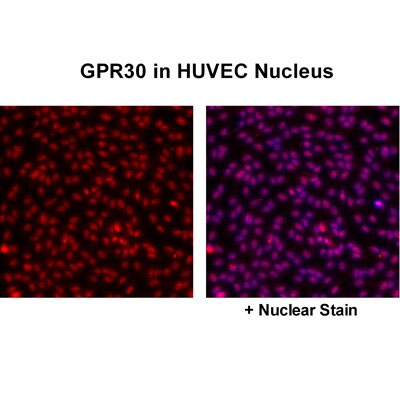
Immunocytochemistry/ Immunofluorescence: GPER/GPR30 Antibody [NBP1-31239]
Immunocytochemistry/Immunofluorescence: GPER/GPR30 Antibody [NBP1-31239] - Detection of GPER/GPR30 in HUVEC nucleus. Image courtesy of a product review by Dr. Subhadeep Chakrabarti of University of Alberta.
Western Blot: GPER/GPR30 Antibody [NBP1-31239] -
Western Blot: GPER/GPR30 Antibody [NBP1-31239] - A. 50 ug 293T whole cell lysate/extract. B. 50 ug whole cell lysate/extract of GFP-human GPR30-transfected 293T cells (No boiling). C. 50 ug whole cell lysate/extract of GFP-human GPR30 and GPR30 siRNA-transfected 293T cells (No boiling). 5 % SDS-PAGE GPR30 antibody ) dilution: 1:1000
Western Blot: GPER/GPR30 Antibody [NBP1-31239] -
Western Blot: GPER/GPR30 Antibody [NBP1-31239] - AhR & GPR30 receptors are present & functional in the MCF10AT1 cells. (A) RT-qPCR analysis of AhR & GPR30 mRNA expression levels represented in arbitrary units (a.u.) in the MCF10AT1 & MCF10CA1a.cl1 cells. MCF-7 cells were used as a control. Values represent mean ± SD of three independent experiments conducted in triplicate. (B) Representative Western blot analyses from three independent experiments of AhR & GPR30 protein expression in MCF10AT1 & MCF10CA1a.cl1 cells. MCF-7 cells were used as a control. (C) XRE-luciferase activity following 8 h exposure of MCF10AT1 cells to ITE at the indicated concentrations. TCDD 10−7 M was used as a control & results were expressed as % of TCDD 10−7 M activity. ***p < 0.001 in Student t-test. (D) XRE-luciferase activity upon 8 h of exposure to ITE 10−10 M alone or in combination with GNF351 at the indicated concentrations. TCDD 10−7 M was used as a control, & results were expressed as % of TCDD 10−7 M activity. Student t-tests revealed the statistically significant differences between unexposed & exposed cells: ***p < 0.001; & between ITE & ITE+GNF351: ###p < 0.001. Values in (C,D) represent mean ± SD of three independent experiments. (E) Representative Western blot analyses from three independent experiments of the phospho-MAPK/MAPK ratio upon exposure of MCF10AT1 cells to G1 (GPR30 agonist) for the times indicated, in the presence or absence of a 2 h pre-treatment with G15 (GPR30 antagonist). Image collected & cropped by CiteAb from the following publication (https://pubmed.ncbi.nlm.nih.gov/32670863), licensed under a CC-BY license. Not internally tested by Novus Biologicals.
Western Blot: GPER/GPR30 Antibody [NBP1-31239] -
Western Blot: GPER/GPR30 Antibody [NBP1-31239] - Effects of short-term exposure of BPA, B[a]P, ITE & G1 10−10 M on AIG & MFE are inhibited by siRNA-AhR & siRNA-GPR30. Representative Western blot analysis from three independent experiments of AhR & GPR30 expression in transfected MCF10AT1 cells with (A) siRNA-AhR, (B) siRNA-GPR30 or their scrambled controls. Quantification of protein expression levels was normalized against tubulin expression. (C,D) Secondary mammospheres formation & (E,F) average number of colonies in soft agar, with the following treatments: BPA and/or B[a]P, G1, or ITE, 10−10 M. Cells were transfected with either siRNA-AhR, siRNA-GPR30 or their scrambled controls before being subjected to the treatments. Treatments were maintained throughout the course of experiments. (mean ± SD of 2 independent experiments, in triplicate). ***p < 0.001, *p < 0.05 vs. their respective unexposed; ###p < 0.001 siRNA vs. scrambled in Student t-test. Image collected & cropped by CiteAb from the following publication (https://pubmed.ncbi.nlm.nih.gov/32670863), licensed under a CC-BY license. Not internally tested by Novus Biologicals.
Western Blot: GPER/GPR30 Antibody [NBP1-31239] -
Western Blot: GPER/GPR30 Antibody [NBP1-31239] - Human endothelial cells express GPR30 protein in the cell nucleus.(A) Confluent monolayers of HUVECs from 3 different cords were lysed & the protein lysates were immunoblotted for GPR30. alpha -tubulin was used as loading control. (B) HUVEC monolayers at 30–40% confluence were treated with 40 nM siRNA (control or GPR30_4) for 48 hours prior to lysis followed by immunoblotting of the cell lysates for GPR30. alpha -tubulin was used as loading control. Data shown are mean ± SEM of 4 independent experiments. ** & ## indicate p<0.01 compared to untreated & control siRNA-treated cells, respectively. (C) Confluent HUVECs grown on glass coverslips were fixed, permeabilized & immunostained with anti-GPR30 antibody. Nuclei were stained with Hoechst33342 dye. The merged image shows GPR30 (red) & nuclei (blue) in pseudocolor. Representative images from 3 independent experiments are shown. Bar, 20 µm. (D) Confluent HUVECs were lysed & fractionated into cytosolic (C) & nuclear (N) fractions prior to western blotting for eNOS, GPR30, p65, alpha -tubulin & c-Jun. A representative set of images (obtained from different membranes) from 3 independent experiments is shown. Image collected & cropped by CiteAb from the following publication (https://pubmed.ncbi.nlm.nih.gov/23285008), licensed under a CC-BY license. Not internally tested by Novus Biologicals.
Western Blot: GPER/GPR30 Antibody [NBP1-31239] -
Western Blot: GPER/GPR30 Antibody [NBP1-31239] - Effects of short-term exposure of BPA, B[a]P, ITE & G1 10−10 M on AIG & MFE are inhibited by siRNA-AhR & siRNA-GPR30. Representative Western blot analysis from three independent experiments of AhR & GPR30 expression in transfected MCF10AT1 cells with (A) siRNA-AhR, (B) siRNA-GPR30 or their scrambled controls. Quantification of protein expression levels was normalized against tubulin expression. (C,D) Secondary mammospheres formation & (E,F) average number of colonies in soft agar, with the following treatments: BPA and/or B[a]P, G1, or ITE, 10−10 M. Cells were transfected with either siRNA-AhR, siRNA-GPR30 or their scrambled controls before being subjected to the treatments. Treatments were maintained throughout the course of experiments. (mean ± SD of 2 independent experiments, in triplicate). ***p < 0.001, *p < 0.05 vs. their respective unexposed; ###p < 0.001 siRNA vs. scrambled in Student t-test. Image collected & cropped by CiteAb from the following publication (https://pubmed.ncbi.nlm.nih.gov/32670863), licensed under a CC-BY license. Not internally tested by Novus Biologicals.
Western Blot: GPER/GPR30 Antibody [NBP1-31239] -
Non-transfected (-) and transfected (+) boiled and unboiled 293T whole cell extracts (30 ug) were separated by 5% SDS-PAGE, and the membrane was blotted with GPER/GPR30 antibody (NBP1-31239) diluted at 1:1000. The HRP-conjugated anti-rabbit IgG antibody was used to detect the primary antibody.Applications for GPER/GPR30 Antibody - BSA Free
Application
Recommended Usage
Flow Cytometry
Reported in scientific literature (PMID 27899250)
Immunocytochemistry/ Immunofluorescence
Validated from a verified customer review
Immunohistochemistry-Paraffin
Assay dependent
Western Blot
1:500-1:3000
Application Notes
WB As is commonly seen with membrane proteins, significant hydrophobicity can lead to aggregation following boiling of samples prior to SDS-PAGE and subsequent western blotting. We recommend to avoid boiling in this case. After harvesting lysate with RIPA buffer, sample loading buffer (including 2-ME) is directly added to the lysate. Samples are then mixed well and added directly without heating.
Reviewed Applications
Read 1 review rated 4 using NBP1-31239 in the following applications:
Flow Cytometry Panel Builder
Bio-Techne Knows Flow Cytometry
Save time and reduce costly mistakes by quickly finding compatible reagents using the Panel Builder Tool.
Advanced Features
- Spectra Viewer - Custom analysis of spectra from multiple fluorochromes
- Spillover Popups - Visualize the spectra of individual fluorochromes
- Antigen Density Selector - Match fluorochrome brightness with antigen density

Formulation, Preparation, and Storage
Purification
Antigen Affinity-purified
Formulation
PBS, 20% Glycerol
Format
BSA Free
Preservative
0.01% Thimerosal
Concentration
Concentrations vary lot to lot. See vial label for concentration. If unlisted please contact technical services.
Shipping
The product is shipped with polar packs. Upon receipt, store it immediately at the temperature recommended below.
Stability & Storage
Store at 4C short term. Aliquot and store at -20C long term. Avoid freeze-thaw cycles.
Background: GPER/GPR30
Long Name
G Protein-coupled Estrogen Receptor 1
Alternate Names
CEPR, CMKRL2, DRY12, FEG-1, GPCR-BR, GPR30, LERGU2, LyGPR
Entrez Gene IDs
2852 (Human)
Gene Symbol
GPER1
OMIM
601805 (Human)
UniProt
Additional GPER/GPR30 Products
Product Documents for GPER/GPR30 Antibody - BSA Free
Certificate of Analysis
To download a Certificate of Analysis, please enter a lot or batch number in the search box below.
Product Specific Notices for GPER/GPR30 Antibody - BSA Free
This product is for research use only and is not approved for use in humans or in clinical diagnosis. Primary Antibodies are guaranteed for 1 year from date of receipt.
⚠ WARNING: This product can expose you to chemicals including mercury, which is known to the State of California to cause reproductive toxicity with developmental effects. For more information go to www.P65Warnings.ca.gov.Related Research Areas
Citations for GPER/GPR30 Antibody - BSA Free
 Powered by Bioz
Customer Reviews for GPER/GPR30 Antibody - BSA Free (1)
4 out of 5
1 Customer Rating
Have you used GPER/GPR30 Antibody - BSA Free?
Submit a review and receive an Amazon gift card!
$25/€18/£15/$25CAN/¥2500 Yen for a review with an image
$10/€7/£6/$10CAN/¥1110 Yen for a review without an image
Submit a review
Customer Images
Showing
1
-
1 的
1 review
Showing All
Filter By:
-
Application: ImmunofluorescenceSample Tested: Human umbilical vein endothelial cellsSpecies: HumanVerified Customer | Posted 03/05/2013

There are no reviews that match your criteria.
Protocols
Find general support by application which include: protocols, troubleshooting, illustrated assays, videos and webinars.
- 7-Amino Actinomycin D (7-AAD) Cell Viability Flow Cytometry Protocol
- Antigen Retrieval Protocol (PIER)
- Antigen Retrieval for Frozen Sections Protocol
- Appropriate Fixation of IHC/ICC Samples
- Cellular Response to Hypoxia Protocols
- Chromogenic IHC Staining of Formalin-Fixed Paraffin-Embedded (FFPE) Tissue Protocol
- Chromogenic Immunohistochemistry Staining of Frozen Tissue
- ClariTSA™ Fluorophore Kits
- Detection & Visualization of Antibody Binding
- Extracellular Membrane Flow Cytometry Protocol
- Flow Cytometry Protocol for Cell Surface Markers
- Flow Cytometry Protocol for Staining Membrane Associated Proteins
- Flow Cytometry Staining Protocols
- Flow Cytometry Troubleshooting Guide
- Fluorescent IHC Staining of Frozen Tissue Protocol
- Graphic Protocol for Heat-induced Epitope Retrieval
- Graphic Protocol for the Preparation and Fluorescent IHC Staining of Frozen Tissue Sections
- Graphic Protocol for the Preparation and Fluorescent IHC Staining of Paraffin-embedded Tissue Sections
- Graphic Protocol for the Preparation of Gelatin-coated Slides for Histological Tissue Sections
- ICC Cell Smear Protocol for Suspension Cells
- ICC Immunocytochemistry Protocol Videos
- ICC for Adherent Cells
- IHC Sample Preparation (Frozen sections vs Paraffin)
- Immunocytochemistry (ICC) Protocol
- Immunocytochemistry Troubleshooting
- Immunofluorescence of Organoids Embedded in Cultrex Basement Membrane Extract
- Immunofluorescent IHC Staining of Formalin-Fixed Paraffin-Embedded (FFPE) Tissue Protocol
- Immunohistochemistry (IHC) and Immunocytochemistry (ICC) Protocols
- Immunohistochemistry Frozen Troubleshooting
- Immunohistochemistry Paraffin Troubleshooting
- Intracellular Flow Cytometry Protocol Using Alcohol (Methanol)
- Intracellular Flow Cytometry Protocol Using Detergents
- Intracellular Nuclear Staining Flow Cytometry Protocol Using Detergents
- Intracellular Staining Flow Cytometry Protocol Using Alcohol Permeabilization
- Intracellular Staining Flow Cytometry Protocol Using Detergents to Permeabilize Cells
- Preparing Samples for IHC/ICC Experiments
- Preventing Non-Specific Staining (Non-Specific Binding)
- Primary Antibody Selection & Optimization
- Propidium Iodide Cell Viability Flow Cytometry Protocol
- Protocol for Heat-Induced Epitope Retrieval (HIER)
- Protocol for Liperfluo
- Protocol for Making a 4% Formaldehyde Solution in PBS
- Protocol for VisUCyte™ HRP Polymer Detection Reagent
- Protocol for the Characterization of Human Th22 Cells
- Protocol for the Characterization of Human Th9 Cells
- Protocol for the Fluorescent ICC Staining of Cell Smears - Graphic
- Protocol for the Fluorescent ICC Staining of Cultured Cells on Coverslips - Graphic
- Protocol for the Preparation & Fixation of Cells on Coverslips
- Protocol for the Preparation and Chromogenic IHC Staining of Frozen Tissue Sections
- Protocol for the Preparation and Chromogenic IHC Staining of Frozen Tissue Sections - Graphic
- Protocol for the Preparation and Chromogenic IHC Staining of Paraffin-embedded Tissue Sections
- Protocol for the Preparation and Chromogenic IHC Staining of Paraffin-embedded Tissue Sections - Graphic
- Protocol for the Preparation and Fluorescent ICC Staining of Cells on Coverslips
- Protocol for the Preparation and Fluorescent ICC Staining of Non-adherent Cells
- Protocol for the Preparation and Fluorescent ICC Staining of Stem Cells on Coverslips
- Protocol for the Preparation and Fluorescent IHC Staining of Frozen Tissue Sections
- Protocol for the Preparation and Fluorescent IHC Staining of Paraffin-embedded Tissue Sections
- Protocol for the Preparation of Gelatin-coated Slides for Histological Tissue Sections
- Protocol for the Preparation of a Cell Smear for Non-adherent Cell ICC - Graphic
- Protocol: Annexin V and PI Staining by Flow Cytometry
- Protocol: Annexin V and PI Staining for Apoptosis by Flow Cytometry
- R&D Systems Quality Control Western Blot Protocol
- TUNEL and Active Caspase-3 Detection by IHC/ICC Protocol
- The Importance of IHC/ICC Controls
- Troubleshooting Guide: Fluorokine Flow Cytometry Kits
- Troubleshooting Guide: Immunohistochemistry
- Troubleshooting Guide: Western Blot Figures
- Western Blot Conditions
- Western Blot Protocol
- Western Blot Protocol for Cell Lysates
- Western Blot Troubleshooting
- Western Blot Troubleshooting Guide
- View all Protocols, Troubleshooting, Illustrated assays and Webinars
Loading...