
Key Product Details
Species Reactivity
Validated:
Human
Cited:
Human
Applications
Validated:
Immunohistochemistry, Western Blot, Intracellular Staining by Flow Cytometry
Cited:
Western Blot, Immunocytochemistry
Label
Unconjugated
Antibody Source
Monoclonal Mouse IgG2B Clone # 971526
Loading...
Product Specifications
Immunogen
Human embryonic kidney cell line HEK-293-derived transfected with human BPI
Val32-Lys487
Accession # P17213
Val32-Lys487
Accession # P17213
Specificity
Detects human BPI in direct ELISAs.
Clonality
Monoclonal
Host
Mouse
Isotype
IgG2B
Scientific Data Images for Human BPI Antibody (971526)
Detection of Human BPI by Western Blot.
Western blot shows lysates of Human bone marrow and human spleen tissue. PVDF membrane was probed with 2 µg/mL of Mouse Anti-Human BPI Monoclonal Antibody (Catalog # MAB74681) followed by HRP-conjugated Anti-Mouse IgG Secondary Antibody (Catalog # HAF018). A specific band was detected for BPI at approximately 55 kDa (as indicated). This experiment was conducted under reducing conditions and using Immunoblot Buffer Group 1.
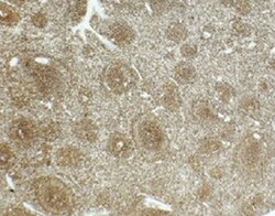
BPI in Human Tonsil.
BPI was detected in immersion fixed paraffin-embedded sections of human tonsil using Mouse Anti-Human BPI Monoclonal Antibody (Catalog # MAB74681) at 5 µg/mL for 1 hour at room temperature followed by incubation with the Anti-Mouse IgG VisUCyte™ HRP Polymer Antibody (Catalog # VC001). Tissue was stained using DAB (brown) and counterstained with hematoxylin (blue). Specific staining was localized to lymphocytes. View our protocol for IHC Staining with VisUCyte HRP Polymer Detection Reagents.
Detection of BPI in HL-60 Human Cell Line by Flow Cytometry.
HL-60 human acute promyelocytic leukemia cell line was stained with Mouse Anti-Human BPI Monoclonal Antibody (Catalog # MAB74681, filled histogram) or isotype control antibody (Catalog # MAB0041, open histogram), followed by Phycoerythrin-conjugated Anti-Mouse IgG Secondary Antibody (Catalog # F0102B). To facilitate intracellular staining, cells were fixed with Flow Cytometry Fixation Buffer (Catalog # FC004) and permeabilized with Flow Cytometry Permeabilization/Wash Buffer I (Catalog # FC005). View our protocol for Staining Intracellular Molecules.Applications for Human BPI Antibody (971526)
Application
Recommended Usage
Immunohistochemistry
5-25 µg/mL
Sample: Immersion fixed paraffin-embedded sections of human tonsil
Sample: Immersion fixed paraffin-embedded sections of human tonsil
Intracellular Staining by Flow Cytometry
0.25 µg/106 cells
Sample: HL-60 Human acute promyelocytic leukemia cell line fixed with Flow Cytometry Fixation Buffer (Catalog # FC004) and permeabilized with Flow Cytometry Permeabilization/Wash Buffer I (Catalog # FC005).
Sample: HL-60 Human acute promyelocytic leukemia cell line fixed with Flow Cytometry Fixation Buffer (Catalog # FC004) and permeabilized with Flow Cytometry Permeabilization/Wash Buffer I (Catalog # FC005).
Western Blot
2 µg/mL
Sample: Human bone marrow and human spleen tissue
Sample: Human bone marrow and human spleen tissue
Reviewed Applications
Read 1 review rated 5 using MAB74681 in the following applications:
Flow Cytometry Panel Builder
Bio-Techne Knows Flow Cytometry
Save time and reduce costly mistakes by quickly finding compatible reagents using the Panel Builder Tool.
Advanced Features
- Spectra Viewer - Custom analysis of spectra from multiple fluorochromes
- Spillover Popups - Visualize the spectra of individual fluorochromes
- Antigen Density Selector - Match fluorochrome brightness with antigen density

Formulation, Preparation, and Storage
Purification
Protein A or G purified from hybridoma culture supernatant
Reconstitution
Reconstitute at 0.5 mg/mL in sterile PBS. For liquid material, refer to CoA for concentration.
Loading...
Formulation
Lyophilized from a 0.2 μm filtered solution in PBS with Trehalose. *Small pack size (SP) is supplied either lyophilized or as a 0.2 µm filtered solution in PBS.
Shipping
Lyophilized product is shipped at ambient temperature. Liquid small pack size (-SP) is shipped with polar packs. Upon receipt, store immediately at the temperature recommended below.
Stability & Storage
Use a manual defrost freezer and avoid repeated freeze-thaw cycles.
- 12 months from date of receipt, -20 to -70 °C as supplied.
- 1 month, 2 to 8 °C under sterile conditions after reconstitution.
- 6 months, -20 to -70 °C under sterile conditions after reconstitution.
Calculators
Background: BPI
References
- Schultz, H. and J.P. Weiss (2007) Clin. Chim. Acta 384:12.
- Holweg, A. et al. (2011) Biochem. Soc. Trans. 39:1045.
- Esteve, E. et al. (2010) Thromb. Haemost. 103:780.
- Horwitz, A.H. et al. (1996) Protein Exp. Purif. 8:28.
- Gray, P.W. et al. (1989) J. Biol. Chem. 264:9505.
- Ooi, C.E. et al. (1987) J. Biol. Chem. 262:14891.
- Tobias, P.S. et al. (1997) J. Biol. Chem. 272:18682.
- Weersink, A.J. et al. (1993) J. Immunol. 150:253.
- Nishimura, H. et al. (2001) Immunology 103:519.
- Canny, G. et al. (2002) Proc. Natl. Acad. Sci. 99:3902.
- Weiss, J. and I. Olsson (1987) Blood 69:652.
- Calafat, J. et al. (1998) Blood 91:4770.
- Lennartsson, A. et al. (2005) J. Leukoc. Biol. 77:369.
- van der Schaft, D.W.J. et al. (2000) Blood 96:176.
- Rauniyar, R.K. et al. (2002) Invest. Ophthalmol. Vis. Sci. 43:503.
Long Name
Bactericidal/Permeability-increasing Protein
Alternate Names
BPIFD1, CAP 57, rBPI
Gene Symbol
BPI
UniProt
Additional BPI Products
Product Documents for Human BPI Antibody (971526)
Certificate of Analysis
To download a Certificate of Analysis, please enter a lot or batch number in the search box below.
Note: Certificate of Analysis not available for kit components.
Product Specific Notices for Human BPI Antibody (971526)
For research use only
Customer Reviews for Human BPI Antibody (971526) (1)
5 out of 5
1 Customer Rating
Have you used Human BPI Antibody (971526)?
Submit a review and receive an Amazon gift card!
$25/€18/£15/$25CAN/¥2500 Yen for a review with an image
$10/€7/£6/$10CAN/¥1110 Yen for a review without an image
Submit a review
Customer Images
Showing
1
-
1 的
1 review
Showing All
Filter By:
-
Application: ImmunohistochemistrySample Tested: Spleen tissueSpecies: HumanVerified Customer | Posted 11/02/2022

There are no reviews that match your criteria.
Protocols
Find general support by application which include: protocols, troubleshooting, illustrated assays, videos and webinars.
- 7-Amino Actinomycin D (7-AAD) Cell Viability Flow Cytometry Protocol
- Antigen Retrieval Protocol (PIER)
- Antigen Retrieval for Frozen Sections Protocol
- Appropriate Fixation of IHC/ICC Samples
- Cellular Response to Hypoxia Protocols
- Chromogenic IHC Staining of Formalin-Fixed Paraffin-Embedded (FFPE) Tissue Protocol
- Chromogenic Immunohistochemistry Staining of Frozen Tissue
- ClariTSA™ Fluorophore Kits
- Detection & Visualization of Antibody Binding
- Extracellular Membrane Flow Cytometry Protocol
- Flow Cytometry Protocol for Cell Surface Markers
- Flow Cytometry Protocol for Staining Membrane Associated Proteins
- Flow Cytometry Staining Protocols
- Flow Cytometry Troubleshooting Guide
- Fluorescent IHC Staining of Frozen Tissue Protocol
- Graphic Protocol for Heat-induced Epitope Retrieval
- Graphic Protocol for the Preparation and Fluorescent IHC Staining of Frozen Tissue Sections
- Graphic Protocol for the Preparation and Fluorescent IHC Staining of Paraffin-embedded Tissue Sections
- Graphic Protocol for the Preparation of Gelatin-coated Slides for Histological Tissue Sections
- IHC Sample Preparation (Frozen sections vs Paraffin)
- Immunofluorescent IHC Staining of Formalin-Fixed Paraffin-Embedded (FFPE) Tissue Protocol
- Immunohistochemistry (IHC) and Immunocytochemistry (ICC) Protocols
- Immunohistochemistry Frozen Troubleshooting
- Immunohistochemistry Paraffin Troubleshooting
- Intracellular Flow Cytometry Protocol Using Alcohol (Methanol)
- Intracellular Flow Cytometry Protocol Using Detergents
- Intracellular Nuclear Staining Flow Cytometry Protocol Using Detergents
- Intracellular Staining Flow Cytometry Protocol Using Alcohol Permeabilization
- Intracellular Staining Flow Cytometry Protocol Using Detergents to Permeabilize Cells
- Preparing Samples for IHC/ICC Experiments
- Preventing Non-Specific Staining (Non-Specific Binding)
- Primary Antibody Selection & Optimization
- Propidium Iodide Cell Viability Flow Cytometry Protocol
- Protocol for Heat-Induced Epitope Retrieval (HIER)
- Protocol for Liperfluo
- Protocol for Making a 4% Formaldehyde Solution in PBS
- Protocol for VisUCyte™ HRP Polymer Detection Reagent
- Protocol for the Characterization of Human Th22 Cells
- Protocol for the Characterization of Human Th9 Cells
- Protocol for the Preparation & Fixation of Cells on Coverslips
- Protocol for the Preparation and Chromogenic IHC Staining of Frozen Tissue Sections
- Protocol for the Preparation and Chromogenic IHC Staining of Frozen Tissue Sections - Graphic
- Protocol for the Preparation and Chromogenic IHC Staining of Paraffin-embedded Tissue Sections
- Protocol for the Preparation and Chromogenic IHC Staining of Paraffin-embedded Tissue Sections - Graphic
- Protocol for the Preparation and Fluorescent IHC Staining of Frozen Tissue Sections
- Protocol for the Preparation and Fluorescent IHC Staining of Paraffin-embedded Tissue Sections
- Protocol for the Preparation of Gelatin-coated Slides for Histological Tissue Sections
- Protocol: Annexin V and PI Staining by Flow Cytometry
- Protocol: Annexin V and PI Staining for Apoptosis by Flow Cytometry
- R&D Systems Quality Control Western Blot Protocol
- TUNEL and Active Caspase-3 Detection by IHC/ICC Protocol
- The Importance of IHC/ICC Controls
- Troubleshooting Guide: Fluorokine Flow Cytometry Kits
- Troubleshooting Guide: Immunohistochemistry
- Troubleshooting Guide: Western Blot Figures
- Western Blot Conditions
- Western Blot Protocol
- Western Blot Protocol for Cell Lysates
- Western Blot Troubleshooting
- Western Blot Troubleshooting Guide
- View all Protocols, Troubleshooting, Illustrated assays and Webinars
Loading...