Human c-Myc is a helix-loop-helix transcription factor which efficiently binds DNA after heterodimerization with the bHLH protein Max. It is often overexpressed and mutated in hematopoietic tumors. Mutations frequently result in truncation around amino acid (aa) 252, before the C-terminal DNA binding, HLH and leucine zipper domains. The 439 aa human c-Myc has one O-glycosylation site and has three Ser/Thr phosphorylation sites near the N-terminus. Human c-Myc shows 92% aa identity with mouse or rat c-Myc.
Human c-Myc Antibody (9E10)
R&D Systems | Catalog # MAB3696

Key Product Details
Validated by
Knockout/Knockdown
Species Reactivity
Validated:
Human
Cited:
Human, Mouse, E. coli
Applications
Validated:
Knockout Validated, Immunohistochemistry, Western Blot, Flow Cytometry, Immunocytochemistry, Simple Western, Immunoprecipitation, CyTOF-ready
Cited:
Immunohistochemistry, Immunohistochemistry-Paraffin, Western Blot
Label
Unconjugated
Antibody Source
Monoclonal Mouse IgG1 Clone # 9E10
Loading...
Product Specifications
Immunogen
C-terminal region peptide of human c-Myc
Ala408-Ala439
Accession # P01106
Ala408-Ala439
Accession # P01106
Specificity
Detects endogenous human c-Myc and c-Myc tagged proteins in Western blots.
Clonality
Monoclonal
Host
Mouse
Isotype
IgG1
Scientific Data Images for Human c-Myc Antibody (9E10)
Detection of Human c‑Myc by Western Blot.
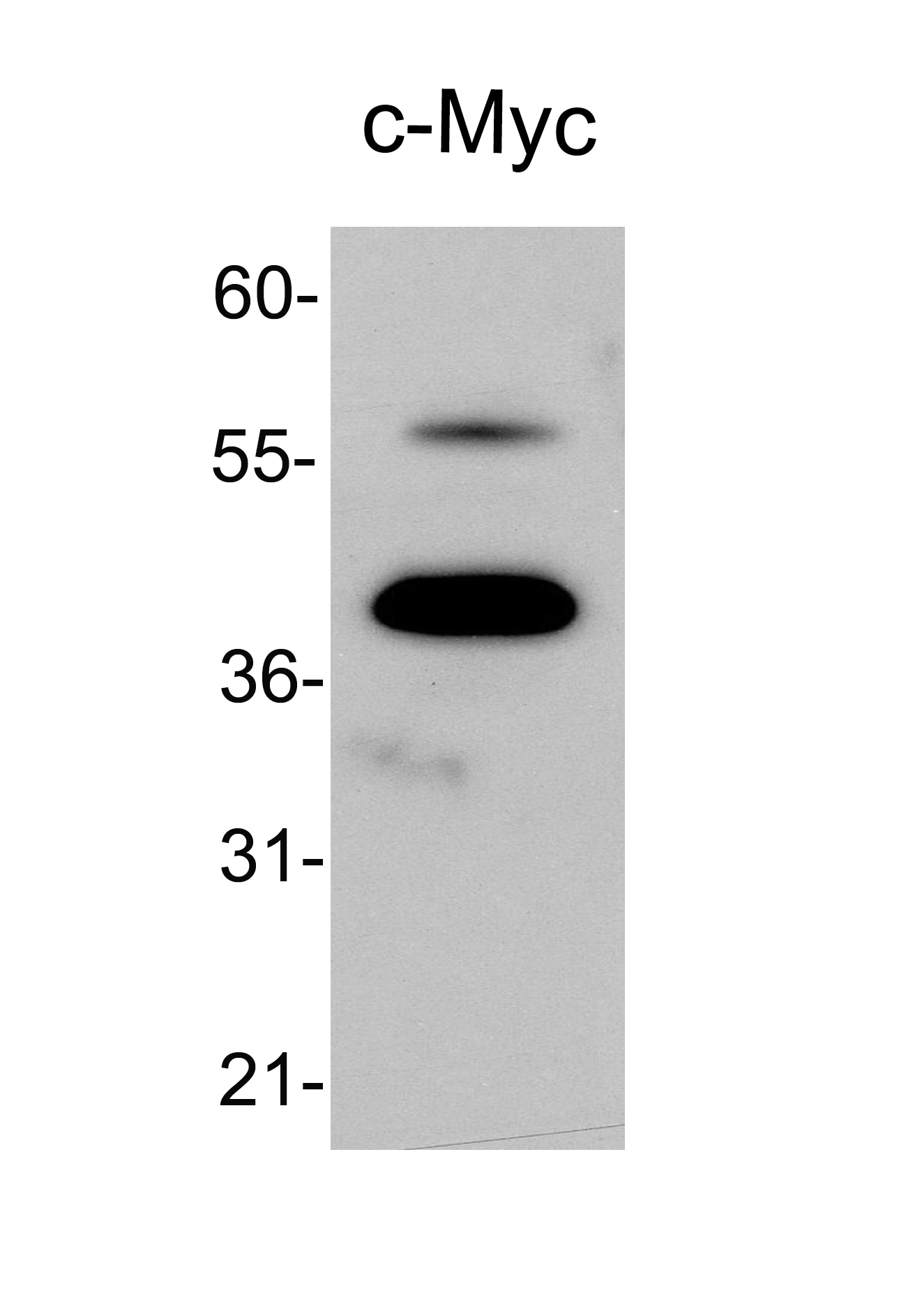
Western blot shows lysates of Daudi human Burkitt's lymphoma cell line and HeLa human cervical epithelial carcinoma cell line. PVDF membrane was probed with 2 µg/mL of Mouse Anti-Human c-Myc Monoclonal Antibody (Catalog # MAB3696) followed by HRP-conjugated Anti-Mouse IgG Secondary Antibody (Catalog # HAF007). A specific band was detected for c-Myc at approximately 52 kDa (as indicated). This experiment was conducted under reducing conditions and using Immunoblot Buffer Group 1.
Detection of c‑Myc-tagged Protein by Western Blot.
Western blot shows lysates of CHO Chinese hamster ovary cell line either mock transfected (-) or transfected with c-Myc-tagged recombinant mouse Wnt-3a. PVDF membrane was probed with 2 µg/mL of Mouse Anti-Human c-Myc Monoclonal Antibody (Catalog # MAB3696) followed by HRP-conjugated Anti-Mouse IgG Secondary Antibody (Catalog # HAF007). A specific band was detected for c-Myc-tagged recombinant mouse Wnt-3a at approximately 41 kDa (as indicated). This experiment was conducted under reducing conditions and using Immunoblot Buffer Group 1.
Detection of c‑Myc in Jurkat Human Cell Line by Flow Cytometry.
Jurkat human acute T cell leukemia cell line was stained with Mouse Anti-Human c-Myc Mono-clonal Antibody (Catalog # MAB3696, filled histogram) or isotype control antibody (Catalog # MAB002, open histogram), followed by Phycoerythrin-conjugated Anti-Mouse IgG Secondary Antibody (Catalog # F0102B). To facilitate intracellular staining, cells were fixed with paraformaldehyde and permeabilized with methanol.
c-Myc in HEK293 Human Cell Line Transfected with c-Myc-tagged Serotonin Receptor.
c-Myc was detected in immersion fixed HEK293 human embryonic kidney cell line transfected with c-Myc-tagged Serotonin Receptor using Mouse Anti-Human c-Myc Monoclonal Antibody (Catalog # MAB3696) at 25 µg/mL for 3 hours at room temperature. Cells were stained using the NorthernLights™ 557-conjugated Anti-Mouse IgG Secondary Antibody (red; Catalog # NL007) and counterstained with DAPI (blue). Specific staining was localized to nuclei. View our protocol for Fluorescent ICC Staining of Cells on Coverslips.
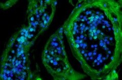
c‑Myc in Human Prostate.
c-Myc was detected in immersion fixed paraffin-embedded sections of human prostate using Mouse Anti-Human c-Myc Monoclonal Antibody (Catalog # MAB3696) at 3 µg/mL overnight at 4 °C. Tissue was stained using the Anti-Mouse HRP-DAB Cell & Tissue Staining Kit (brown; Catalog # CTS002) and counterstained with hematoxylin (blue). Specific staining was localized to nuclei of epithelial cells. View our protocol for Chromogenic IHC Staining of Paraffin-embedded Tissue Sections.
Detection of Human c‑Myc by Simple WesternTM.
Simple Western lane view shows lysates of HeLa human cervical epithelial carcinoma cell line, loaded at 0.2 mg/mL. A specific band was detected for c‑Myc at approximately 74 kDa (as indicated) using 20 µg/mL of Mouse Anti-Human c‑Myc Monoclonal Antibody (Catalog # MAB3696). This experiment was conducted under reducing conditions and using the 12-230 kDa separation system.
Detection of Human c‑Myc by Simple WesternTM.
Simple Western lane view shows c-Myc-tagged recombinant human ANGPTL6, loaded at 0.2 mg/mL. A specific band was detected for c‑Myc at approximately 161 kDa (as indicated) using 20 µg/mL of Mouse Anti-Human c‑Myc Monoclonal Antibody (Catalog # MAB3696). This experiment was conducted under reducing conditions and using the 12-230 kDa separation system.
Western Blot Shows Human c‑Myc Specificity by Using Knockout Cell Line.
Western blot shows lysates of HEK293T human embryonic kidney parental cell line and c-Myc knockout HEK293T cell line (KO). PVDF membrane was probed with 2 µg/mL of Mouse Anti-Human c-Myc Monoclonal Antibody (Catalog # MAB3696) followed by HRP-conjugated Anti-Mouse IgG Secondary Antibody (Catalog # HAF018). A specific band was detected for c-Myc at approximately 52 kDa (as indicated) in the parental HEK293T cell line, but is not detectable in knockout HEK293T cell line. GAPDH (Catalog # MAB5718) is shown as a loading control. This experiment was conducted under reducing conditions and using Immunoblot Buffer Group 1.
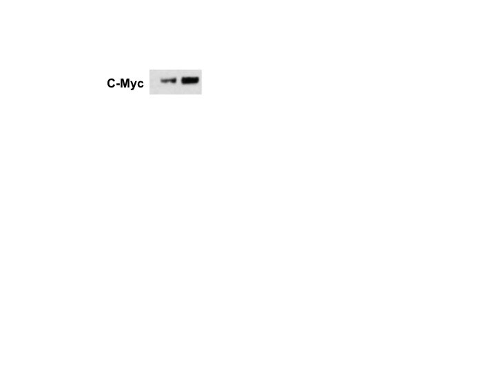
Detection of c-Myc by Western Blot
Effect of mtp53 knockdown on cell proliferation and Akt/mTOR pathway. (A) AN3CA shLuc and shp53 cells (2 × 10⁵ cells/35 mm dish) were seeded on a 35 mm dish. After 24 (Day 0), 48 (Day 1), 72 (Day 2), and 96 h (Day 3) incubation, the cells were harvested by trypsin and counted under an inverted microscope. (B) HEC-59 shLuc and shp53 cells (2.5 × 10⁵ cells/35 mm dish) were seeded on a 35 mm dish. After 24 (Day 0), 48 (Day 1), 72 (Day 2), and 96 h (Day 3) incubation, the cells were harvested by trypsin and counted under an inverted microscope. (C) Protein expressions of p53, p-Akt, Akt, p- p-p85S6K/p70S6K, and p85S6K/p70S6K were determined by Western blot. ImageJ software was used to quantify the band intensities of p53, p-Akt, Akt, p-p85S6K/p70S6K, and p85S6K/p70S6K. Data shown are the relative expression standardized by the beta -actin protein level. The ratio of shLuc cells was set at 1. (D) Protein expressions of p53, hTERT, c-Myc, CDK6, p27, LC3B, and survivin were determined by Western blot. The arrow indicated the nonspecific band. ImageJ software was used to quantify the band intensities of indicated protein. Data shown are the relative expression standardized by the beta -actin protein level. The ratio of shLuc cells was set at 1. The symbol ∗ indicates p < 0.05. N.D., not detected. Image collected and cropped by CiteAb from the following open publication (https://pubmed.ncbi.nlm.nih.gov/34831139), licensed under a CC-BY license. Not internally tested by R&D Systems.
Detection of c-Myc by Western Blot
Effect of PQR309 on the expression of c-Myc and its possible target genes. (A) After treating with PQR309 for 48 h in HEC-59 cells, Proteome Profiler Human XL Oncology Array was performed according to the manufacturer protocol. The dot intensities were quantified by ImageJ software. The ratio of cells without treatment was set at 1. (B) After treating with PQR309 in AN3CA and HEC-59 cells for 48 h, protein expressions of endoglin, KLK6, p53, and survivin were determined by Western blot. ImageJ software was used to quantify the band intensities of endoglin, KLK6, p53, and survivin. Data shown are the relative expression standardized by the beta -actin protein level. The ratio of cells without treatment was set at 1. (C) Western blot assay was performed to detect the expressions of hTERT and c-Myc in AN3CA and HEC-59 cells after PQR309 treatment for 48 h. ImageJ software was used to quantify the band intensities of hTERT and c-Myc. Data shown are the relative expression standardized by the beta -actin protein level. The ratio of cells without treatment was set at 1. (D) AN3CA (5 × 10³ cells/well of 96-well plate) and HEC-59 (6 × 10³ cells/well of 96-well plate) were treated with KJ-Pyr-9 (0, 2.5, 5, 10 and 20 μM), an inhibitor of c-Myc, for 72 h. Cell viability was analyzed by MTT assay. (E) Protein expressions of c-Myc, p53, KLK6, and survivin were determined by Western blot. ImageJ software was used to quantify the band intensities of c-Myc, p53, KLK6, and survivin. Data shown are the relative expression standardized by the beta -actin protein level. The ratio of cells without treatment was set at 1. N.D., not detected. Image collected and cropped by CiteAb from the following open publication (https://pubmed.ncbi.nlm.nih.gov/34831139), licensed under a CC-BY license. Not internally tested by R&D Systems.
Detection of c-Myc by Western Blot
HJURP promotes proliferation and chemoresistance of GC cell by activating the TOP2A. (A) The relationship between the expression of TOP2A in Kaplan–Meier Plotter database and OS in GC patients. (B) The relationship between the expression of TOP2A in Kaplan–Meier Plotter database and FP in GC patients. (C, D) HGC27 and AGS were transfected with NC siRNA or HJURP siRNA for 48 h and then cell samples were collected, and the levels of TOP2A were determined by qRT-PCR and Western blot. (E, F) HGC27 and AGS were transfected with Flag-con or Flag-HJURP for 48 h and then cell samples were collected, and the levels of TOP2A were determined by qRT-PCR and Western blot. (G) CCK-8 assays were employed to evaluate the effect of HJURP knockdown with or without TOP2A overexpression on the proliferation of HGC27 GC cells. (H) Colony formation assays were employed to evaluate the effect of HJURP overexpression with or without TOP2A knockdown on the chemoresistance of HGC27 and AGS. **P < 0.01, ***P < 0.001, ****P < 0.0001. GC: gastric cancer, HJURP: Holliday Junction Recognition Protein, OS: overall survival, FP: first progression survival, DDP: cisplatin, TOP2A: Topoisomerase II alpha. Image collected and cropped by CiteAb from the following open publication (https://pubmed.ncbi.nlm.nih.gov/40290723), licensed under a CC-BY license. Not internally tested by R&D Systems.
Detection of c-Myc by Western Blot
HJURP improves transcriptional activity of TOP2A via the promotion of MYC signaling. (A) The Venn diagram displays the potential transcription factors of TOP2A. (B, C) AGS were transfected with HJURP siRNA or His-MYC for 48 h and then cell samples were collected, and the levels of TOP2A, MYC and HJURP were determined by qRT-PCR and Western blot. (D–F) Genes are divided into 4 classes (Positive, Moderate, Weak, Negative) according to the expression level (visualized by contingency table heat map, and the depth of color represents the number of samples. The Pearson correlation of the two genes is calculated and Fisher’s exact test is performed. (G) The difference of MYC expression in TCGA-STAD dataset. (H) The ROC analysis of diagnostic accuracy for GC with MYC expression in TCGA databases. (I) The difference of TOP2A expression in TCGA-STAD dataset. (J) The ROC analysis of diagnostic accuracy for GC with TOP2A expression in TCGA databases. TCGA: The Cancer Genome Atlas, HJURP: Holliday Junction Recognition Protein, TOP2A: Topoisomerase II alpha. Image collected and cropped by CiteAb from the following open publication (https://pubmed.ncbi.nlm.nih.gov/40290723), licensed under a CC-BY license. Not internally tested by R&D Systems.
Detection of c-Myc by Western Blot
HJURP promotes proliferation and chemoresistance of GC cell by activating the TOP2A. (A) The relationship between the expression of TOP2A in Kaplan–Meier Plotter database and OS in GC patients. (B) The relationship between the expression of TOP2A in Kaplan–Meier Plotter database and FP in GC patients. (C, D) HGC27 and AGS were transfected with NC siRNA or HJURP siRNA for 48 h and then cell samples were collected, and the levels of TOP2A were determined by qRT-PCR and Western blot. (E, F) HGC27 and AGS were transfected with Flag-con or Flag-HJURP for 48 h and then cell samples were collected, and the levels of TOP2A were determined by qRT-PCR and Western blot. (G) CCK-8 assays were employed to evaluate the effect of HJURP knockdown with or without TOP2A overexpression on the proliferation of HGC27 GC cells. (H) Colony formation assays were employed to evaluate the effect of HJURP overexpression with or without TOP2A knockdown on the chemoresistance of HGC27 and AGS. **P < 0.01, ***P < 0.001, ****P < 0.0001. GC: gastric cancer, HJURP: Holliday Junction Recognition Protein, OS: overall survival, FP: first progression survival, DDP: cisplatin, TOP2A: Topoisomerase II alpha. Image collected and cropped by CiteAb from the following open publication (https://pubmed.ncbi.nlm.nih.gov/40290723), licensed under a CC-BY license. Not internally tested by R&D Systems.Applications for Human c-Myc Antibody (9E10)
Application
Recommended Usage
CyTOF-ready
Ready to be labeled using established conjugation methods. No BSA or other carrier proteins that could interfere with conjugation.
Flow Cytometry
2.5 µg/106 cells
Sample: Jurkat human acute T cell leukemia cell line fixed with paraformaldehyde and permeabilized with methanol.
Sample: Jurkat human acute T cell leukemia cell line fixed with paraformaldehyde and permeabilized with methanol.
Immunocytochemistry
8-25 µg/mL
Sample: Immersion fixed HEK293 human embryonic kidney cell line
Sample: Immersion fixed HEK293 human embryonic kidney cell line
Immunohistochemistry
8-25 µg/mL
Sample: Immersion fixed paraffin-embedded sections of human prostate
Sample: Immersion fixed paraffin-embedded sections of human prostate
Immunoprecipitation
Fan H, et al. (1998) Biochem. Cell. Biol. 76:125.
Knockout Validated
c‑Myc
is specifically detected in HEK293T human embryonic kidney parental cell line but is not detectable in
c‑Myc knockout HEK293T cell line.
Simple Western
20 µg/mL
Sample: HeLa human cervical epithelial carcinoma cell line and c-Myc-tagged recombinant human ANGPTL6
Sample: HeLa human cervical epithelial carcinoma cell line and c-Myc-tagged recombinant human ANGPTL6
Western Blot
2 µg/mL
Sample: Daudi human Burkitt's lymphoma cell line, HeLa human cervical epithelial carcinoma cell line, and CHO Chinese hamster ovary cell line either transfected with c-Myc-tagged recombinant mouse Wnt-3a
Sample: Daudi human Burkitt's lymphoma cell line, HeLa human cervical epithelial carcinoma cell line, and CHO Chinese hamster ovary cell line either transfected with c-Myc-tagged recombinant mouse Wnt-3a
Reviewed Applications
Read 3 reviews rated 4.3 using MAB3696 in the following applications:
Flow Cytometry Panel Builder
Bio-Techne Knows Flow Cytometry
Save time and reduce costly mistakes by quickly finding compatible reagents using the Panel Builder Tool.
Advanced Features
- Spectra Viewer - Custom analysis of spectra from multiple fluorochromes
- Spillover Popups - Visualize the spectra of individual fluorochromes
- Antigen Density Selector - Match fluorochrome brightness with antigen density

Formulation, Preparation, and Storage
Purification
Protein A or G purified from hybridoma culture supernatant
Reconstitution
Sterile PBS to a final concentration of 0.5 mg/mL. For liquid material, refer to CoA for concentration.
Loading...
Formulation
Lyophilized from a 0.2 μm filtered solution in PBS with Trehalose. *Small pack size (SP) is supplied either lyophilized or as a 0.2 µm filtered solution in PBS.
Shipping
Lyophilized product is shipped at ambient temperature. Liquid small pack size (-SP) is shipped with polar packs. Upon receipt, store immediately at the temperature recommended below.
Stability & Storage
Use a manual defrost freezer and avoid repeated freeze-thaw cycles.
- 12 months from date of receipt, -20 to -70 °C as supplied.
- 1 month, 2 to 8 °C under sterile conditions after reconstitution.
- 6 months, -20 to -70 °C under sterile conditions after reconstitution.
Calculators
Background: c-Myc
Long Name
v-Myc Avian Myelocytomatosis Viral Oncogene Homolog (Avian)
Alternate Names
cMyc, Myc, Myc2, Niard, Nird
Gene Symbol
MYC
UniProt
Additional c-Myc Products
Product Documents for Human c-Myc Antibody (9E10)
Certificate of Analysis
To download a Certificate of Analysis, please enter a lot or batch number in the search box below.
Note: Certificate of Analysis not available for kit components.
Product Specific Notices for Human c-Myc Antibody (9E10)
For research use only
Citations for Human c-Myc Antibody (9E10)
 Powered by Bioz
Customer Reviews for Human c-Myc Antibody (9E10) (3)
4.3 out of 5
3 Customer Ratings
Have you used Human c-Myc Antibody (9E10)?
Submit a review and receive an Amazon gift card!
$25/€18/£15/$25CAN/¥2500 Yen for a review with an image
$10/€7/£6/$10CAN/¥1110 Yen for a review without an image
Submit a review
Customer Images
Showing
1
-
3 的
3 reviews
Showing All
Filter By:
-
Application: ImmunohistochemistrySample Tested: intravascular B cell lymphomaSpecies: HumanVerified Customer | Posted 09/05/2021

-
Application: Western BlotSample Tested: HEK293 human embryonic kidney cell lineSpecies: HumanVerified Customer | Posted 01/25/2018
-
Application: Western BlotSample Tested: Whole cell lysate from 293T cells, in lysis bufferSpecies: HumanVerified Customer | Posted 12/18/2014Whole cell lysate from 293T cells, in lysis buffer
There are no reviews that match your criteria.
Protocols
Find general support by application which include: protocols, troubleshooting, illustrated assays, videos and webinars.
- 7-Amino Actinomycin D (7-AAD) Cell Viability Flow Cytometry Protocol
- Antigen Retrieval Protocol (PIER)
- Antigen Retrieval for Frozen Sections Protocol
- Appropriate Fixation of IHC/ICC Samples
- Cellular Response to Hypoxia Protocols
- Chromogenic IHC Staining of Formalin-Fixed Paraffin-Embedded (FFPE) Tissue Protocol
- Chromogenic Immunohistochemistry Staining of Frozen Tissue
- ClariTSA™ Fluorophore Kits
- Detection & Visualization of Antibody Binding
- Extracellular Membrane Flow Cytometry Protocol
- Flow Cytometry Protocol for Cell Surface Markers
- Flow Cytometry Protocol for Staining Membrane Associated Proteins
- Flow Cytometry Staining Protocols
- Flow Cytometry Troubleshooting Guide
- Fluorescent IHC Staining of Frozen Tissue Protocol
- Graphic Protocol for Heat-induced Epitope Retrieval
- Graphic Protocol for the Preparation and Fluorescent IHC Staining of Frozen Tissue Sections
- Graphic Protocol for the Preparation and Fluorescent IHC Staining of Paraffin-embedded Tissue Sections
- Graphic Protocol for the Preparation of Gelatin-coated Slides for Histological Tissue Sections
- ICC Cell Smear Protocol for Suspension Cells
- ICC Immunocytochemistry Protocol Videos
- ICC for Adherent Cells
- IHC Sample Preparation (Frozen sections vs Paraffin)
- Immunocytochemistry (ICC) Protocol
- Immunocytochemistry Troubleshooting
- Immunofluorescence of Organoids Embedded in Cultrex Basement Membrane Extract
- Immunofluorescent IHC Staining of Formalin-Fixed Paraffin-Embedded (FFPE) Tissue Protocol
- Immunohistochemistry (IHC) and Immunocytochemistry (ICC) Protocols
- Immunohistochemistry Frozen Troubleshooting
- Immunohistochemistry Paraffin Troubleshooting
- Immunoprecipitation Protocol
- Intracellular Flow Cytometry Protocol Using Alcohol (Methanol)
- Intracellular Flow Cytometry Protocol Using Detergents
- Intracellular Nuclear Staining Flow Cytometry Protocol Using Detergents
- Intracellular Staining Flow Cytometry Protocol Using Alcohol Permeabilization
- Intracellular Staining Flow Cytometry Protocol Using Detergents to Permeabilize Cells
- Preparing Samples for IHC/ICC Experiments
- Preventing Non-Specific Staining (Non-Specific Binding)
- Primary Antibody Selection & Optimization
- Propidium Iodide Cell Viability Flow Cytometry Protocol
- Protocol for Heat-Induced Epitope Retrieval (HIER)
- Protocol for Liperfluo
- Protocol for Making a 4% Formaldehyde Solution in PBS
- Protocol for VisUCyte™ HRP Polymer Detection Reagent
- Protocol for the Characterization of Human Th22 Cells
- Protocol for the Characterization of Human Th9 Cells
- Protocol for the Fluorescent ICC Staining of Cell Smears - Graphic
- Protocol for the Fluorescent ICC Staining of Cultured Cells on Coverslips - Graphic
- Protocol for the Preparation & Fixation of Cells on Coverslips
- Protocol for the Preparation and Chromogenic IHC Staining of Frozen Tissue Sections
- Protocol for the Preparation and Chromogenic IHC Staining of Frozen Tissue Sections - Graphic
- Protocol for the Preparation and Chromogenic IHC Staining of Paraffin-embedded Tissue Sections
- Protocol for the Preparation and Chromogenic IHC Staining of Paraffin-embedded Tissue Sections - Graphic
- Protocol for the Preparation and Fluorescent ICC Staining of Cells on Coverslips
- Protocol for the Preparation and Fluorescent ICC Staining of Non-adherent Cells
- Protocol for the Preparation and Fluorescent ICC Staining of Stem Cells on Coverslips
- Protocol for the Preparation and Fluorescent IHC Staining of Frozen Tissue Sections
- Protocol for the Preparation and Fluorescent IHC Staining of Paraffin-embedded Tissue Sections
- Protocol for the Preparation of Gelatin-coated Slides for Histological Tissue Sections
- Protocol for the Preparation of a Cell Smear for Non-adherent Cell ICC - Graphic
- Protocol: Annexin V and PI Staining by Flow Cytometry
- Protocol: Annexin V and PI Staining for Apoptosis by Flow Cytometry
- R&D Systems Quality Control Western Blot Protocol
- TUNEL and Active Caspase-3 Detection by IHC/ICC Protocol
- The Importance of IHC/ICC Controls
- Troubleshooting Guide: Fluorokine Flow Cytometry Kits
- Troubleshooting Guide: Immunohistochemistry
- Troubleshooting Guide: Western Blot Figures
- Western Blot Conditions
- Western Blot Protocol
- Western Blot Protocol for Cell Lysates
- Western Blot Troubleshooting
- Western Blot Troubleshooting Guide
- View all Protocols, Troubleshooting, Illustrated assays and Webinars


