
HGF, also known as scatter factor and hepatopoietin A, is a pleiotropic protein in the plasminogen subfamily of S1 peptidases. It is a multidomain molecule that includes an N-terminal PAN/APPLE-like domain, four Kringle domains, and a serine proteinase-like domain that has no detectable protease activity. Human HGF is secreted as an inactive 728 amino acid (aa) single chain propeptide. It is cleaved after the fourth Kringle domain by a serine protease to form bioactive disulfide‑linked HGF with a 60 kDa alpha and 30 kDa beta chain. Alternate splicing generates human HGF isoforms that lack the proteinase-like domain and different numbers of the Kringle domains. Human HGF shares 91%‑94% aa sequence identity with bovine, canine, feline, mouse, and rat HGF. HGF binds heparan-sulfate proteoglycans and the widely expressed receptor tyrosine kinase, HGF R/c-MET. HGF-dependent c-MET activation is implicated in the development of many human cancers. HGF regulates epithelial morphogenesis by inducing cell scattering and branching tubulogenesis. HGF induces the upregulation of integrin alpha 2 beta 1 in epithelial cells by a selective increase in alpha 2 gene transcription. This integrin serves as a collagen I receptor, and its blockade disrupts epithelial cell branching tubulogenesis. HGF can also alter epithelium morphology by the induction of nectin-1 alpha ectodomain shedding, an adhesion protein component of adherens junctions. In the thyroid, HGF induces the proliferation, motility, and loss of differentiation markers of thyrocytes and inhibits TSH-stimulated iodine uptake. HGF promotes the motility of cardiac stem cells in damaged myocardium.

Scientific images may be resized, cropped, or adjusted for brightness or contrast for presentation purposes. These updates are limited to presentation and do not affect the underlying scientific interpretation of the data. Where available, additional source data may be provided upon request.
Key Product Details
Species Reactivity
Validated:
Cited:
Applications
Validated:
Cited:
Label
Antibody Source
Product Specifications
Immunogen
Gln32-Ser728 (Asp384Asn, Asp416Asn, Leu622Ser, His645Arg, Val678Ile)
Accession # P14210
Specificity
Clonality
Host
Isotype
Endotoxin Level
Scientific Data Images for Human HGF Antibody
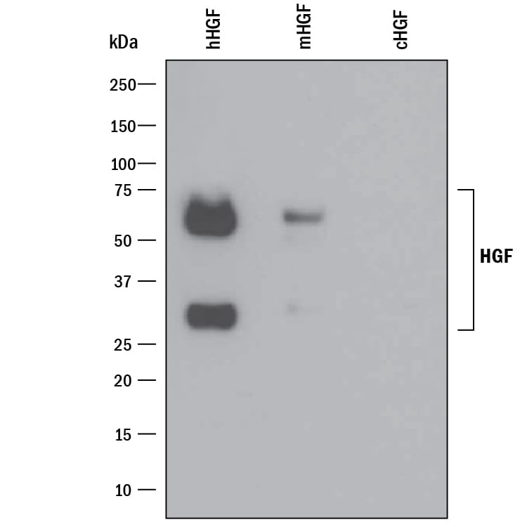
Detection of Recombinant Human HGF by Western Blot.
Western blot shows 25 ng of Recombinant Human HGF (Catalog # 294-HG), Recombinant Mouse HGF (Catalog # 2207-HG) and Recombinant Canine HGF (Catalog # 3386-HG). PVDF Membrane was probed with 0.1 µg/mL of Goat Anti-Human HGF Antigen Affinity-purified Polyclonal Antibody (Catalog # AF-294-NA) followed by HRP-conjugated Anti-Goat IgG Secondary Antibody (Catalog # HAF109). Specific bands were detected for HGF at approximately 30 and 60 kDa (as indicated). This experiment was conducted under reducing conditions and using Immunoblot Buffer Group 3.
HGF in Human Liver.
HGF was detected in formalin fixed paraffin-embedded sections of human liver using 15 µg/mL Goat Anti-Human HGF Antigen Affinity-purified Polyclonal Antibody (Catalog # AF-294-NA) overnight at 4 °C. Tissue was stained with the Anti-Goat HRP-DAB Cell & Tissue Staining Kit (brown; Catalog # CTS008) and counterstained with hematoxylin (blue). View our protocol for Chromogenic IHC Staining of Paraffin-embedded Tissue Sections.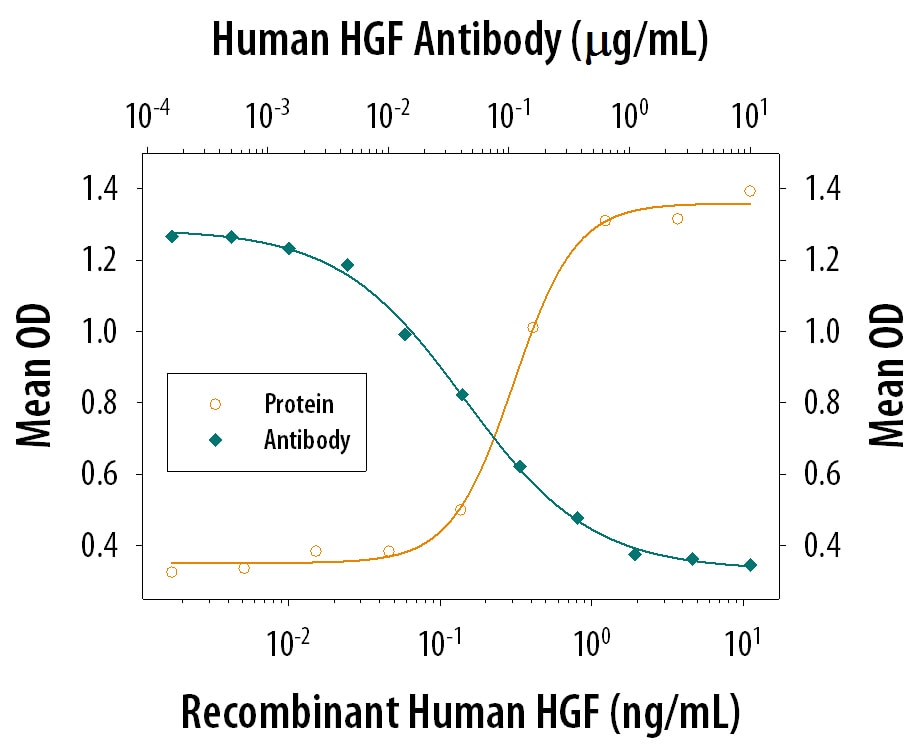
IL-11 Secretion Induced by HGF and Neutralization by Human HGF Antibody.
Recombinant Human HGF induces IL-11 secretion in the Saos-2 human osteosarcoma cell line. The dose-dependent response (orange line) is measured by the Human IL-11 Duoset (Catalog # DY218). Under these conditions, IL-11 secretion elicited by HGF is neutralized (green line) by increasing concentrations of Goat Anti-Human HGF Antigen Affinity-purified Polyclonal Antibod (Catalog # AF-294-NA). The ND50 is typically 0.01-0.06 µg/mL.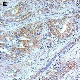
Detection of Mouse HGF by Immunohistochemistry-Paraffin
Characterization of CB42.A) Histology (H&E staining) of CB42 primary tumor. B). Histology (H&E) of CB42 after subcutaneous propagation (CB42P1). C). Immunohistochemical staining for beta -Catenin. D). Immunohistochemical staining for pan Cytokeratin. E). Immunohistochemical staining for HGF. F). Immunohistochemical staining for phospho-Met. Image collected and cropped by CiteAb from the following open publication (https://dx.plos.org/10.1371/journal.pone.0105886), licensed under a CC-BY license. Not internally tested by R&D Systems.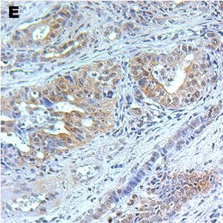
Detection of Mouse HGF by Immunohistochemistry-Paraffin
Characterization of CB42.A) Histology (H&E staining) of CB42 primary tumor. B). Histology (H&E) of CB42 after subcutaneous propagation (CB42P1). C). Immunohistochemical staining for beta -Catenin. D). Immunohistochemical staining for pan Cytokeratin. E). Immunohistochemical staining for HGF. F). Immunohistochemical staining for phospho-Met. Image collected and cropped by CiteAb from the following open publication (https://dx.plos.org/10.1371/journal.pone.0105886), licensed under a CC-BY license. Not internally tested by R&D Systems.
Human HGF ELISA Standard Curve
Recombinant Human HGF (Catalog # 294-HG) was serially diluted and captured by Mouse Anti-Human HGF Monoclonal Antibody (Catalog # MAB694) coated on a Clear Polystyrene Microplate (Catalog # DY990). Goat Anti-Human HGF Antigen Affinity-purified Polyclonal Antibody (Catalog # AF-294-NA) was biotinylated and incubated with the protein captured on the plate. Detection of the standard curve was achieved by incubating Streptavidin-HRP (Catalog # DY998)Applications for Human HGF Antibody
Immunohistochemistry
Sample: Immersion fixed paraffin-embedded sections of human liver
Western Blot
Sample: Recombinant Human HGF (Catalog # 294-HG)
Neutralization
Reviewed Applications
Read 1 review rated 5 using AF-294-NA in the following applications:
Formulation, Preparation, and Storage
Purification
Reconstitution
Reconstitute at 0.2 mg/mL in sterile PBS. For liquid material, refer to CoA for concentration.
Formulation
Shipping
Stability & Storage
- 12 months from date of receipt, -20 to -70 °C as supplied.
- 1 month, 2 to 8 °C under sterile conditions after reconstitution.
- 6 months, -20 to -70 °C under sterile conditions after reconstitution.
Calculators
Background: HGF
Long Name
Alternate Names
Entrez Gene IDs
Gene Symbol
UniProt
Additional HGF Products
Product Documents for Human HGF Antibody
Certificate of Analysis
To download a Certificate of Analysis, please enter a lot or batch number in the search box below.
Note: Certificate of Analysis not available for kit components.
Product Specific Notices for Human HGF Antibody
For research use only
Related Research Areas
Citations for Human HGF Antibody
 Powered by Bioz
Customer Reviews for Human HGF Antibody (1)
Have you used Human HGF Antibody?
Submit a review and receive an Amazon gift card!
$25/€18/£15/$25CAN/¥2500 for a review with an image
$10/€7/£6/$10CAN/¥1110 for a review without an image
Submit a review
Customer Images
-
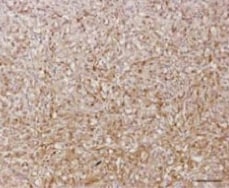
Application: ImmunohistochemistrySample Tested: Breast tissueSpecies: HumanVerified Customer | Posted 04/01/2021primary antibody concentration is 1 to 500 dilution.

There are no reviews that match your criteria.
Protocols
Find general support by application which include: protocols, troubleshooting, illustrated assays, videos and webinars.
- Antigen Retrieval Protocol (PIER)
- Antigen Retrieval for Frozen Sections Protocol
- Appropriate Fixation of IHC/ICC Samples
- Cellular Response to Hypoxia Protocols
- Chromogenic IHC Staining of Formalin-Fixed Paraffin-Embedded (FFPE) Tissue Protocol
- Chromogenic Immunohistochemistry Staining of Frozen Tissue
- ClariTSA™ Fluorophore Kits
- Detection & Visualization of Antibody Binding
- Fluorescent IHC Staining of Frozen Tissue Protocol
- Graphic Protocol for Heat-induced Epitope Retrieval
- Graphic Protocol for the Preparation and Fluorescent IHC Staining of Frozen Tissue Sections
- Graphic Protocol for the Preparation and Fluorescent IHC Staining of Paraffin-embedded Tissue Sections
- Graphic Protocol for the Preparation of Gelatin-coated Slides for Histological Tissue Sections
- IHC Sample Preparation (Frozen sections vs Paraffin)
- Immunofluorescent IHC Staining of Formalin-Fixed Paraffin-Embedded (FFPE) Tissue Protocol
- Immunohistochemistry (IHC) and Immunocytochemistry (ICC) Protocols
- Immunohistochemistry Frozen Troubleshooting
- Immunohistochemistry Paraffin Troubleshooting
- Preparing Samples for IHC/ICC Experiments
- Preventing Non-Specific Staining (Non-Specific Binding)
- Primary Antibody Selection & Optimization
- Protocol for Heat-Induced Epitope Retrieval (HIER)
- Protocol for Making a 4% Formaldehyde Solution in PBS
- Protocol for VisUCyte™ HRP Polymer Detection Reagent
- Protocol for the Preparation & Fixation of Cells on Coverslips
- Protocol for the Preparation and Chromogenic IHC Staining of Frozen Tissue Sections
- Protocol for the Preparation and Chromogenic IHC Staining of Frozen Tissue Sections - Graphic
- Protocol for the Preparation and Chromogenic IHC Staining of Paraffin-embedded Tissue Sections
- Protocol for the Preparation and Chromogenic IHC Staining of Paraffin-embedded Tissue Sections - Graphic
- Protocol for the Preparation and Fluorescent IHC Staining of Frozen Tissue Sections
- Protocol for the Preparation and Fluorescent IHC Staining of Paraffin-embedded Tissue Sections
- Protocol for the Preparation of Gelatin-coated Slides for Histological Tissue Sections
- R&D Systems Quality Control Western Blot Protocol
- TUNEL and Active Caspase-3 Detection by IHC/ICC Protocol
- The Importance of IHC/ICC Controls
- Troubleshooting Guide: Immunohistochemistry
- Troubleshooting Guide: Western Blot Figures
- Western Blot Conditions
- Western Blot Protocol
- Western Blot Protocol for Cell Lysates
- Western Blot Troubleshooting
- Western Blot Troubleshooting Guide
- View all Protocols, Troubleshooting, Illustrated assays and Webinars
Associated Pathways
