
Tie-2 (also known as TEK) is an angiogenic receptor tyrosine kinase required for the later stage of blood vessel maturation. Ligand binding induces receptor dimerization and autophosphorylation on multiple tyrosine residues. Y992 is located on the putative activation loop of Tie-2 and is a major autophosphorylation site.

Scientific images may be resized, cropped, or adjusted for brightness or contrast for presentation purposes. These updates are limited to presentation and do not affect the underlying scientific interpretation of the data. Where available, additional source data may be provided upon request.
Loading...
Key Product Details
Validated by
Biological Validation
Species Reactivity
Validated:
Human, Mouse
Cited:
Human, Mouse, Rat
Applications
Validated:
Immunohistochemistry, Western Blot, Intracellular Staining by Flow Cytometry, CyTOF-ready
Cited:
Immunohistochemistry, Western Blot, Immunocytochemistry, FACS
Label
Unconjugated
Antibody Source
Polyclonal Rabbit IgG
Loading...
Product Specifications
Immunogen
Phosphopeptide containing human Tie-2 Y992 site
Specificity
Detects human and mouse Tie-2 when phosphorylated at Y992 in Western blots.
Clonality
Polyclonal
Host
Rabbit
Isotype
IgG
Scientific Data Images for phospho-Tie-2 (Y992) Antibody
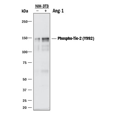
Detection of Mouse Phospho-Tie‑2 (Y992) by Western Blot.
Western blot shows lysates of NIH-3T3 mouse embryonic fibroblast cell line transfected with mouse Tie-2 and untreated (-) or treated (+) with 600 ng/mL Recombinant Human Angiopoietin-1 (Catalog # 923-AN) for 5 minutes. PVDF membrane was probed with 0.5 µg/mL of Rabbit Anti-Human/Mouse Phospho-Tie-2 (Y992) Antigen Affinity-purified Polyclonal Antibody (Catalog # AF2720) followed by HRP-conjugated Anti-Rabbit IgG Secondary Antibody (Catalog # HAF008). A specific band was detected for Phospho-Tie-2 (Y992) at approximately 150 kDa (as indicated). This experiment was conducted under reducing conditions and using Immunoblot Buffer Group 1.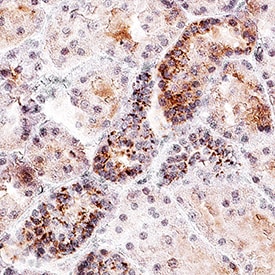
Phospho-Tie-2 (Y992) in Human Kidney Tissue.
Tie-2 was detected in immersion fixed paraffin-embedded sections of human kidney tissue using Rabbit Anti-Human/Mouse Phospho-Tie-2 (Y992) Antigen Affinity-purified Polyclonal Antibody (Catalog # AF2720) at 1 µg/mL overnight at 4 °C. Before incubation with the primary antibody, tissue was subjected to heat-induced epitope retrieval using Antigen Retrieval Reagent-Basic (Catalog # CTS013). Tissue was stained using the Anti-Rabbit IgG VisUCyte™ HRP Polymer Antibody (brown; Catalog # VC003) and counterstained with hematoxylin (blue). Specific staining was localized to cytoplasm in convoluted tubules. View our protocol for IHC Staining with VisUCyte HRP Polymer Detection Reagents.
Detection of Tie‑2 in HUVEC Human Cells by Flow Cytometry.
HUVEC human umbilical vein endothelial cells were unstimulated (light orange filled histogram) or treated with 100 µM pervanadate for 15 minutes (dark orange filled histogram), then stained with Human/Mouse Phospho- Tie-2 (Y992) Antigen Affinity-purified Polyclonal Antibody (Catalog # AF2720) or control antibody (Catalog # AB-105-C, open histogram), followed by Phycoerythrin-conjugated Anti-Rabbit IgG Secondary Antibody (Catalog # F0110). To facilitate intracellular staining, cells were fixed with paraformaldehyde and permeabilized with methanol.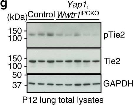
Detection of Mouse Tie-2 by Western Blot
Pericyte-derived Angpt1 controls alveologenesis. a RT-qPCR analysis of Angpt1 and Tie2/Tek expression in freshly sorted lung GFP+, CD31+ or EpCAM+ cells from P7 Pdgfrb(BAC)-CreERT2 R26-mT/mG mice. Data represents mean ± s.e.m. (n = 4 mice). b High magnification images of P10 Angpt1GFP lungs stained for GFP (green), PDGFR beta (red), and PDGFR alpha (blue). Arrows indicate GFP and PDGFR beta double positive pericytes. Scale bar, 15 µm. c RT-qPCR analysis of Angpt1 expression in freshly sorted PDGFR beta + cells from P7 Yap1,Wwtr1iPCKO and control lungs. Data represents mean ± s.e.m. (n = 4 mice, two-tailed unpaired t-test). dAngpt1 expression in cultured Verteporfin (VP)-treated (48 h) and control pericytes. Data represents mean ± s.e.m. (n = 4, Welch’s t-test). e Expression of the indicated transcripts in freshly sorted CD31+ cells from P7 Yap1,Wwtr1iPCKO and control lungs. Data represents mean ± s.e.m. (n = 4 mice, NS not significant, two-tailed unpaired t-test). f–h Western blot analysis of Angpt1 protein (f; n = 2 controls and 4 mutant mice) and of total and phospho-Tie2 (pTie2) in P12 Yap1,Wwtr1iPCKO and control total lung lysates (g, n = 3 controls and 5 mutants). Molecular weight marker (kDa) is indicated. Relative quantification of signals is shown in h. Two-tailed unpaired t-test. i Scheme showing the time points of tamoxifen administration and analysis for Angpt1iPCKO mice. j, k 3D reconstruction confocal images of P12 Angpt1iPCKO and littermate control lungs stained for AQP5 (green), PDGFR beta (red), and PECAM1 (blue). Panels in k show higher magnification of PECAM1 staining. Scale bar, 50 µm (j) and 30 µm (k). l Quantitation of airspace volume in P12 Angpt1iPCKO and littermate control lung sections with 3D reconstruction surface images. Data represents mean ± s.e.m. (n = 4 mice; p < 0.0001, two-tailed unpaired t-test). m 3D reconstruction confocal images of P12 Angpt1iPCKO and littermate control lungs stained for alpha SMA (red) and tropoelastin (blue). Scale bar, 50 µm. n Quantit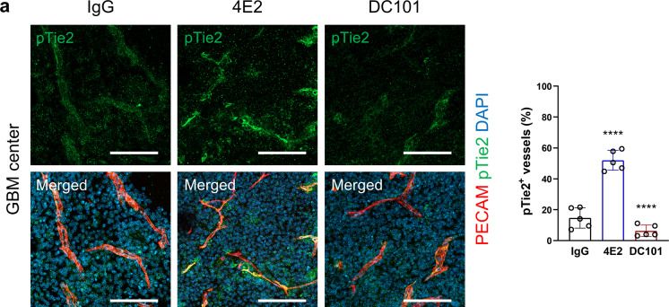
Detection of Phospho-Tie-2 (Y992) by Immunohistochemistry
Tie2 activation reduces the level of phosphorylated VEGFR2 in GBM vessels.a Increase in phospho-Tie2 in GBM vessels by 4E2 treatment, with quantification (n = 5 per group). b Decrease in phospho-VEGFR2 in GBM vessels by 4E2 or DC101 treatment, with quantification (n = 5 per group). c Decrease in phospho-Akt in GBM vessels by 4E2 or DC101 treatment, with quantification (n = 5 per group). Scale bars: 100 µm (a–c). ns not significant; ****, P < 0.0001 vs. IgG. Image collected and cropped by CiteAb from the following open publication (https://pubmed.ncbi.nlm.nih.gov/36828931), licensed under a CC-BY license. Not internally tested by R&D Systems.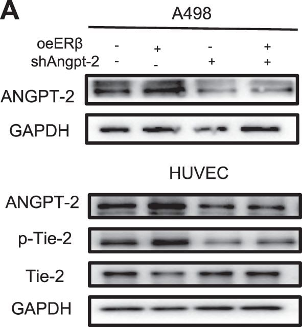
Detection of Phospho-Tie-2 (Y992) by Western Blot
ER beta signals could increase Angiopoietin-2 and up-regulate the Angiopoietin-2/Tie-2 axis.a Protein expressions of ANGPT-2 of RCC cells and ANGPT-2, Tie-2 and p-Tie-2 of HUVEC cells. Western blot assays were performed on A498 cells transfected with pWPI+pLKO, oeER beta + pLKO, pWPI+shANGPT-2, oeER beta + shANGPT-2 (left panels), and on 786-O cells transfected with pLKO+pWPI, shER beta + pWPI, pLKO+oeANGPT-2, shER beta + oeANGPT-2 (right panels). b–e Tube formation assays were performed using (b) A498 cells transfected with pWPI+pLKO, oeER beta + pLKO, pWPI+shANGPT-2, oeER beta + shANGPT-2, using (c) A498 cells transfected with pWPI+pLKO, oeER beta + pLKO, pWPI+shANGPT-2, oeER beta + shANGPT-2, and treated with1 μM Sunitinib, using (d) 786-O cells transfected with pLKO+pWPI, shER beta + pWPI, pLKO+oeANGPT-2, shER beta + oeANGPT-2 and (e) using 786-O cells transfected with pLKO+pWPI, shER beta + pWPI, pLKO+oeANGPT-2, shER beta + oeANGPT-2, and treated with1 μM sunitinib. For b–e, quantitation is at the right of the images. Data are presented as mean ± SD, *p < 0.05; **p < 0.01, compared with the controls, and Scale bar: 5 μm. Image collected and cropped by CiteAb from the following open publication (https://pubmed.ncbi.nlm.nih.gov/32409702), licensed under a CC-BY license. Not internally tested by R&D Systems.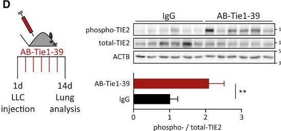
Detection of Phospho-Tie-2 (Y992) by Western Blot
AB‐Tie1‐39 treatment stabilizes lung vessels to restrict extravasation of disseminated tumor cellsAMice were pretreated with IgG or AB‐Tie1‐39. Thereafter, B16F10 cells were intravenously injected to initiate experimental metastasis independent of the primary tumor.BRepresentative images of the whole lungs are shown on the left. Scale bars = 5 mm. Quantitation of the number of lung metastatic foci is plotted on the right (mean ± SD, nIgG = 9, nAB‐Tie1‐39 = 8 mice). *P < 0.05 (two‐tailed Mann–Whitney U‐test).CECs were seeded on gelatin‐coated Transwell inserts. RFP‐labeled LLC cells were allowed to transmigrate for 8 h through the endothelial layer. Thereafter, transmigrated tumor cells were counted. The dot plot presents the normalized data (mean ± SD, n = 9 inserts from three independent experiments). ***P < 0.001 (two‐tailed Mann–Whitney U‐test).DLLC tumor‐bearing mice were pretreated with IgG or AB‐Tie1‐39. On day 14, lung tissue lysates were immunoblotted with anti‐phospho‐TIE2, total‐TIE2, and ACTB (upper panel; n = 6 mice). Densitometric quantitation of the blots presented in the upper panel is shown (mean ± SD, n = 6 mice). **P < 0.01 (two‐tailed Mann–Whitney U‐test).ELLC tumor‐bearing mice were pretreated with IgG or AB‐Tie1‐39. On day 14, lung ECs were FACS‐sorted and used for global gene expression (n = 4 mice). On the left, a heatmap shows significantly regulated genes (red outline = AB‐Tie1‐39‐treated; black outline = IgG‐treated). On the right, top significantly regulated bio‐functions in AB‐Tie1‐39‐treated as compared to IgG‐treated mice in ingenuity pathway analysis.Source data are available online for this figure. Image collected and cropped by CiteAb from the following open publication (https://pubmed.ncbi.nlm.nih.gov/32302470), licensed under a CC-BY license. Not internally tested by R&D Systems.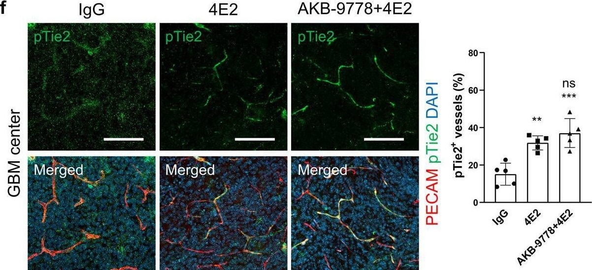
Detection of Phospho-Tie-2 (Y992) by Immunohistochemistry
Antibody-based Tie2 activation attenuates VEGFR2 phosphorylation via VE-PTP.a–c Immunoblots (a) showing VE-PTP-mediated VEGFR2 dephosphorylation by 4E2 and quantification of the phospho-VEGFR2 (b, n = 3 per group) and phospho-Tie2 (c, n = 3 per group) levels. AKB-9778, inhibitor of VE-PTP. d Quantification of VEGF-induced dextran leakage in HUVECs (n = 3 per group). e–g Immunostaining and quantification of phospho-VEGFR2 (e), phospho-Tie2 (f), and VE-cadherin (g) in GBM vessels cotreated with 4E2 and AKB-9778 (n = 5 per group). h Increased immunoprecipitation of VE-PTP with VEGFR2 but not Tie2 by 4E2. i, j Quantification of the normalized VEGFR2/VE-PTP (i, n = 3 per group) and normalized Tie2/VE-PTP (j, n = 3 per group) levels. k Schematic showing the regulatory mechanisms of tumor vascular normalization by 4E2. EC, endothelial cell; PC, pericyte; BM, basement membrane. Scale bars: 100 µm (e–g). ns, not significant; *, P < 0.05; **, P < 0.01; ***, P < 0.001; ****, P < 0.0001 vs. Ctrl (c, h-i) or IgG (e-g). ns not significant; #, P < 0.05; ##, P < 0.01 vs. VEGF (b, d) or 4E2 (e–g). Image collected and cropped by CiteAb from the following open publication (https://pubmed.ncbi.nlm.nih.gov/36828931), licensed under a CC-BY license. Not internally tested by R&D Systems.Applications for phospho-Tie-2 (Y992) Antibody
Application
Recommended Usage
CyTOF-ready
Ready to be labeled using established conjugation methods. No BSA or other carrier proteins that could interfere with conjugation.
Immunohistochemistry
1-15 µg/mL
Sample: Immersion fixed paraffin-embedded sections of human kidney tissue
Sample: Immersion fixed paraffin-embedded sections of human kidney tissue
Intracellular Staining by Flow Cytometry
0.25 µg/106 cells
Sample: HUVEC human umbilical vein endothelial cells treated with pervanadate, fixed with paraformaldehyde, and permeabilized with methanol
Sample: HUVEC human umbilical vein endothelial cells treated with pervanadate, fixed with paraformaldehyde, and permeabilized with methanol
Western Blot
0.5 µg/mL
Sample: NIH‑3T3 mouse embryonic fibroblast cell line transfected with mouse Tie-2 and treated Recombinant Human Angiopoietin‑1 (Catalog # 923-AN)
Sample: NIH‑3T3 mouse embryonic fibroblast cell line transfected with mouse Tie-2 and treated Recombinant Human Angiopoietin‑1 (Catalog # 923-AN)
Reviewed Applications
Read 3 reviews rated 3.7 using AF2720 in the following applications:
Flow Cytometry Panel Builder
Bio-Techne Knows Flow Cytometry
Save time and reduce costly mistakes by quickly finding compatible reagents using the Panel Builder Tool.
Advanced Features
- Spectra Viewer - Custom analysis of spectra from multiple fluorochromes
- Spillover Popups - Visualize the spectra of individual fluorochromes
- Antigen Density Selector - Match fluorochrome brightness with antigen density

Formulation, Preparation, and Storage
Purification
Antigen Affinity-purified
Reconstitution
Reconstitute at 0.2 mg/mL in sterile PBS. For liquid material, refer to CoA for concentration.
Loading...
Formulation
Lyophilized from a 0.2 μm filtered solution in PBS with Trehalose. *Small pack size (SP) is supplied either lyophilized or as a 0.2 µm filtered solution in PBS.
Shipping
Lyophilized product is shipped at ambient temperature. Liquid small pack size (-SP) is shipped with polar packs. Upon receipt, store immediately at the temperature recommended below.
Stability & Storage
Use a manual defrost freezer and avoid repeated freeze-thaw cycles.
- 12 months from date of receipt, -20 to -70 °C as supplied.
- 1 month, 2 to 8 °C under sterile conditions after reconstitution.
- 6 months, -20 to -70 °C under sterile conditions after reconstitution.
Calculators
Background: Tie-2
Long Name
Tyrosine Kinase with Immunoglobulin and Epidermal Growth Factor Homology Domains 2
Alternate Names
CD202b, TEK, Tie2
Entrez Gene IDs
Gene Symbol
TEK
Additional Tie-2 Products
Product Documents for phospho-Tie-2 (Y992) Antibody
Certificate of Analysis
To download a Certificate of Analysis, please enter a lot or batch number in the search box below.
Note: Certificate of Analysis not available for kit components.
Product Specific Notices for phospho-Tie-2 (Y992) Antibody
For research use only
Citations for phospho-Tie-2 (Y992) Antibody
 Powered by Bioz
Customer Reviews for phospho-Tie-2 (Y992) Antibody (3)
3.7 out of 5
3 Customer Ratings
Have you used phospho-Tie-2 (Y992) Antibody?
Submit a review and receive an Amazon gift card!
$25/€18/£15/$25CAN/¥2500 for a review with an image
$10/€7/£6/$10CAN/¥1110 for a review without an image
Submit a review
Customer Images
Showing
1
-
3 的
3 reviews
Showing All
Filter By:
-
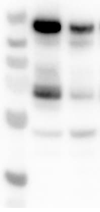
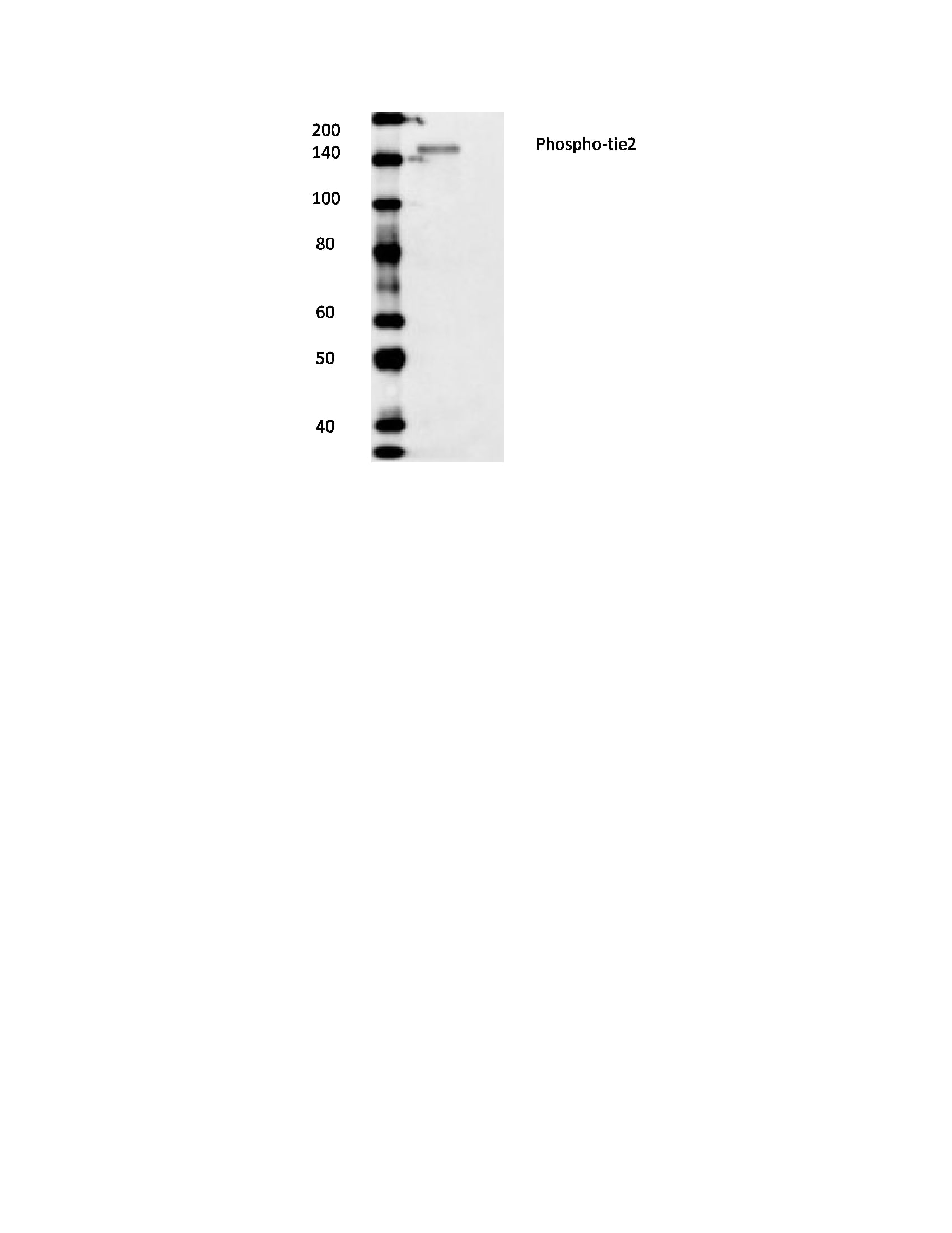
Application: Western BlotSample Tested: HUVEC human umbilical vein endothelial cellsSpecies: HumanVerified Customer | Posted 01/06/2026After trying several pTIE2 Y992 antibodies I have realized that this antibody works on HUVEC and its variant cells. It would usually give some other bands, but the top upper band at around 160 kDa usually corresponds to pTIE2 on my HUVEC/L914F cells.

-
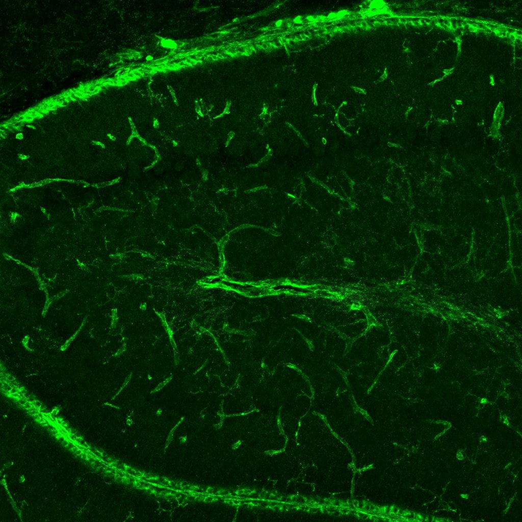
Application: Immunofluorescence in mouse brain tissueSample Tested: Cerebellum tissueSpecies: MouseVerified Customer | Posted 10/02/2020Immunofluorescence with phospho Tie2Postnatal mouse cerebellum at P14 was used to determine the levels of phospho-Tie2 in the blood vessels. The blocking buffer and the antibody buffers were prepared in 0.2% gelatin in PBST (In primary and secondary solutions were used 0.025% of Saponin was added). The sections were antigen retrieved by boiling in a microwave for 5 min using 10mM Sodium citrate (pH6).
-
Application: Western BlotSample Tested: HASMC human aortic smooth muscle cellsSpecies: HumanVerified Customer | Posted 10/17/2018
There are no reviews that match your criteria.
Protocols
Find general support by application which include: protocols, troubleshooting, illustrated assays, videos and webinars.
- 7-Amino Actinomycin D (7-AAD) Cell Viability Flow Cytometry Protocol
- Antigen Retrieval Protocol (PIER)
- Antigen Retrieval for Frozen Sections Protocol
- Appropriate Fixation of IHC/ICC Samples
- Cellular Response to Hypoxia Protocols
- Chromogenic IHC Staining of Formalin-Fixed Paraffin-Embedded (FFPE) Tissue Protocol
- Chromogenic Immunohistochemistry Staining of Frozen Tissue
- ClariTSA™ Fluorophore Kits
- Detection & Visualization of Antibody Binding
- Extracellular Membrane Flow Cytometry Protocol
- Flow Cytometry Protocol for Cell Surface Markers
- Flow Cytometry Protocol for Staining Membrane Associated Proteins
- Flow Cytometry Staining Protocols
- Flow Cytometry Troubleshooting Guide
- Fluorescent IHC Staining of Frozen Tissue Protocol
- Graphic Protocol for Heat-induced Epitope Retrieval
- Graphic Protocol for the Preparation and Fluorescent IHC Staining of Frozen Tissue Sections
- Graphic Protocol for the Preparation and Fluorescent IHC Staining of Paraffin-embedded Tissue Sections
- Graphic Protocol for the Preparation of Gelatin-coated Slides for Histological Tissue Sections
- IHC Sample Preparation (Frozen sections vs Paraffin)
- Immunofluorescent IHC Staining of Formalin-Fixed Paraffin-Embedded (FFPE) Tissue Protocol
- Immunohistochemistry (IHC) and Immunocytochemistry (ICC) Protocols
- Immunohistochemistry Frozen Troubleshooting
- Immunohistochemistry Paraffin Troubleshooting
- Intracellular Flow Cytometry Protocol Using Alcohol (Methanol)
- Intracellular Flow Cytometry Protocol Using Detergents
- Intracellular Nuclear Staining Flow Cytometry Protocol Using Detergents
- Intracellular Staining Flow Cytometry Protocol Using Alcohol Permeabilization
- Intracellular Staining Flow Cytometry Protocol Using Detergents to Permeabilize Cells
- Preparing Samples for IHC/ICC Experiments
- Preventing Non-Specific Staining (Non-Specific Binding)
- Primary Antibody Selection & Optimization
- Propidium Iodide Cell Viability Flow Cytometry Protocol
- Protocol for Heat-Induced Epitope Retrieval (HIER)
- Protocol for Liperfluo
- Protocol for Making a 4% Formaldehyde Solution in PBS
- Protocol for VisUCyte™ HRP Polymer Detection Reagent
- Protocol for the Characterization of Human Th22 Cells
- Protocol for the Characterization of Human Th9 Cells
- Protocol for the Preparation & Fixation of Cells on Coverslips
- Protocol for the Preparation and Chromogenic IHC Staining of Frozen Tissue Sections
- Protocol for the Preparation and Chromogenic IHC Staining of Frozen Tissue Sections - Graphic
- Protocol for the Preparation and Chromogenic IHC Staining of Paraffin-embedded Tissue Sections
- Protocol for the Preparation and Chromogenic IHC Staining of Paraffin-embedded Tissue Sections - Graphic
- Protocol for the Preparation and Fluorescent IHC Staining of Frozen Tissue Sections
- Protocol for the Preparation and Fluorescent IHC Staining of Paraffin-embedded Tissue Sections
- Protocol for the Preparation of Gelatin-coated Slides for Histological Tissue Sections
- Protocol: Annexin V and PI Staining by Flow Cytometry
- Protocol: Annexin V and PI Staining for Apoptosis by Flow Cytometry
- R&D Systems Quality Control Western Blot Protocol
- TUNEL and Active Caspase-3 Detection by IHC/ICC Protocol
- The Importance of IHC/ICC Controls
- Troubleshooting Guide: Fluorokine Flow Cytometry Kits
- Troubleshooting Guide: Immunohistochemistry
- Troubleshooting Guide: Western Blot Figures
- Western Blot Conditions
- Western Blot Protocol
- Western Blot Protocol for Cell Lysates
- Western Blot Troubleshooting
- Western Blot Troubleshooting Guide
- View all Protocols, Troubleshooting, Illustrated assays and Webinars
Loading...
Associated Pathways

