Sortilin (neurotensin receptor 3, glycoprotein 95) is a 95 kDa Type I transmembrane monomeric glycoprotein that is one of five known members of the mammalian vacuolar protein sorting 10p domain (Vps10p-D) family of sorting receptors (1, 2). Human preprosortilin is processed by signal sequence cleavage followed by propeptide cleavage at a furin recognition site. The cationic propeptide exhibits pH-dependent high affinity binding that blocks the Sortilin ligand binding site both pre‑ and post-cleavage (3). The extracellular/luminal sequence comprises the Vps10p domain, including 10 conserved cysteines (10 CC) essential for ligand binding (2). The cytoplasmic domain sorting motifs confer all trafficking during synthesis, targeting to lysosomes, endocytosis and Golgi-endosome transport; as little as 10% may be found on the cell surface (4). Mature human Sortilin shares 91% aa identity with mouse and rat Sortilin and 93% aa identity with dog. During murine development, sortilin is mainly expressed in the nervous system (5) where it is a receptor for neuropeptides including neurotensin, nerve growth factor (NGF) and brain‑derived neurotrophic factor (BDNF) (6-9). ProNGF (or the NGF propeptide alone) binds sortilin with much higher affinity (Kd ~5-8 nM) than does mature NGF (Kd ~90 nM). The complex of sortilin, pro-NGF and the receptor p75ntr results in endocytosis of proNGF and induction of apoptosis (7). Similar results have been obtained with pro-BDNF and BDNF (8, 9). Sortilin is expressed in other tissues including testis, skeletal muscle and fat (1, 10). It is essential and sufficient for biogenesis of Glut4 storage vesicles necessary for insulin responsiveness in adipocytes (10). Sortilin also binds lipoprotein lipase (11), apoE (2) and RAP (1, 11). Binding is competitive, indicating that although unrelated, targets likely bind the same site.

Key Product Details
Species Reactivity
Validated:
Human
Cited:
Human, Mouse, Primate, Transgenic Mouse
Applications
Validated:
Immunohistochemistry, Western Blot, Blockade of Receptor-ligand Interaction, Flow Cytometry, Simple Western
Cited:
Immunohistochemistry, Immunohistochemistry-Paraffin, Western Blot, Neutralization, Immunocytochemistry, Bioassay
Label
Unconjugated
Antibody Source
Polyclonal Goat IgG
Loading...
Product Specifications
Immunogen
Mouse myeloma cell line NS0-derived recombinant human Sortilin
Ser78-Asn755
Accession # Q99523
Ser78-Asn755
Accession # Q99523
Specificity
Detects human and mouse Sortilin in direct ELISAs and Western blots.
Clonality
Polyclonal
Host
Goat
Isotype
IgG
Endotoxin Level
<0.10 EU per 1 μg of the antibody by the LAL method.
Scientific Data Images for Human Sortilin Antibody
Detection of Human Sortilin by Western Blot.
Western blot shows lysates of human motor cortex. PVDF membrane was probed with 2 µg/mL of Goat Anti-Human Sortilin Antigen Affinity-purified Polyclonal Antibody (Catalog # AF3154) followed by HRP-conjugated Anti-Goat IgG Secondary Antibody (Catalog # HAF017). A specific band was detected for Sortilin at approximately 95-105 kDa (as indicated). This experiment was conducted under reducing conditions and using Immunoblot Buffer Group 1.
Detection of Sortilin in Human Blood Monocytes by Flow Cytometry.
Human peripheral blood monocytes were stained with Goat Anti-Human Sortilin Antigen Affinity-purified Polyclonal Antibody (Catalog # AF3154, filled histogram) or isotype control antibody (Catalog # AB-108-C, open histogram), followed by Allophycocyanin-conjugated Anti-Goat IgG Secondary Antibody (Catalog # F0108). View our protocol for Staining Membrane-associated Proteins.
Detection of Sortilin in K562 Human Cell Line by Flow Cytometry.
K562 human chronic myelogenous leukemia cell line was stained with Goat Anti-Human Sortilin Antigen Affinity-purified Polyclonal Antibody (Catalog # AF3154, filled histogram) or isotype control antibody (Catalog # AB-108-C, open histogram), followed by Allophycocyanin-conjugated Anti-Goat IgG Secondary Antibody (Catalog # F0108). View our protocol for Staining Membrane-associated Proteins.
Sortilin in Human Brain.
Sortilin was detected in immersion fixed paraffin-embedded sections of human brain using Goat Anti-Human Sortilin Antigen Affinity-purified Polyclonal Antibody (Catalog # AF3154) at 1.7 µg/mL overnight at 4 °C. Tissue was stained using the Anti-Goat HRP-DAB Cell & Tissue Staining Kit (brown; Catalog # CTS008) and counterstained with hematoxylin (blue). View our protocol for Chromogenic IHC Staining of Paraffin-embedded Tissue Sections.
Detection of Human Sortilin by Simple WesternTM.
Simple Western lane view shows lysates of human brain (cerebellum) tissue, loaded at 0.2 mg/mL. A specific band was detected for Sortilin at approximately 82 kDa (as indicated) using 10 µg/mL of Goat Anti-Human Sortilin Antigen Affinity-purified Polyclonal Antibody (Catalog # AF3154). This experiment was conducted under reducing conditions and using the 12-230 kDa separation system.Applications for Human Sortilin Antibody
Application
Recommended Usage
Blockade of Receptor-ligand Interaction
Flow Cytometry
0.25 µg/106 cells
Sample: Human peripheral blood monocytes and K562 human chronic myelogenous leukemia cell line
Sample: Human peripheral blood monocytes and K562 human chronic myelogenous leukemia cell line
Immunohistochemistry
5-15 µg/mL
Sample: Immersion fixed paraffin-embedded sections of human brain (cerebellum and cortex)
Sample: Immersion fixed paraffin-embedded sections of human brain (cerebellum and cortex)
Simple Western
10 µg/mL
Sample: Human brain (cerebellum) tissue
Sample: Human brain (cerebellum) tissue
Western Blot
2 µg/mL
Sample: Human motor cortex
Sample: Human motor cortex
Reviewed Applications
Read 2 reviews rated 4.5 using AF3154 in the following applications:
Flow Cytometry Panel Builder
Bio-Techne Knows Flow Cytometry
Save time and reduce costly mistakes by quickly finding compatible reagents using the Panel Builder Tool.
Advanced Features
- Spectra Viewer - Custom analysis of spectra from multiple fluorochromes
- Spillover Popups - Visualize the spectra of individual fluorochromes
- Antigen Density Selector - Match fluorochrome brightness with antigen density

Formulation, Preparation, and Storage
Purification
Antigen Affinity-purified
Reconstitution
Reconstitute at 0.2 mg/mL in sterile PBS. For liquid material, refer to CoA for concentration.
Loading...
Formulation
Lyophilized from a 0.2 μm filtered solution in PBS with Trehalose. *Small pack size (SP) is supplied either lyophilized or as a 0.2 µm filtered solution in PBS.
Shipping
Lyophilized product is shipped at ambient temperature. Liquid small pack size (-SP) is shipped with polar packs. Upon receipt, store immediately at the temperature recommended below.
Stability & Storage
Use a manual defrost freezer and avoid repeated freeze-thaw cycles.
- 12 months from date of receipt, -20 to -70 °C as supplied.
- 1 month, 2 to 8 °C under sterile conditions after reconstitution.
- 6 months, -20 to -70 °C under sterile conditions after reconstitution.
Calculators
Background: Sortilin
References
- Petersen, C.M. et al. (1997) J. Biol. Chem. 272:3599.
- Westergaard, U.B. et al. (2004) J. Biol. Chem. 279:50221.
- Petersen, C.M. et al. (1998) EMBO J. 18:595.
- Nielsen, M.S. et al. (2001) EMBO J. 20:2180.
- Hermans-Borgmeyer, I. et al. (1999) Mol. Brain Res. 65:216.
- Mazella, J. et al. (1998) J. Biol. Chem. 273:26273.
- Nykjaer, A. et al. (2004) Nature 427:843.
- Teng, H.K. et al. (2005) J. Neurosci. 25:5455.
- Chen, Z-Y. et al. (2004) J. Neurosci. 25:6156.
- Shi, J. and K.V. Kandror (2005) Dev. Cell 9:99.
- Nielsen, M.S. et al. (1999) J. Biol. Chem. 274:8832.
Alternate Names
Gp95, Ntr3, SORT1
Gene Symbol
SORT1
UniProt
Additional Sortilin Products
Product Documents for Human Sortilin Antibody
Certificate of Analysis
To download a Certificate of Analysis, please enter a lot or batch number in the search box below.
Note: Certificate of Analysis not available for kit components.
Product Specific Notices for Human Sortilin Antibody
For research use only
Related Research Areas
Citations for Human Sortilin Antibody
 Powered by Bioz
Customer Reviews for Human Sortilin Antibody (2)
4.5 out of 5
2 Customer Ratings
Have you used Human Sortilin Antibody?
Submit a review and receive an Amazon gift card!
$25/€18/£15/$25CAN/¥2500 Yen for a review with an image
$10/€7/£6/$10CAN/¥1110 Yen for a review without an image
Submit a review
Customer Images
Showing
1
-
2 的
2 reviews
Showing All
Filter By:
-
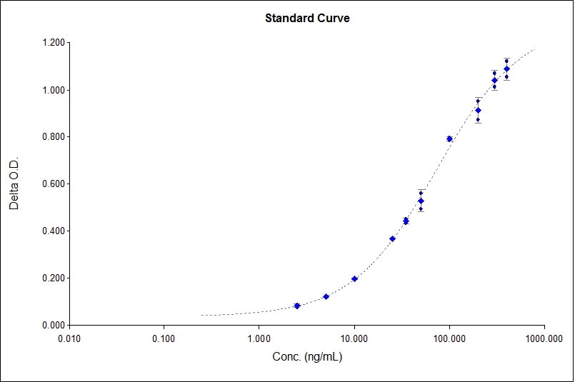
Application: ELISASample Tested: SerumSpecies: HumanVerified Customer | Posted 04/05/2022

-
Application: ImmunofluorescenceSample Tested: See PMID 22579764Species: OtherVerified Customer | Posted 01/07/2015
There are no reviews that match your criteria.
Protocols
Find general support by application which include: protocols, troubleshooting, illustrated assays, videos and webinars.
- 7-Amino Actinomycin D (7-AAD) Cell Viability Flow Cytometry Protocol
- Antigen Retrieval Protocol (PIER)
- Antigen Retrieval for Frozen Sections Protocol
- Appropriate Fixation of IHC/ICC Samples
- Cellular Response to Hypoxia Protocols
- Chromogenic IHC Staining of Formalin-Fixed Paraffin-Embedded (FFPE) Tissue Protocol
- Chromogenic Immunohistochemistry Staining of Frozen Tissue
- ClariTSA™ Fluorophore Kits
- Detection & Visualization of Antibody Binding
- Extracellular Membrane Flow Cytometry Protocol
- Flow Cytometry Protocol for Cell Surface Markers
- Flow Cytometry Protocol for Staining Membrane Associated Proteins
- Flow Cytometry Staining Protocols
- Flow Cytometry Troubleshooting Guide
- Fluorescent IHC Staining of Frozen Tissue Protocol
- Graphic Protocol for Heat-induced Epitope Retrieval
- Graphic Protocol for the Preparation and Fluorescent IHC Staining of Frozen Tissue Sections
- Graphic Protocol for the Preparation and Fluorescent IHC Staining of Paraffin-embedded Tissue Sections
- Graphic Protocol for the Preparation of Gelatin-coated Slides for Histological Tissue Sections
- IHC Sample Preparation (Frozen sections vs Paraffin)
- Immunofluorescent IHC Staining of Formalin-Fixed Paraffin-Embedded (FFPE) Tissue Protocol
- Immunohistochemistry (IHC) and Immunocytochemistry (ICC) Protocols
- Immunohistochemistry Frozen Troubleshooting
- Immunohistochemistry Paraffin Troubleshooting
- Intracellular Flow Cytometry Protocol Using Alcohol (Methanol)
- Intracellular Flow Cytometry Protocol Using Detergents
- Intracellular Nuclear Staining Flow Cytometry Protocol Using Detergents
- Intracellular Staining Flow Cytometry Protocol Using Alcohol Permeabilization
- Intracellular Staining Flow Cytometry Protocol Using Detergents to Permeabilize Cells
- Preparing Samples for IHC/ICC Experiments
- Preventing Non-Specific Staining (Non-Specific Binding)
- Primary Antibody Selection & Optimization
- Propidium Iodide Cell Viability Flow Cytometry Protocol
- Protocol for Heat-Induced Epitope Retrieval (HIER)
- Protocol for Liperfluo
- Protocol for Making a 4% Formaldehyde Solution in PBS
- Protocol for VisUCyte™ HRP Polymer Detection Reagent
- Protocol for the Characterization of Human Th22 Cells
- Protocol for the Characterization of Human Th9 Cells
- Protocol for the Preparation & Fixation of Cells on Coverslips
- Protocol for the Preparation and Chromogenic IHC Staining of Frozen Tissue Sections
- Protocol for the Preparation and Chromogenic IHC Staining of Frozen Tissue Sections - Graphic
- Protocol for the Preparation and Chromogenic IHC Staining of Paraffin-embedded Tissue Sections
- Protocol for the Preparation and Chromogenic IHC Staining of Paraffin-embedded Tissue Sections - Graphic
- Protocol for the Preparation and Fluorescent IHC Staining of Frozen Tissue Sections
- Protocol for the Preparation and Fluorescent IHC Staining of Paraffin-embedded Tissue Sections
- Protocol for the Preparation of Gelatin-coated Slides for Histological Tissue Sections
- Protocol: Annexin V and PI Staining by Flow Cytometry
- Protocol: Annexin V and PI Staining for Apoptosis by Flow Cytometry
- R&D Systems Quality Control Western Blot Protocol
- TUNEL and Active Caspase-3 Detection by IHC/ICC Protocol
- The Importance of IHC/ICC Controls
- Troubleshooting Guide: Fluorokine Flow Cytometry Kits
- Troubleshooting Guide: Immunohistochemistry
- Troubleshooting Guide: Western Blot Figures
- Western Blot Conditions
- Western Blot Protocol
- Western Blot Protocol for Cell Lysates
- Western Blot Troubleshooting
- Western Blot Troubleshooting Guide
- View all Protocols, Troubleshooting, Illustrated assays and Webinars
Loading...