Mouse CD68/SR-D1 Antibody (2449D)
R&D Systems | Catalog # MAB101141
Recombinant Monoclonal Antibody.

Key Product Details
Species Reactivity
Validated:
Mouse
Cited:
Human, Mouse, Transgenic Mouse
Applications
Validated:
Multiplex Immunofluorescence, Immunohistochemistry, Intracellular Staining by Flow Cytometry, COMET, CyTOF-ready
Cited:
Immunohistochemistry, In Situ Hybridization, CyTOF-reported
Label
Unconjugated
Antibody Source
Recombinant Monoclonal Rabbit IgG Clone # 2449D
Loading...
Product Specifications
Immunogen
Chinese Hamster Ovary cell line, CHO-derived mouse CD68
Asp21-Pro282
Accession # P31996
Asp21-Pro282
Accession # P31996
Specificity
Detects mouse CD68 in direct ELISAs.
Clonality
Monoclonal
Host
Rabbit
Isotype
IgG
Scientific Data Images for Mouse CD68/SR-D1 Antibody (2449D)
Detection of CD68 in Mouse Liver via seqIF™ staining on COMET™
CD68 was detected in immersion fixed paraffin-embedded sections of mouse Liver using Rabbit Anti-Mouse CD68, Monoclonal Antibody (Catalog #MAB101141) at 5ug/mL at 37 ° Celsius for 4 minutes. Before incubation with the primary antibody, tissue underwent an all-in-one dewaxing and antigen retrieval preprocessing using PreTreatment Module (PT Module) and Dewax and HIER Buffer H (pH 9; Epredia Catalog # TA-999-DHBH). Tissue was stained using the Alexa Fluor™ Plus 555 Goat anti-Rabbit IgG Secondary Antibody at 1:100 at 37 ° Celsius for 2 minutes. (Yellow; Lunaphore Catalog # DR555RB) and counterstained with DAPI (blue; Lunaphore Catalog # DR100). Specific staining was localized to the cytoplasm of Kupffer cells. Protocol available in COMET™ Panel Builder.
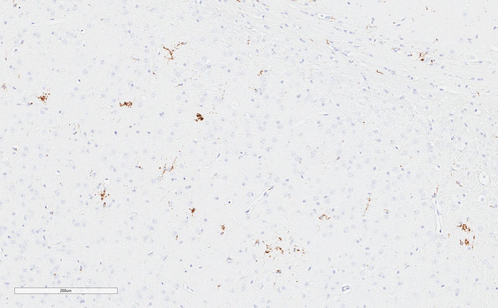
Detection of CD68 in Mouse Brain Medulla via seqIF™ staining on COMET™
CD68 was detected in perfusion fixed-frozen sections of mouse Brain Medulla using Rabbit Anti-Mouse CD68, Monoclonal Antibody (Catalog#MAB101141) at 20ug/mL at 27° Celsius for 8minutes. Before incubation with the primary antibody, tissue underwent preprocessing by incubating tissue with Multi Staining Buffer (Lunaphore Catalog # BU06) for 5 minutes at room temperature followed by a 20-minute incubation in Tris-Buffered Saline + 0.2% Triton at room temperature. Tissue was stained using the Alexa Fluor™ Plus 647 Goat anti-Rabbit IgG Secondary Antibody at 1:200 at 37 ° Celsius for 2 minutes. (Yellow; Lunaphore Catalog # DR647RB) and counterstained with DAPI (blue; Lunaphore Catalog # DR100). Specific staining was localized to the cytoplasm. Protocol available in COMET™ Panel Builder.
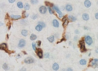
CD68/SR‑D1 in Mouse Liver.
CD68/SR-D1 was detected in immersion fixed paraffin-embedded sections of mouse liver using Rabbit Anti-Mouse CD68/SR-D1 Monoclonal Antibody (Catalog # MAB101141) at 3 µg/mL for 1 hour at room temperature followed by incubation with the Anti-Rabbit IgG VisUCyte™ HRP Polymer Antibody (Catalog # VC003). Before incubation with the primary antibody, tissue was subjected to heat-induced epitope retrieval using Antigen Retrieval Reagent-Basic (Catalog # CTS013). Tissue was stained using DAB (brown) and counterstained with hematoxylin (blue). Specific staining was localized to Kupffer cells. View our protocol for IHC Staining with VisUCyte HRP Polymer Detection Reagents.
Detection of CD68 in Raw264.7 Mouse Cell line by Flow Cytometry.
Raw264.7 mouse cell line was stained with Rabbit Anti-Mouse CD68 Monoclonal Antibody (Catalog # MAB101141, filled histogram) or Normal Rabbit IgG Control (Catalog # MAB1050, open histogram) followed by Phycoerythrin-conjugated Anti-Rabbit IgG Secondary Antibody (Catalog # F0110). To facilitate intracellular staining, cells were fixed with Flow Cytometry Fixation Buffer (1x) (Catalog # FC004) and permeabilized with ice-cold methanol.Applications for Mouse CD68/SR-D1 Antibody (2449D)
Application
Recommended Usage
COMET
Optimal dilutions of this antibody should be experimentally determined.
CyTOF-ready
Ready to be labeled using established conjugation methods. No BSA or other carrier proteins that could interfere with conjugation.
Immunohistochemistry
3-25 µg/mL
Sample: Immersion fixed paraffin-embedded sections of mouse liver
Sample: Immersion fixed paraffin-embedded sections of mouse liver
Intracellular Staining by Flow Cytometry
0.25 µg/106 cells
Sample: RAW264.7 mouse monocyte/macrophage cell line fixed with Flow Cytometry Fixation Buffer (Catalog # FC004) and permeabilized with ice-cold methanol
Sample: RAW264.7 mouse monocyte/macrophage cell line fixed with Flow Cytometry Fixation Buffer (Catalog # FC004) and permeabilized with ice-cold methanol
Multiplex Immunofluorescence
5-20 µg/mL
Sample: Immersion fixed paraffin-embedded sections of mouse liver and perfusion fixed-frozen sections of mouse brain medulla
Sample: Immersion fixed paraffin-embedded sections of mouse liver and perfusion fixed-frozen sections of mouse brain medulla
Reviewed Applications
Read 3 reviews rated 5 using MAB101141 in the following applications:
Flow Cytometry Panel Builder
Bio-Techne Knows Flow Cytometry
Save time and reduce costly mistakes by quickly finding compatible reagents using the Panel Builder Tool.
Advanced Features
- Spectra Viewer - Custom analysis of spectra from multiple fluorochromes
- Spillover Popups - Visualize the spectra of individual fluorochromes
- Antigen Density Selector - Match fluorochrome brightness with antigen density

Formulation, Preparation, and Storage
Purification
Protein A or G purified from cell culture supernatant
Reconstitution
Reconstitute at 0.5 mg/mL in sterile PBS. For liquid material, refer to CoA for concentration.
Loading...
Formulation
Lyophilized from a 0.2 μm filtered solution in PBS with Trehalose. See Certificate of Analysis for details.
*Small pack size (-SP) is supplied either lyophilized or as a 0.2 µm filtered solution in PBS.
*Small pack size (-SP) is supplied either lyophilized or as a 0.2 µm filtered solution in PBS.
Shipping
Lyophilized product is shipped at ambient temperature. Liquid small pack size (-SP) is shipped with polar packs. Upon receipt, store immediately at the temperature recommended below.
Stability & Storage
Use a manual defrost freezer and avoid repeated freeze-thaw cycles.
- 12 months from date of receipt, -20 to -70 °C as supplied.
- 1 month, 2 to 8 °C under sterile conditions after reconstitution.
- 6 months, -20 to -70 °C under sterile conditions after reconstitution.
Calculators
Background: CD68/SR-D1
Alternate Names
CD68, gp110, Macrosialin, SCARD1, SR-D1, SRD1
Gene Symbol
CD68
UniProt
Additional CD68/SR-D1 Products
Product Documents for Mouse CD68/SR-D1 Antibody (2449D)
Certificate of Analysis
To download a Certificate of Analysis, please enter a lot or batch number in the search box below.
Note: Certificate of Analysis not available for kit components.
Product Specific Notices for Mouse CD68/SR-D1 Antibody (2449D)
For research use only
Citations for Mouse CD68/SR-D1 Antibody (2449D)
 Powered by Bioz
Customer Reviews for Mouse CD68/SR-D1 Antibody (2449D) (3)
5 out of 5
3 Customer Ratings
Have you used Mouse CD68/SR-D1 Antibody (2449D)?
Submit a review and receive an Amazon gift card!
$25/€18/£15/$25CAN/¥2500 Yen for a review with an image
$10/€7/£6/$10CAN/¥1110 Yen for a review without an image
Submit a review
Customer Images

Showing
1
-
3 的
3 reviews
Showing All
Filter By:
-
Application: ImmunohistochemistrySample Tested: Brain (cerebral cortex)Species: MouseVerified Customer | Posted 06/23/2022Deparaffin; Citrate acid (6.0) 30min while steaming; 1:100. Works very well! :)

-
Application: ImmunohistochemistrySample Tested: Liver tissueSpecies: MouseVerified Customer | Posted 03/09/2022
-
Application: ImmunohistochemistrySample Tested: mouse oral tissueSpecies: MouseVerified Customer | Posted 11/23/2020
There are no reviews that match your criteria.
Protocols
Find general support by application which include: protocols, troubleshooting, illustrated assays, videos and webinars.
- 7-Amino Actinomycin D (7-AAD) Cell Viability Flow Cytometry Protocol
- Antigen Retrieval Protocol (PIER)
- Antigen Retrieval for Frozen Sections Protocol
- Appropriate Fixation of IHC/ICC Samples
- Cellular Response to Hypoxia Protocols
- Chromogenic IHC Staining of Formalin-Fixed Paraffin-Embedded (FFPE) Tissue Protocol
- Chromogenic Immunohistochemistry Staining of Frozen Tissue
- ClariTSA™ Fluorophore Kits
- Detection & Visualization of Antibody Binding
- Extracellular Membrane Flow Cytometry Protocol
- Flow Cytometry Protocol for Cell Surface Markers
- Flow Cytometry Protocol for Staining Membrane Associated Proteins
- Flow Cytometry Staining Protocols
- Flow Cytometry Troubleshooting Guide
- Fluorescent IHC Staining of Frozen Tissue Protocol
- Graphic Protocol for Heat-induced Epitope Retrieval
- Graphic Protocol for the Preparation and Fluorescent IHC Staining of Frozen Tissue Sections
- Graphic Protocol for the Preparation and Fluorescent IHC Staining of Paraffin-embedded Tissue Sections
- Graphic Protocol for the Preparation of Gelatin-coated Slides for Histological Tissue Sections
- IHC Sample Preparation (Frozen sections vs Paraffin)
- Immunofluorescent IHC Staining of Formalin-Fixed Paraffin-Embedded (FFPE) Tissue Protocol
- Immunohistochemistry (IHC) and Immunocytochemistry (ICC) Protocols
- Immunohistochemistry Frozen Troubleshooting
- Immunohistochemistry Paraffin Troubleshooting
- Intracellular Flow Cytometry Protocol Using Alcohol (Methanol)
- Intracellular Flow Cytometry Protocol Using Detergents
- Intracellular Nuclear Staining Flow Cytometry Protocol Using Detergents
- Intracellular Staining Flow Cytometry Protocol Using Alcohol Permeabilization
- Intracellular Staining Flow Cytometry Protocol Using Detergents to Permeabilize Cells
- Preparing Samples for IHC/ICC Experiments
- Preventing Non-Specific Staining (Non-Specific Binding)
- Primary Antibody Selection & Optimization
- Propidium Iodide Cell Viability Flow Cytometry Protocol
- Protocol for Heat-Induced Epitope Retrieval (HIER)
- Protocol for Liperfluo
- Protocol for Making a 4% Formaldehyde Solution in PBS
- Protocol for VisUCyte™ HRP Polymer Detection Reagent
- Protocol for the Characterization of Human Th22 Cells
- Protocol for the Characterization of Human Th9 Cells
- Protocol for the Preparation & Fixation of Cells on Coverslips
- Protocol for the Preparation and Chromogenic IHC Staining of Frozen Tissue Sections
- Protocol for the Preparation and Chromogenic IHC Staining of Frozen Tissue Sections - Graphic
- Protocol for the Preparation and Chromogenic IHC Staining of Paraffin-embedded Tissue Sections
- Protocol for the Preparation and Chromogenic IHC Staining of Paraffin-embedded Tissue Sections - Graphic
- Protocol for the Preparation and Fluorescent IHC Staining of Frozen Tissue Sections
- Protocol for the Preparation and Fluorescent IHC Staining of Paraffin-embedded Tissue Sections
- Protocol for the Preparation of Gelatin-coated Slides for Histological Tissue Sections
- Protocol: Annexin V and PI Staining by Flow Cytometry
- Protocol: Annexin V and PI Staining for Apoptosis by Flow Cytometry
- TUNEL and Active Caspase-3 Detection by IHC/ICC Protocol
- The Importance of IHC/ICC Controls
- Troubleshooting Guide: Fluorokine Flow Cytometry Kits
- Troubleshooting Guide: Immunohistochemistry
- View all Protocols, Troubleshooting, Illustrated assays and Webinars
Loading...
Associated Pathways
