
The EGFR subfamily of receptor tyrosine kinases comprises four members: EGFR (also known as Her1, ErbB1, or ErbB), ErbB2 (Neu, Her2), ErbB3 (Her3), and ErbB4 (Her4). All family members are type I transmembrane glycoproteins. They contain an extracellular ligand binding domain containing two cysteine-rich domains and a cytoplasmic domain containing a membrane-proximal tyrosine kinase domain followed by multiple tyrosine autophosphorylation sites (1, 2). The mouse EGFR cDNA encodes a 1210 amino acid (aa) precursor with a 24 aa signal peptide, a 623 aa extracellular domain (ECD), a 23 aa transmembrane segment, and a 540 aa cytoplasmic domain (3). Soluble receptors consisting of the extracellular ligand binding domain are generated by alternate splicing in human and mouse (4-6). Within the ECD, mouse EGFR shares 88% and 93% aa sequence identity with human and rat EGFR, respectively. It shares 44-48% aa sequence identity with the ECD of mouse ErbB2, ErbB3, and ErbB4. EGFR binds a subset of the EGF family ligands, including EGF, amphiregulin, TGF-alpha, betacellulin, epiregulin, HB-EGF, and epigen (1, 2). Ligand binding induces EGFR homodimerization as well as heterodimerization with ErbB2, resulting in kinase activation, heterodimerization tyrosine phosphorylation and cell signaling (7-11). EGFR can also be recruited to form heterodimers with the ligand-activated ErbB3 or ErbB4. EGFR signaling regulates multiple biological functions including cell proliferation, differentiation, motility, and apoptosis (12, 13). EGFR is over-expressed in a wide variety of tumors and is the target of several anti-cancer drugs (14).

Scientific images may be resized, cropped, or adjusted for brightness or contrast for presentation purposes. These updates are limited to presentation and do not affect the underlying scientific interpretation of the data. Where available, additional source data may be provided upon request.
Key Product Details
Species Reactivity
Validated:
Cited:
Applications
Validated:
Cited:
Label
Antibody Source
Product Specifications
Immunogen
Leu25-Ser647
Accession # Q01279
Specificity
Clonality
Host
Isotype
Scientific Data Images for Mouse EGFR Antibody [Biotin]
EGFR in Mouse Embryo.
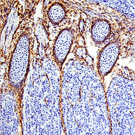
EGFR was detected in immersion fixed frozen sections of mouse embryo (13 d.p.c.) using Goat Anti-Mouse EGFR Biotinylated Antigen Affinity-purified Polyclonal Antibody (Catalog # BAF1280) at 15 µg/mL overnight at 4 °C. Tissue was stained using the Anti-Goat HRP-DAB Cell & Tissue Staining Kit (brown; Catalog # CTS008) and counterstained with hematoxylin (blue). Specific staining was localized to developing muscle. View our protocol for Chromogenic IHC Staining of Frozen Tissue Sections.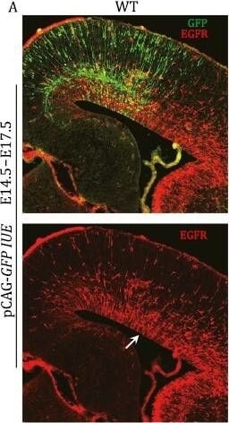
Detection of Mouse EGFR by Immunocytochemistry/ Immunofluorescence
BMP7-SMAD signaling inhibits EGFR expression. (A and B) IUE of vectors that express either GFP (control) or Bmp7-GFP in E14.5 WT mouse cortex. At E17.5, EGFR expression was significantly downregulated and pSMAD1/5/9 expression was strongly upregulated in the Bmp7-IUE cortex (arrows). (C) In the hGFAP-Cre;Smad4F/F cortex, Bmp7-IUE showed less EGFR inhibition. (D) Quantification of numbers of EGFR-expressing cells in the cortex. (E) Bmp7 was overexpressed in the E14.5 or E15.5 cortex using IUE. The cortical GLI3R to GLI3FL ratio was significantly increased 48 h later (see Western blot in Fig. S8). Image collected and cropped by CiteAb from the following open publication (https://academic.oup.com/proteincell/article/15/1/21/7193717), licensed under a CC-BY license. Not internally tested by R&D Systems.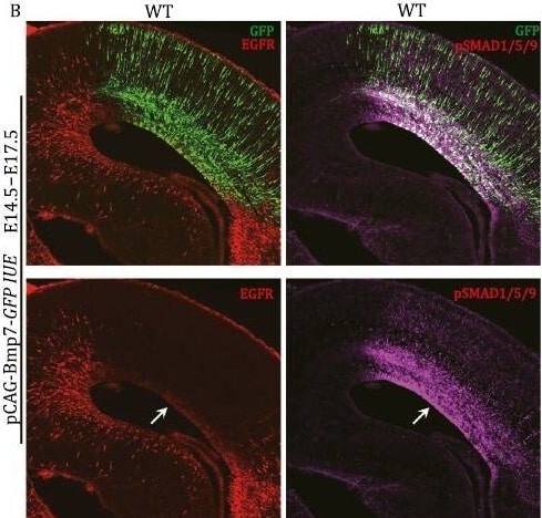
Detection of Mouse EGFR by Immunocytochemistry/ Immunofluorescence
BMP7-SMAD signaling inhibits EGFR expression. (A and B) IUE of vectors that express either GFP (control) or Bmp7-GFP in E14.5 WT mouse cortex. At E17.5, EGFR expression was significantly downregulated and pSMAD1/5/9 expression was strongly upregulated in the Bmp7-IUE cortex (arrows). (C) In the hGFAP-Cre;Smad4F/F cortex, Bmp7-IUE showed less EGFR inhibition. (D) Quantification of numbers of EGFR-expressing cells in the cortex. (E) Bmp7 was overexpressed in the E14.5 or E15.5 cortex using IUE. The cortical GLI3R to GLI3FL ratio was significantly increased 48 h later (see Western blot in Fig. S8). Image collected and cropped by CiteAb from the following open publication (https://academic.oup.com/proteincell/article/15/1/21/7193717), licensed under a CC-BY license. Not internally tested by R&D Systems.Applications for Mouse EGFR Antibody [Biotin]
Immunohistochemistry
Sample: Immersion fixed frozen sections of mouse embryo (13 d.p.c.)
Western Blot
Sample: Recombinant Mouse EGFR Fc Chimera (Catalog # 1280-ER)
Reviewed Applications
Read 1 review rated 4 using BAF1280 in the following applications:
Formulation, Preparation, and Storage
Purification
Reconstitution
Reconstitute at 0.2 mg/mL in sterile PBS.
Formulation
Shipping
Stability & Storage
- 12 months from date of receipt, -20 to -70 °C as supplied.
- 1 month, 2 to 8 °C under sterile conditions after reconstitution.
- 6 months, -20 to -70 °C under sterile conditions after reconstitution.
Calculators
Background: EGFR
References
- Singh, A.B. and R.C. Harris (2005) Cell. Signal. 17:1183.
- Shilo, B.Z. (2005) Development 132:4017.
- Avivi, A. et al. (1991) Oncogene 6:673.
- Reiter, J.L. and N.J. Maihle (1996) Nucleic Acids Res. 24:4050.
- Reiter J.L. et al. (2001) Genomics 71:1.
- Xu, Y.H. et al. (1984) Nature 309:806.
- Graus-Porta, D. et al. (1997) EMBO J. 16:1647.
- Yarden, Y. et al. (1987) Biochemistry 26:1434.
- Burgess, A.W. et al. (2003) Mol. Cell 12:541.
- Lemmon, M.A. et al. (1997) EMBO J. 16:281.
- Cohen, S. et al. (1982) J. Biol. Chem. 257:1523.
- Sibilia, M. and E.F. Wagner (1995) Science 269:234.
- Miettinen, P.J. et al. (1995) Nature 376:337.
- Roskoski Jr., R. (2004) Biochem. Biophys. Res. Commun. 319:1.
Long Name
Alternate Names
Gene Symbol
UniProt
Additional EGFR Products
Product Documents for Mouse EGFR Antibody [Biotin]
Certificate of Analysis
To download a Certificate of Analysis, please enter a lot or batch number in the search box below.
Note: Certificate of Analysis not available for kit components.
Product Specific Notices for Mouse EGFR Antibody [Biotin]
For research use only
Related Research Areas
Citations for Mouse EGFR Antibody [Biotin]
 Powered by Bioz
Customer Reviews for Mouse EGFR Antibody [Biotin] (1)
Have you used Mouse EGFR Antibody [Biotin]?
Submit a review and receive an Amazon gift card!
$25/€18/£15/$25CAN/¥2500 for a review with an image
$10/€7/£6/$10CAN/¥1110 for a review without an image
Submit a review
Customer Images
![Mouse EGFR Antibody [Biotin] BAF1280](https://resources.rndsystems.com/images/reviews/review_baf1280_926.png)
-
Application: Flow CytometrySample Tested: Neural progenitor cellsSpecies: MouseVerified Customer | Posted 02/23/2016I used this antibody with 1:200 dilution (each sample included 10x106 cells).
![Mouse EGFR Antibody [Biotin] BAF1280](data:image/png;base64,R0lGODlhAQABAAD/ACwAAAAAAQABAAACADs=)
There are no reviews that match your criteria.
Protocols
Find general support by application which include: protocols, troubleshooting, illustrated assays, videos and webinars.
- Antigen Retrieval Protocol (PIER)
- Antigen Retrieval for Frozen Sections Protocol
- Appropriate Fixation of IHC/ICC Samples
- Cellular Response to Hypoxia Protocols
- Chromogenic IHC Staining of Formalin-Fixed Paraffin-Embedded (FFPE) Tissue Protocol
- Chromogenic Immunohistochemistry Staining of Frozen Tissue
- ClariTSA™ Fluorophore Kits
- Detection & Visualization of Antibody Binding
- Fluorescent IHC Staining of Frozen Tissue Protocol
- Graphic Protocol for Heat-induced Epitope Retrieval
- Graphic Protocol for the Preparation and Fluorescent IHC Staining of Frozen Tissue Sections
- Graphic Protocol for the Preparation and Fluorescent IHC Staining of Paraffin-embedded Tissue Sections
- Graphic Protocol for the Preparation of Gelatin-coated Slides for Histological Tissue Sections
- IHC Sample Preparation (Frozen sections vs Paraffin)
- Immunofluorescent IHC Staining of Formalin-Fixed Paraffin-Embedded (FFPE) Tissue Protocol
- Immunohistochemistry (IHC) and Immunocytochemistry (ICC) Protocols
- Immunohistochemistry Frozen Troubleshooting
- Immunohistochemistry Paraffin Troubleshooting
- Preparing Samples for IHC/ICC Experiments
- Preventing Non-Specific Staining (Non-Specific Binding)
- Primary Antibody Selection & Optimization
- Protocol for Heat-Induced Epitope Retrieval (HIER)
- Protocol for Making a 4% Formaldehyde Solution in PBS
- Protocol for VisUCyte™ HRP Polymer Detection Reagent
- Protocol for the Preparation & Fixation of Cells on Coverslips
- Protocol for the Preparation and Chromogenic IHC Staining of Frozen Tissue Sections
- Protocol for the Preparation and Chromogenic IHC Staining of Frozen Tissue Sections - Graphic
- Protocol for the Preparation and Chromogenic IHC Staining of Paraffin-embedded Tissue Sections
- Protocol for the Preparation and Chromogenic IHC Staining of Paraffin-embedded Tissue Sections - Graphic
- Protocol for the Preparation and Fluorescent IHC Staining of Frozen Tissue Sections
- Protocol for the Preparation and Fluorescent IHC Staining of Paraffin-embedded Tissue Sections
- Protocol for the Preparation of Gelatin-coated Slides for Histological Tissue Sections
- R&D Systems Quality Control Western Blot Protocol
- TUNEL and Active Caspase-3 Detection by IHC/ICC Protocol
- The Importance of IHC/ICC Controls
- Troubleshooting Guide: Immunohistochemistry
- Troubleshooting Guide: Western Blot Figures
- Western Blot Conditions
- Western Blot Protocol
- Western Blot Protocol for Cell Lysates
- Western Blot Troubleshooting
- Western Blot Troubleshooting Guide
- View all Protocols, Troubleshooting, Illustrated assays and Webinars
Associated Pathways


