
SPARC, an acronym for “secreted protein, acidic and rich in cysteine”, is also known as osteonectin or BM-40 (1-5). It is the founding member of a family of secreted matricellular proteins with similar domain structure. The 302 amino acid (aa), 43 kDa protein contains a 17 aa signal sequence, an N-terminal acidic region that binds calcium, a follistatin domain containing Kazal-like sequences, and a C-terminal extracellular calcium (EC) binding domain with two EF-hand motifs (1-5). Crystal structure shows that residues implicated in cell binding, inhibition of cell spreading and disassembly of focal adhesions cluster on one face of SPARC, while a collagen binding epitope and an N-glycosylation site are opposite this face (6). SPARC is produced by fibroblasts, capillary endothelial cells, platelets, and macrophages, especially in areas of tissue morphogenesis and remodeling (3, 7). SPARC shows context-specific effects, but generally inhibits adhesion, spreading and proliferation, and promotes collagen matrix formation (3-5). For endothelial cells, SPARC disrupts focal adhesions and binds and sequesters PDGF and VEGF (3-5). SPARC is abundantly expressed in bone, where it promotes osteoblast differentiation and inhibits adipogenesis (5, 8). SPARC is potentially cleaved by metalloproteinases, producing an angiogenic peptide that includes the copper-binding sequence KGHK (7). Paradoxically, SPARC is highly expressed in many tumor types, yet expression mainly decreases the likelihood of metastasis and confers sensitivity to chemotherapy and radiation (4, 9, 10). Stabilin-1, which is expressed on alternately activated macrophages, is the first SPARC receptor to be identified. It binds the SPARC EC domain and mediates endocytosis for degradation (11). Mature mouse SPARC shows 97%, 92%, 92%, 92%, and 83% aa identity with rat, human, dog, cow, and chick SPARC, respectively.

Scientific images may be resized, cropped, or adjusted for brightness or contrast for presentation purposes. These updates are limited to presentation and do not affect the underlying scientific interpretation of the data. Where available, additional source data may be provided upon request.
Key Product Details
Species Reactivity
Validated:
Cited:
Applications
Validated:
Cited:
Label
Antibody Source
Product Specifications
Immunogen
Ala18-Ile302
Accession # P07214
Specificity
Clonality
Host
Isotype
Scientific Data Images for Mouse SPARC Antibody
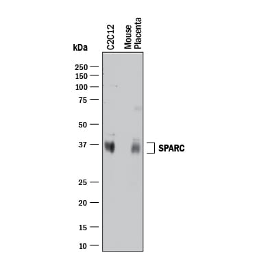
Detection of Mouse SPARC by Western Blot.
Western blot shows lysates of C2C12 mouse myoblast cell line and mouse placenta tissue. PVDF membrane was probed with 0.2 µg/mL of Goat Anti-Mouse SPARC Antigen Affinity-purified Polyclonal Antibody (Catalog # AF942) followed by HRP-conjugated Anti-Goat IgG Secondary Antibody (Catalog # HAF017). A specific band was detected for SPARC at approximately 35-37 kDa (as indicated). This experiment was conducted under reducing conditions and using Immunoblot Buffer Group 1.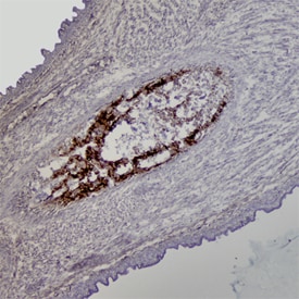
SPARC/Osteonectin in Mouse Embryo.
SPARC/Osteonectin was detected in immersion fixed frozen sections of mouse embryo (E15) using Mouse SPARC/ Osteonectin Antigen Affinity-purified Polyclonal Antibody (Catalog # AF942) at 1.7 µg/mL overnight at 4 °C. Tissue was stained using the Anti-Goat HRP-DAB Cell & Tissue Staining Kit (brown; Catalog # CTS008) and counterstained with hematoxylin (blue). Specific staining was localized to developing cartilage. View our protocol for Chromogenic IHC Staining of Frozen Tissue Sections.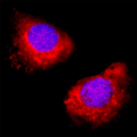
SPARC in C2C12 Mouse Cell Line.
SPARC was detected in immersion fixed C2C12 mouse myoblast cell line using Goat Anti-Mouse SPARC Antigen Affinity-purified Polyclonal Antibody (Catalog # AF942) at 5 µg/mL for 3 hours at room temperature. Cells were stained using the NorthernLights™ 557-conjugated Anti-Goat IgG Secondary Antibody (red; Catalog # NL001) and counterstained with DAPI (blue). Specific staining was localized to endoplasmic reticuli. View our protocol for Fluorescent ICC Staining of Cells on Coverslips.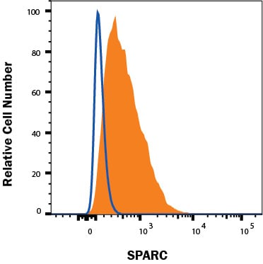
Detection of SPARC in Balb/C-3T3 Mouse Cell Line by Flow Cytometry.
Balb/C-3T3 mouse fibroblast cell line was stained with Goat Anti-Mouse SPARC Polyclonal Antibody (Catalog # AF942, filled histogram) or Goat IgG control antibody (AB-108-C, open histogram), followed by Phycoerythrin-conjugated anti-Goat IgG (F0107). To facilitate intracellular staining, cells were fixed with Flow Cytometry Fixation Buffer (FC004) and permeabilized with Flow Cytometry Permeabilization/Wash Buffer I (FC005). Staining was performed using our Staining Intracellular Molecules protocol.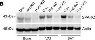
Detection of SPARC by Western Blot
SPARC controls adiposity.(A) Schematic of experiments with inducible global Sparc KO mice. (B) Immunoblot analysis of SPARC protein in bone, VAT, and SAT in control Sparcfl/fl (Con), heterozygote (Sparcfl/+;CAG-CreER, Het iKO), and homozygote (Sparcfl/fl;CAG-CreER, Hom iKO) KO mice 6 weeks after tamoxifen injection. (C and D) Percentage of weight change of male (C) and female (D) littermate control (n = 10, 10), Het iKO (n = 10, 10), and Hom iKO (n = 5, 4) mice after tamoxifen injection. (E and F) Glucose tolerance test (GTT) (E), and insulin tolerance test (ITT) (F) of 14-month old (8 months after tamoxifen injection) male Con, Het iKO, and Hom iKO mice (n = 10, 10, 5). The blue star indicates statistical significance between Con and Het iKO mice, and the red star indicates statistical significance between Con and Hom iKO mice. Error bars represent the mean ± SEM. 2-way ANOVA test with Dunnett’s multiple comparisons test for adjusted P values (C–F) were performed for statistical analysis. *P < 0.05; **P < 0.01; ***P < 0.001. Image collected and cropped by CiteAb from the following open publication (https://pubmed.ncbi.nlm.nih.gov/37781916), licensed under a CC-BY license. Not internally tested by R&D Systems.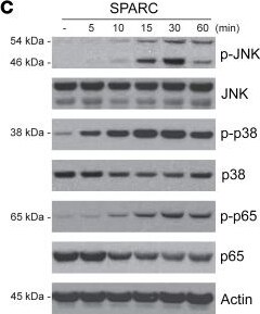
Detection of SPARC by Western Blot
SPARC activates inflammation in macrophages via JNK signaling.(A) Human SPARC or mock vector was overexpressed in RAW 264.7 cells by transient transfection (0.5 and 2.5 μg) and Western-blot analyses of SPARC, JNK and p38 MAPK are shown. The experiment was repeated in triplicate and performed twice. (B) q-PCR analysis of Il1b, Tnf, Nos2, and Il6 in RAW 264.7 cells with SPARC or mock vector overexpression. (C) Primary BMDMs were treated with SPARC (5–60 minutes) and JNK, p65 NF-kappa B, and p38 MPAK were quantified by immunoblot analysis. (D) Representative immunoblot of p-p65 NF-kappa B in BMDMs pretreated with STAT1, p38, and JNK inhibitor and in presence of SPARC (20 μg/mL). (E) q-PCR analysis of Il1b, Tnf, Nos2, and Il6 in BMDMs pretreated with p38 or JNK inhibitor followed by SPARC treatment (20 μg/mL). (F) Primary BMDMs were transfected with JNK and STAT1 siRNA and immunoblot analysis was performed to quantify Pro-IL-1 beta protein levels. The experiment was repeated in triplicate and performed twice. Error bars represent the mean ± SEM. 2-tailed unpaired t tests (B) and 1-way ANOVA test with Bonferroni’s multiple comparisons test for adjusted P values (E) were performed for statistical analysis. *P < 0.05; **P < 0.01; ***P < 0.001; ****P < 0.0001. Image collected and cropped by CiteAb from the following open publication (https://pubmed.ncbi.nlm.nih.gov/37781916), licensed under a CC-BY license. Not internally tested by R&D Systems.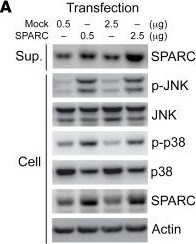
Detection of SPARC by Western Blot
SPARC activates inflammation in macrophages via JNK signaling.(A) Human SPARC or mock vector was overexpressed in RAW 264.7 cells by transient transfection (0.5 and 2.5 μg) and Western-blot analyses of SPARC, JNK and p38 MAPK are shown. The experiment was repeated in triplicate and performed twice. (B) q-PCR analysis of Il1b, Tnf, Nos2, and Il6 in RAW 264.7 cells with SPARC or mock vector overexpression. (C) Primary BMDMs were treated with SPARC (5–60 minutes) and JNK, p65 NF-kappa B, and p38 MPAK were quantified by immunoblot analysis. (D) Representative immunoblot of p-p65 NF-kappa B in BMDMs pretreated with STAT1, p38, and JNK inhibitor and in presence of SPARC (20 μg/mL). (E) q-PCR analysis of Il1b, Tnf, Nos2, and Il6 in BMDMs pretreated with p38 or JNK inhibitor followed by SPARC treatment (20 μg/mL). (F) Primary BMDMs were transfected with JNK and STAT1 siRNA and immunoblot analysis was performed to quantify Pro-IL-1 beta protein levels. The experiment was repeated in triplicate and performed twice. Error bars represent the mean ± SEM. 2-tailed unpaired t tests (B) and 1-way ANOVA test with Bonferroni’s multiple comparisons test for adjusted P values (E) were performed for statistical analysis. *P < 0.05; **P < 0.01; ***P < 0.001; ****P < 0.0001. Image collected and cropped by CiteAb from the following open publication (https://pubmed.ncbi.nlm.nih.gov/37781916), licensed under a CC-BY license. Not internally tested by R&D Systems.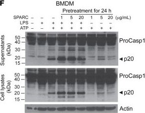
Detection of SPARC by Western Blot
Adipocyte-derived SPARC controls macrophage inflammation.(A and B) Schematic and weight change of Con and Adip-KO mice with 21 weeks of HFD followed by 13 weeks of chow diet (n = 5, 5, respectively). (C) Body composition analysis of female Con and Adip-KO mice before and after diet change from HFD to chow diet (n = 4, 4, respectively). (D and E) q-PCR analysis of inflammatory gene (D) and components of inflammasome (E) in VAT macrophages (F4/80+) in obese mice switched to chow diet (n = 4, 4, respectively). (F) Inflammasome activation after pretreatment of SPARC protein for 24 hours following ATP (5mM) treatment with or without LPS (1 μg/mL) measured by caspase-1 Western blot analysis in cell lysate (lower) and supernatant (upper). Error bars represent the mean ± SEM. 2-tailed unpaired t tests were performed for statistical analysis. *P < 0.05; **P < 0.01; ***P < 0.001. Image collected and cropped by CiteAb from the following open publication (https://pubmed.ncbi.nlm.nih.gov/37781916), licensed under a CC-BY license. Not internally tested by R&D Systems.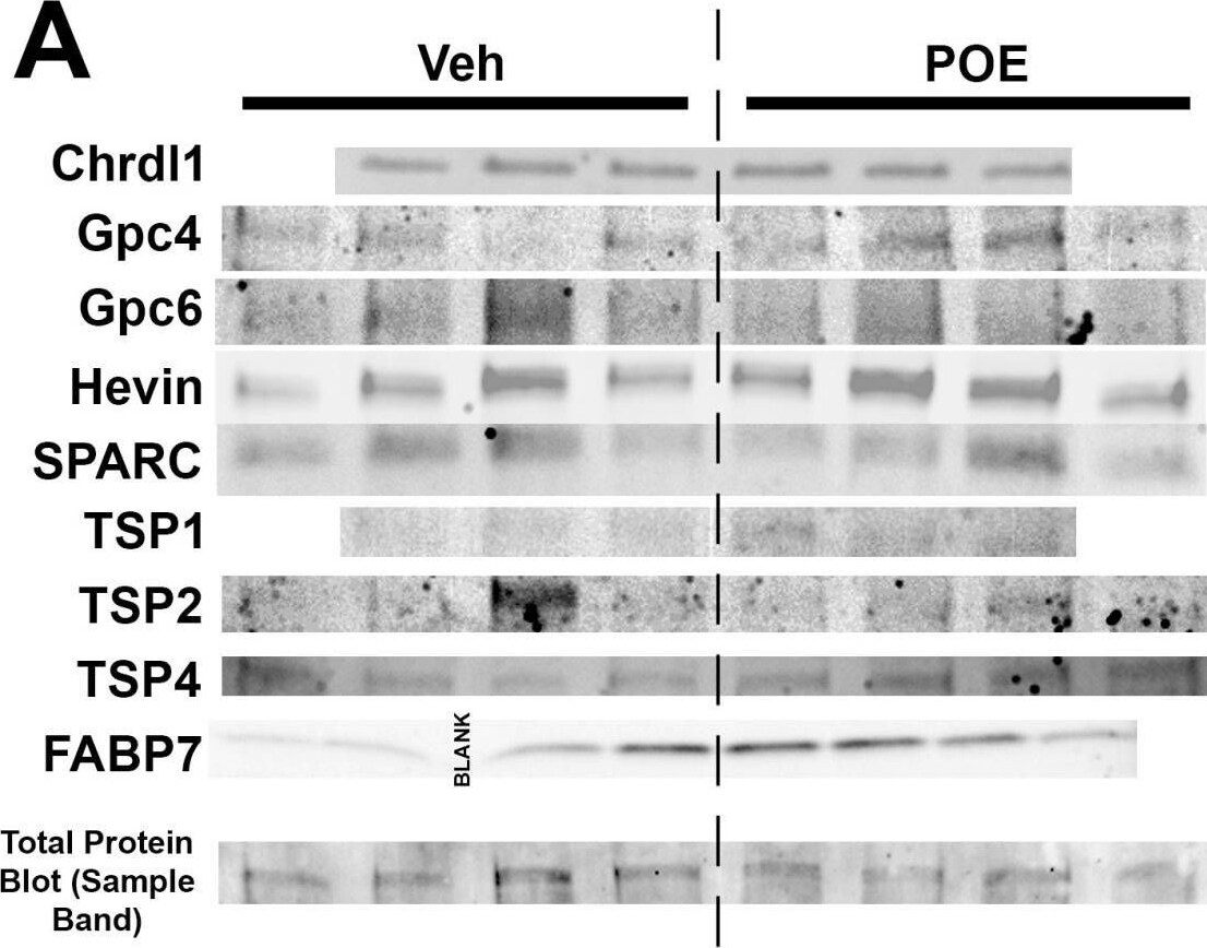
Detection of SPARC by Western Blot
Changes in synapse-related protein expression in POE astrocytes. (A) Western blot images reveal expression patterns of prominent synapse-associated proteins from the DIV10 vehicle and POE astrocyte lysates. Bottom row shows a representative band from one of the total protein blot images that was used for normalization purposes (full image shown in Figure S1). (B) Quantification of Western blots for proteins indicated in (A). Data shown as fold-change in band intensity values for POE astrocyte lysates compared to vehicle (n = 3–4 experimental replicates; unpaired t-test (unlabeled p values were not significant)). Image collected and cropped by CiteAb from the following open publication (https://pubmed.ncbi.nlm.nih.gov/38786059), licensed under a CC-BY license. Not internally tested by R&D Systems.Applications for Mouse SPARC Antibody
CyTOF-ready
Immunocytochemistry
Sample: Immersion fixed C2C12 mouse myoblast cell line
Immunohistochemistry
Sample: Immersion fixed frozen sections of mouse embryo (E15)
Intracellular Staining by Flow Cytometry
Sample: Balb/C-3T3 mouse embryonic fibroblast cell line fixed with paraformaldehyde and permeabilized with saponin
Western Blot
Sample: C2C12 mouse myoblast cell line and mouse placenta tissue
Reviewed Applications
Read 3 reviews rated 4.7 using AF942 in the following applications:
Flow Cytometry Panel Builder
Bio-Techne Knows Flow Cytometry
Save time and reduce costly mistakes by quickly finding compatible reagents using the Panel Builder Tool.
Advanced Features
- Spectra Viewer - Custom analysis of spectra from multiple fluorochromes
- Spillover Popups - Visualize the spectra of individual fluorochromes
- Antigen Density Selector - Match fluorochrome brightness with antigen density

Formulation, Preparation, and Storage
Purification
Reconstitution
Reconstitute at 0.2 mg/mL in sterile PBS. For liquid material, refer to CoA for concentration.
Formulation
Shipping
Stability & Storage
- 12 months from date of receipt, -20 to -70 °C as supplied.
- 1 month, 2 to 8 °C under sterile conditions after reconstitution.
- 6 months, -20 to -70 °C under sterile conditions after reconstitution.
Calculators
Background: SPARC
References
- Lankat-Buttgereit, B. et al. (1988) FEBS Lett. 236:352.
- McVey, J.H. et al. (1988) J. Biol. Chem. 263:11111.
- Sage, H. et al. (1989) J. Cell Biol. 109:341.
- Framson, P.E. and E.H. Sage (2004) J. Cell. Biochem. 92:679.
- Alford, A.I. and K.D. Hankenson (2006) Bone 38:749.
- Hohenester, E. et al. (1997) EMBO J. 16:3778.
- Sage, E.H. et al. (2003) J. Biol. Chem. 278:37849.
- Delany, A.M. et al. (2003) Endocrinology 144:2588.
- Koblinski, J.E. et al. (2005) Cancer Res. 65:7370.
- Tai, I.T. et al. (2005) J. Clin. Invest. 115:1492.
- Kzhyshkowska, J. et al. (2006) J. Immunol. 176:5825.
Long Name
Alternate Names
Gene Symbol
UniProt
Additional SPARC Products
Product Documents for Mouse SPARC Antibody
Certificate of Analysis
To download a Certificate of Analysis, please enter a lot or batch number in the search box below.
Note: Certificate of Analysis not available for kit components.
Product Specific Notices for Mouse SPARC Antibody
For research use only
Citations for Mouse SPARC Antibody
 Powered by Bioz
Customer Reviews for Mouse SPARC Antibody (3)
Have you used Mouse SPARC Antibody?
Submit a review and receive an Amazon gift card!
$25/€18/£15/$25CAN/¥2500 for a review with an image
$10/€7/£6/$10CAN/¥1110 for a review without an image
Submit a review
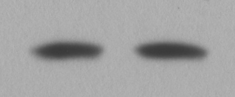
Customer Images
-
Application: Western BlotSample Tested: Mouse hippocampal tissueSpecies: MouseVerified Customer | Posted 02/02/2018Used at 1:3000 in 5% BSA and secondary goat-HRP antibody (R&D) at 1:5000

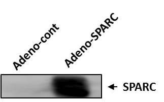
-
Application: Western BlotSample Tested: trabecular meshworkSpecies: MouseVerified Customer | Posted 01/12/2018mouse SPARC was overexpressed by adeno-SPARC in mouse trabecular meshork (TM) cells.
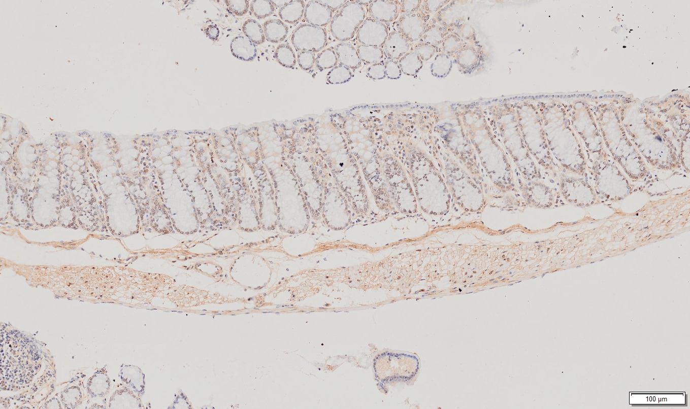
-
Application: ImmunohistochemistrySample Tested: Colon cancer tissue and Colon tissueSpecies: MouseVerified Customer | Posted 05/18/2017Worked well in methacarn fixed tissues
There are no reviews that match your criteria.
Protocols
Find general support by application which include: protocols, troubleshooting, illustrated assays, videos and webinars.
- 7-Amino Actinomycin D (7-AAD) Cell Viability Flow Cytometry Protocol
- Antigen Retrieval Protocol (PIER)
- Antigen Retrieval for Frozen Sections Protocol
- Appropriate Fixation of IHC/ICC Samples
- Cellular Response to Hypoxia Protocols
- Chromogenic IHC Staining of Formalin-Fixed Paraffin-Embedded (FFPE) Tissue Protocol
- Chromogenic Immunohistochemistry Staining of Frozen Tissue
- ClariTSA™ Fluorophore Kits
- Detection & Visualization of Antibody Binding
- Extracellular Membrane Flow Cytometry Protocol
- Flow Cytometry Protocol for Cell Surface Markers
- Flow Cytometry Protocol for Staining Membrane Associated Proteins
- Flow Cytometry Staining Protocols
- Flow Cytometry Troubleshooting Guide
- Fluorescent IHC Staining of Frozen Tissue Protocol
- Graphic Protocol for Heat-induced Epitope Retrieval
- Graphic Protocol for the Preparation and Fluorescent IHC Staining of Frozen Tissue Sections
- Graphic Protocol for the Preparation and Fluorescent IHC Staining of Paraffin-embedded Tissue Sections
- Graphic Protocol for the Preparation of Gelatin-coated Slides for Histological Tissue Sections
- ICC Cell Smear Protocol for Suspension Cells
- ICC Immunocytochemistry Protocol Videos
- ICC for Adherent Cells
- IHC Sample Preparation (Frozen sections vs Paraffin)
- Immunocytochemistry (ICC) Protocol
- Immunocytochemistry Troubleshooting
- Immunofluorescence of Organoids Embedded in Cultrex Basement Membrane Extract
- Immunofluorescent IHC Staining of Formalin-Fixed Paraffin-Embedded (FFPE) Tissue Protocol
- Immunohistochemistry (IHC) and Immunocytochemistry (ICC) Protocols
- Immunohistochemistry Frozen Troubleshooting
- Immunohistochemistry Paraffin Troubleshooting
- Intracellular Flow Cytometry Protocol Using Alcohol (Methanol)
- Intracellular Flow Cytometry Protocol Using Detergents
- Intracellular Nuclear Staining Flow Cytometry Protocol Using Detergents
- Intracellular Staining Flow Cytometry Protocol Using Alcohol Permeabilization
- Intracellular Staining Flow Cytometry Protocol Using Detergents to Permeabilize Cells
- Preparing Samples for IHC/ICC Experiments
- Preventing Non-Specific Staining (Non-Specific Binding)
- Primary Antibody Selection & Optimization
- Propidium Iodide Cell Viability Flow Cytometry Protocol
- Protocol for Heat-Induced Epitope Retrieval (HIER)
- Protocol for Liperfluo
- Protocol for Making a 4% Formaldehyde Solution in PBS
- Protocol for VisUCyte™ HRP Polymer Detection Reagent
- Protocol for the Characterization of Human Th22 Cells
- Protocol for the Characterization of Human Th9 Cells
- Protocol for the Fluorescent ICC Staining of Cell Smears - Graphic
- Protocol for the Fluorescent ICC Staining of Cultured Cells on Coverslips - Graphic
- Protocol for the Preparation & Fixation of Cells on Coverslips
- Protocol for the Preparation and Chromogenic IHC Staining of Frozen Tissue Sections
- Protocol for the Preparation and Chromogenic IHC Staining of Frozen Tissue Sections - Graphic
- Protocol for the Preparation and Chromogenic IHC Staining of Paraffin-embedded Tissue Sections
- Protocol for the Preparation and Chromogenic IHC Staining of Paraffin-embedded Tissue Sections - Graphic
- Protocol for the Preparation and Fluorescent ICC Staining of Cells on Coverslips
- Protocol for the Preparation and Fluorescent ICC Staining of Non-adherent Cells
- Protocol for the Preparation and Fluorescent ICC Staining of Stem Cells on Coverslips
- Protocol for the Preparation and Fluorescent IHC Staining of Frozen Tissue Sections
- Protocol for the Preparation and Fluorescent IHC Staining of Paraffin-embedded Tissue Sections
- Protocol for the Preparation of Gelatin-coated Slides for Histological Tissue Sections
- Protocol for the Preparation of a Cell Smear for Non-adherent Cell ICC - Graphic
- Protocol: Annexin V and PI Staining by Flow Cytometry
- Protocol: Annexin V and PI Staining for Apoptosis by Flow Cytometry
- R&D Systems Quality Control Western Blot Protocol
- TUNEL and Active Caspase-3 Detection by IHC/ICC Protocol
- The Importance of IHC/ICC Controls
- Troubleshooting Guide: Fluorokine Flow Cytometry Kits
- Troubleshooting Guide: Immunohistochemistry
- Troubleshooting Guide: Western Blot Figures
- Western Blot Conditions
- Western Blot Protocol
- Western Blot Protocol for Cell Lysates
- Western Blot Troubleshooting
- Western Blot Troubleshooting Guide
- View all Protocols, Troubleshooting, Illustrated assays and Webinars
Associated Pathways
