WT1 Antibody (6F-H2) - BSA Free
Novus Biologicals | Catalog # NB110-60011
Key Product Details
Species Reactivity
Validated:
Cited:
Applications
Validated:
Cited:
Label
Antibody Source
Format
Product Specifications
Immunogen
Localization
Clonality
Host
Isotype
Theoretical MW
Disclaimer note: The observed molecular weight of the protein may vary from the listed predicted molecular weight due to post translational modifications, post translation cleavages, relative charges, and other experimental factors.
Scientific Data Images for WT1 Antibody (6F-H2) - BSA Free
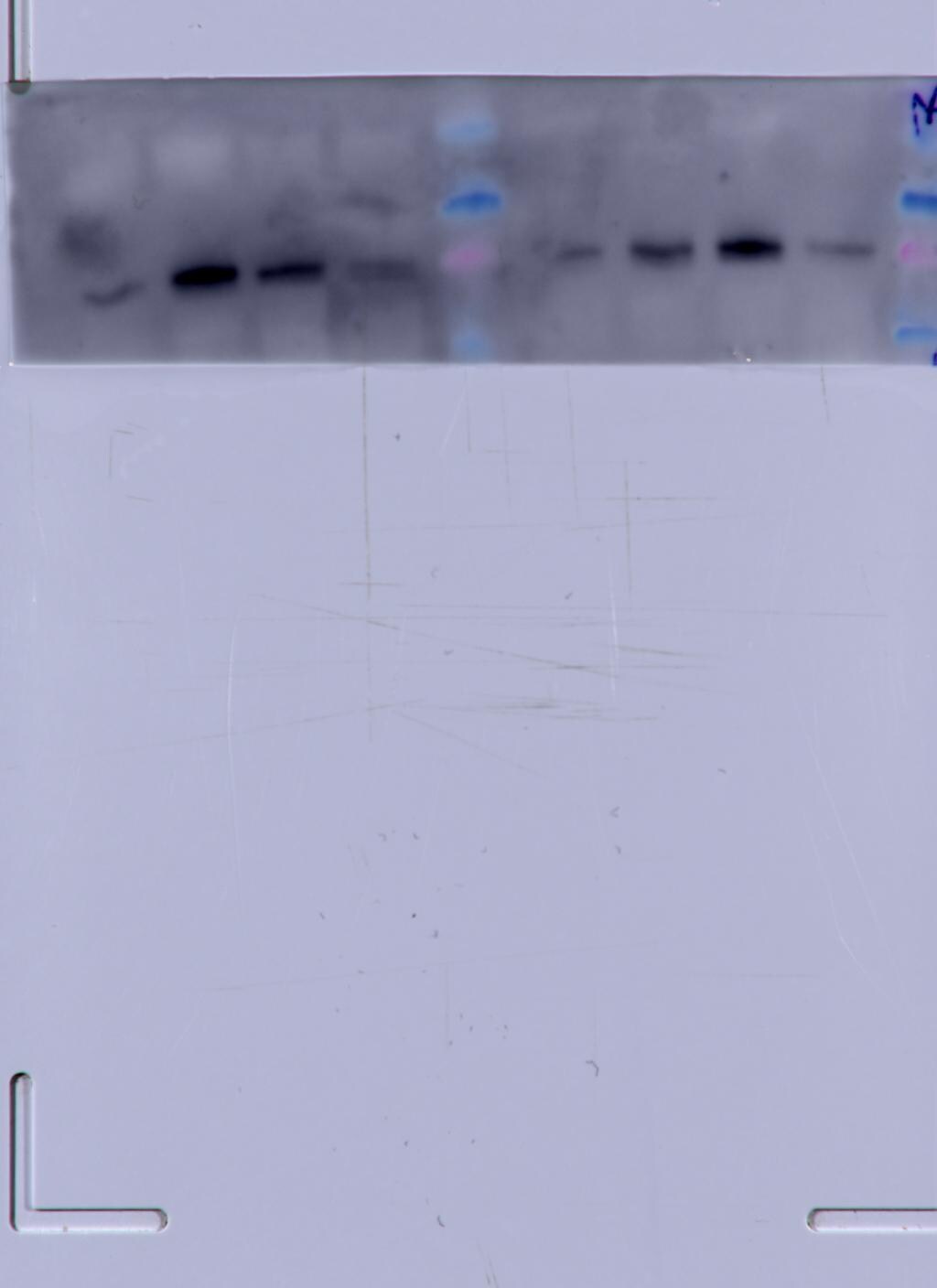
![Western Blot: WT1 Antibody (6F-H2)BSA Free [NB110-60011]](https://resources.rndsystems.com/images/products/WT1-Antibody-6F-H2-Western-Blot-NB110-60011-img0007.jpg "Western Blot: WT1 Antibody (6F-H2)BSA Free [NB110-60011]")
Western Blot: WT1 Antibody (6F-H2)BSA Free [NB110-60011]
WT1-Antibody-6F-H2-Western-Blot-NB110-60011-img0007.jpg![Immunohistochemistry-Paraffin: WT1 Antibody (6F-H2) - BSA Free [NB110-60011]](https://resources.rndsystems.com/images/products/WT1-Antibody-6F-H2-Immunohistochemistry-Paraffin-NB110-60011-img0006.jpg "Immunohistochemistry-Paraffin: WT1 Antibody (6F-H2) - BSA Free [NB110-60011]")
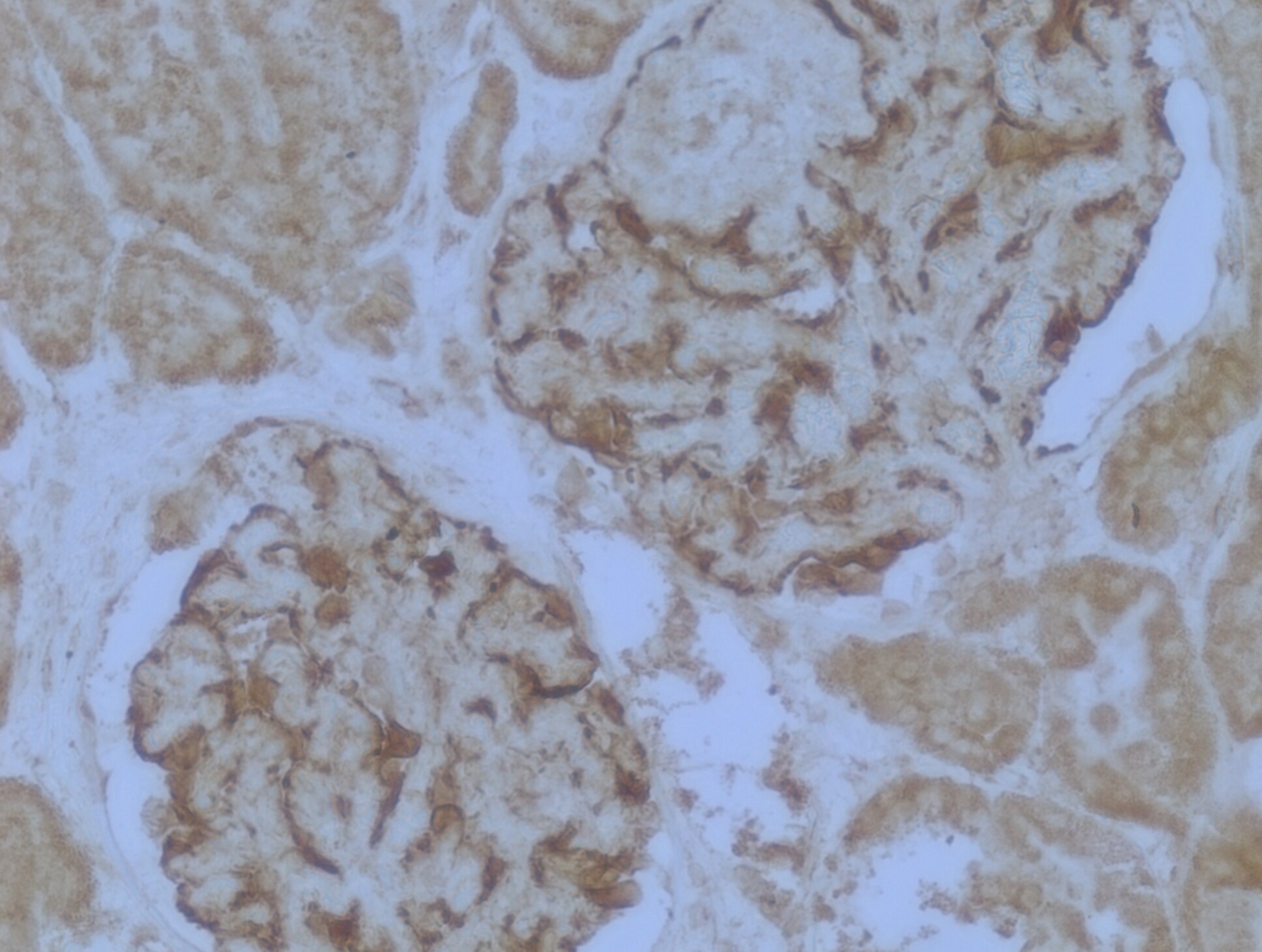
Immunohistochemistry-Paraffin: WT1 Antibody (6F-H2) - BSA Free [NB110-60011]
Immunohistochemistry-Paraffin: WT1 Antibody (6F-H2) [NB110-60011] - Staining of human kidney glomerulus tissue. Heat mediated antigen retrieval was performed by heating in citrate buffer (pH 6) at 95C for 20 minutes. Image from verified customer review.![Western Blot: WT1 Antibody (6F-H2)BSA Free [NB110-60011]](https://resources.rndsystems.com/images/products/WT1-Antibody-6F-H2-Western-Blot-NB110-60011-img0005.jpg "Western Blot: WT1 Antibody (6F-H2)BSA Free [NB110-60011]")
Western Blot: WT1 Antibody (6F-H2)BSA Free [NB110-60011]
Western Blot: WT1 Antibody (6F-H2) [NB110-60011] - Whole cell protein from human K562, HEK293 and kidney tissue was separated on a 12% gel by SDS-PAGE, transferred to PVDF membrane and blocked in 5% non-fat milk in TBST. The membrane was probed with 2.0 ug/ml anti-WT1 in block buffer and detected with an anti-mouse HRP secondary antibody using chemiluminescence.![Immunohistochemistry: WT1 Antibody (6F-H2) - BSA Free [NB110-60011]](https://resources.rndsystems.com/images/products/WT1-Antibody-6F-H2-Immunohistochemistry-NB110-60011-img0003.jpg "Immunohistochemistry: WT1 Antibody (6F-H2) - BSA Free [NB110-60011]")
Immunohistochemistry: WT1 Antibody (6F-H2) - BSA Free [NB110-60011]
Immunohistochemistry: WT1 Antibody (6F-H2) [NB110-60011] - Analysis of Wilms Tumor 1 in human renal cancer using DAB with hematoxylin counterstain.![Simple Western: WT1 Antibody (6F-H2)BSA Free [NB110-60011]](https://resources.rndsystems.com/images/products/WT1-Antibody-6F-H2-Simple-Western-NB110-60011-img0004.jpg "Simple Western: WT1 Antibody (6F-H2)BSA Free [NB110-60011]")
Simple Western: WT1 Antibody (6F-H2)BSA Free [NB110-60011]
Simple Western: WT1 Antibody (6F-H2) [NB110-60011] - Lane view shows a specific band for WT1 in 0.5 mg/ml of Hek293 lysate. This experiment was performed under reducing conditions using the 12-230 kDa separation system. - BSA Free [NB110-60011] -")
Western Blot: WT1 Antibody (6F-H2) - BSA Free [NB110-60011] -
Western Blot: WT1 Antibody (6F-H2) - BSA Free [NB110-60011] - Hypomethylation of Intron 1 CpG island results in WT1 expression. (C) Cell lysates from K562, U937, & HL60 cells treated with CoCl2, 5-azacytidine, or untreated controls analyzed by WB using an antibody against WT1. Histone H3 used as a loading control. Size of molecular weight markers is in kDa. Image collected & cropped by CiteAb from the following publication (https://dx.plos.org/10.1371/journal.pone.0119837), licensed under a CC-BY license. Not internally tested by Novus Biologicals.Applications for WT1 Antibody (6F-H2) - BSA Free
Immunocytochemistry/ Immunofluorescence
Immunohistochemistry
Immunohistochemistry-Frozen
Immunohistochemistry-Paraffin
Immunoprecipitation
Simple Western
Western Blot
See Simple Western Antibody Database for Simple Western validation: Tested in Hek293 lysate 0.5 mg/mL, separated by Size, antibody dilution of 1:50, apparent MW was 68 kDa. Separated by Size-Wes, Sally Sue/Peggy Sue.
Reviewed Applications
Read 3 reviews rated 4.3 using NB110-60011 in the following applications:
Formulation, Preparation, and Storage
Purification
Formulation
Format
Preservative
Concentration
Shipping
Stability & Storage
Background: WT1
Long Name
Alternate Names
Gene Symbol
Additional WT1 Products
Product Documents for WT1 Antibody (6F-H2) - BSA Free
Certificate of Analysis
To download a Certificate of Analysis, please enter a lot or batch number in the search box below.
Product Specific Notices for WT1 Antibody (6F-H2) - BSA Free
This product is for research use only and is not approved for use in humans or in clinical diagnosis. Primary Antibodies are guaranteed for 1 year from date of receipt.
Related Research Areas
Citations for WT1 Antibody (6F-H2) - BSA Free
 Powered by Bioz
Customer Reviews for WT1 Antibody (6F-H2) - BSA Free (3)
Have you used WT1 Antibody (6F-H2) - BSA Free?
Submit a review and receive an Amazon gift card!
$25/€18/£15/$25CAN/¥2500 Yen for a review with an image
$10/€7/£6/$10CAN/¥1110 Yen for a review without an image
Submit a review
Customer Images
-(01-ml)_NB110-60011_7116.png)
-
Application: Western BlotSample Tested: Liver and 293T whole cell lysateSpecies: Mouse and HumanVerified Customer | Posted 10/17/2019WT1 expression in Mouse Liver.

-
Application: Immunohistochemistry-ParaffinSample Tested: Human kidney tissueSpecies: HumanVerified Customer | Posted 05/22/2018Staining in kidney glomerulus.Heat mediated antigen retrieval was performed by heating in citrate buffer (pH6) at 95C for 20 minutes
-
Application: Western BlotSample Tested: Immortalized podocytesSpecies: HumanVerified Customer | Posted 04/28/2014Western blot, immortalized human podocytes (10ug); 1:1000, overnight 4C
There are no reviews that match your criteria.
Protocols
View specific protocols for WT1 Antibody (6F-H2) - BSA Free (NB110-60011):
Western Blot Protocol
1. Perform SDS-PAGE (4-12% MOPS) on samples to be analyzed, loading 28 ug of total protein per lane.
2. Transfer proteins to Nitrocellulose according to the instructions provided by the manufacturer of the transfer
apparatus.
3. Rinse membrane with dH2O and then stain the blot using Ponceau S for 1-2 minutes to access the transfer of proteins onto the nitrocellulose membrane. Rinse the blot in water to remove excess stain and mark the lane locations and locations of molecular weight markers using a pencil.
4. Rinse the blot in TBS for approximately 5 minutes.
5. Block the membrane using 5% non-fat dry milk + 1% BSA in TBS, 1 hour at room temperature.
6. Rinse the membrane in dH2O and then wash the membrane in wash buffer [TBS + 0.1% Tween] 3 times for 10 minutes each.
7. Dilute the mouse anti-Wilm's Tumor 1 primary antibody (NB 110-60011) in blocking buffer and incubate 2 hours at room temperature.
8. Rinse the membrane in dH2O and then wash the membrane in wash buffer [TBS + 0.1% Tween] 3 times for 10 minutes each.
9. Apply the diluted mouse-IgG HRP-conjugated secondary antibody in blocking buffer (as per manufacturers
instructions) and incubate 1 hour at room temperature.
10. Wash the blot in wash buffer [TBS + 0.1% Tween] 3 times for 10 minutes each (this step can be repeated as required to reduce background).
11. Apply the detection reagent of choice in accordance with the manufacturers instructions (Pierce, ECL).
Note: Tween-20 can be added to the blocking or antibody dilution buffer at a final concentration of 0.05-0.2%, provided it does not interfere with antibody-antigen binding.
Find general support by application which include: protocols, troubleshooting, illustrated assays, videos and webinars.
- Antigen Retrieval Protocol (PIER)
- Antigen Retrieval for Frozen Sections Protocol
- Appropriate Fixation of IHC/ICC Samples
- Cellular Response to Hypoxia Protocols
- Chromogenic IHC Staining of Formalin-Fixed Paraffin-Embedded (FFPE) Tissue Protocol
- Chromogenic Immunohistochemistry Staining of Frozen Tissue
- ClariTSA™ Fluorophore Kits
- Detection & Visualization of Antibody Binding
- Fluorescent IHC Staining of Frozen Tissue Protocol
- Graphic Protocol for Heat-induced Epitope Retrieval
- Graphic Protocol for the Preparation and Fluorescent IHC Staining of Frozen Tissue Sections
- Graphic Protocol for the Preparation and Fluorescent IHC Staining of Paraffin-embedded Tissue Sections
- Graphic Protocol for the Preparation of Gelatin-coated Slides for Histological Tissue Sections
- ICC Cell Smear Protocol for Suspension Cells
- ICC Immunocytochemistry Protocol Videos
- ICC for Adherent Cells
- IHC Sample Preparation (Frozen sections vs Paraffin)
- Immunocytochemistry (ICC) Protocol
- Immunocytochemistry Troubleshooting
- Immunofluorescence of Organoids Embedded in Cultrex Basement Membrane Extract
- Immunofluorescent IHC Staining of Formalin-Fixed Paraffin-Embedded (FFPE) Tissue Protocol
- Immunohistochemistry (IHC) and Immunocytochemistry (ICC) Protocols
- Immunohistochemistry Frozen Troubleshooting
- Immunohistochemistry Paraffin Troubleshooting
- Immunoprecipitation Protocol
- Preparing Samples for IHC/ICC Experiments
- Preventing Non-Specific Staining (Non-Specific Binding)
- Primary Antibody Selection & Optimization
- Protocol for Heat-Induced Epitope Retrieval (HIER)
- Protocol for Making a 4% Formaldehyde Solution in PBS
- Protocol for VisUCyte™ HRP Polymer Detection Reagent
- Protocol for the Fluorescent ICC Staining of Cell Smears - Graphic
- Protocol for the Fluorescent ICC Staining of Cultured Cells on Coverslips - Graphic
- Protocol for the Preparation & Fixation of Cells on Coverslips
- Protocol for the Preparation and Chromogenic IHC Staining of Frozen Tissue Sections
- Protocol for the Preparation and Chromogenic IHC Staining of Frozen Tissue Sections - Graphic
- Protocol for the Preparation and Chromogenic IHC Staining of Paraffin-embedded Tissue Sections
- Protocol for the Preparation and Chromogenic IHC Staining of Paraffin-embedded Tissue Sections - Graphic
- Protocol for the Preparation and Fluorescent ICC Staining of Cells on Coverslips
- Protocol for the Preparation and Fluorescent ICC Staining of Non-adherent Cells
- Protocol for the Preparation and Fluorescent ICC Staining of Stem Cells on Coverslips
- Protocol for the Preparation and Fluorescent IHC Staining of Frozen Tissue Sections
- Protocol for the Preparation and Fluorescent IHC Staining of Paraffin-embedded Tissue Sections
- Protocol for the Preparation of Gelatin-coated Slides for Histological Tissue Sections
- Protocol for the Preparation of a Cell Smear for Non-adherent Cell ICC - Graphic
- R&D Systems Quality Control Western Blot Protocol
- TUNEL and Active Caspase-3 Detection by IHC/ICC Protocol
- The Importance of IHC/ICC Controls
- Troubleshooting Guide: Immunohistochemistry
- Troubleshooting Guide: Western Blot Figures
- Western Blot Conditions
- Western Blot Protocol
- Western Blot Protocol for Cell Lysates
- Western Blot Troubleshooting
- Western Blot Troubleshooting Guide
- View all Protocols, Troubleshooting, Illustrated assays and Webinars
FAQs for WT1 Antibody (6F-H2) - BSA Free
-
Q: I just wondering this antibody (WT1 antibody (6F-H2)[Alexa Flour488], when I use it to staining tissue do I need incubate in second antibody or not?
A: Our product NB110-60011AF488 is directly conjugated with an Alexa Fluor 488 dye, so you do not need to use any secondary antibody with this product to detect the antibody. The dye labeled to this antibody excites at 490nm and emits at 525nm.
-
Q: In NB800-PC6, were the 293 cells transfected with WT1 expression plasmids? We have 293 cell lysates and it showed negative WT1 expression. Just want to make sure if our antibody is not working.
A: This protein lysate was derived from human HEK293 cells (without any transfection) and it is a whole cell lysate. WT1's mRNA is expressed significantly in HEK294 cells protein atlas site and given the fact that our Wilms Tumor 1 antibody (6F-H2) [NB110-60011] showed positive bands on HEK293 cells lysate, you should be good for considering this lysate as a positive control for WT1.
-
Q: I just wondering this antibody (WT1 antibody (6F-H2)[Alexa Flour488], when I use it to staining tissue do I need incubate in second antibody or not?
A: Our product NB110-60011AF488 is directly conjugated with an Alexa Fluor 488 dye, so you do not need to use any secondary antibody with this product to detect the antibody. The dye labeled to this antibody excites at 490nm and emits at 525nm.
-
Q: In NB800-PC6, were the 293 cells transfected with WT1 expression plasmids? We have 293 cell lysates and it showed negative WT1 expression. Just want to make sure if our antibody is not working.
A: This protein lysate was derived from human HEK293 cells (without any transfection) and it is a whole cell lysate. WT1's mRNA is expressed significantly in HEK294 cells protein atlas site and given the fact that our Wilms Tumor 1 antibody (6F-H2) [NB110-60011] showed positive bands on HEK293 cells lysate, you should be good for considering this lysate as a positive control for WT1.
Associated Pathways
