15-PGDH/HPGD Antibody - BSA Free
Novus Biologicals | Catalog # NB200-179
![Simple Western: 15-PGDH/HPGD Antibody [NB200-179]](https://resources.rndsystems.com/images/products/15-PGDH-HPGD-Antibody-Simple-Western-NB200-179-img0005.jpg "Simple Western: 15-PGDH/HPGD Antibody [NB200-179]")
Key Product Details
Validated by
Knockout/Knockdown, Biological Validation
Species Reactivity
Validated:
Human, Mouse, Rat
Cited:
Human, Mouse, Rat
Applications
Validated:
Immunohistochemistry, Immunohistochemistry-Paraffin, Immunohistochemistry-Frozen, Western Blot, Immunocytochemistry/ Immunofluorescence, Simple Western, Knockdown Validated
Cited:
Immunohistochemistry-Paraffin, Immunohistochemistry-Frozen, Western Blot, Immunocytochemistry/ Immunofluorescence, IF/IHC
Label
Unconjugated
Antibody Source
Polyclonal Rabbit IgG
Format
BSA Free
Loading...
Product Specifications
Immunogen
Purified type I human placental 15-PGDH protein [UniProt# P15428]
Reactivity Notes
Mouse reactivity reported in scientific literature (PMID: 27140190) Human reactivity reported in scientific literature (PMID:33210098).
Localization
Cytoplasmic
Clonality
Polyclonal
Host
Rabbit
Isotype
IgG
Scientific Data Images for 15-PGDH/HPGD Antibody - BSA Free
Simple Western: 15-PGDH/HPGD Antibody [NB200-179]
Simple Western: 15-PGDH/HPGD Antibody [NB200-179] - Simple Western lane view shows a specific band for 15-PGDH/HPGD in 0.5 mg/ml of Lovo (left) and HeLa (right) lysate. This experiment was performed under reducing conditions using the 12-230 kDa separation system.![Immunohistochemistry: 15-PGDH/HPGD Antibody [NB200-179]](https://resources.rndsystems.com/images/products/15-PGDH-HPGD-Antibody-Immunohistochemistry-NB200-179-img0015.jpg "Immunohistochemistry: 15-PGDH/HPGD Antibody [NB200-179]")
Immunohistochemistry: 15-PGDH/HPGD Antibody [NB200-179]
15-PGDH-HPGD-Antibody-Immunohistochemistry-NB200-179-img0015.jpg![Immunohistochemistry: 15-PGDH/HPGD Antibody [NB200-179]](https://resources.rndsystems.com/images/products/15-PGDH-HPGD-Antibody-Immunohistochemistry-NB200-179-img0016.jpg "Immunohistochemistry: 15-PGDH/HPGD Antibody [NB200-179]")
![Western Blot: 15-PGDH/HPGD Antibody [NB200-179]](https://resources.rndsystems.com/images/products/15-PGDH-HPGD-Antibody-Western-Blot-NB200-179-img0001.jpg "Western Blot: 15-PGDH/HPGD Antibody [NB200-179]")
Western Blot: 15-PGDH/HPGD Antibody [NB200-179]
Western Blot: 15-PGDH/HPGD Antibody [NB200-179] - Western blot analysis of 15-PGDH in Lovo cell lysate.![Immunocytochemistry/ Immunofluorescence: 15-PGDH/HPGD Antibody [NB200-179]](https://resources.rndsystems.com/images/products/15-PGDH-HPGD-Antibody-Immunocytochemistry-Immunofluorescence-NB200-179-img0017.jpg "Immunocytochemistry/ Immunofluorescence: 15-PGDH/HPGD Antibody [NB200-179]")
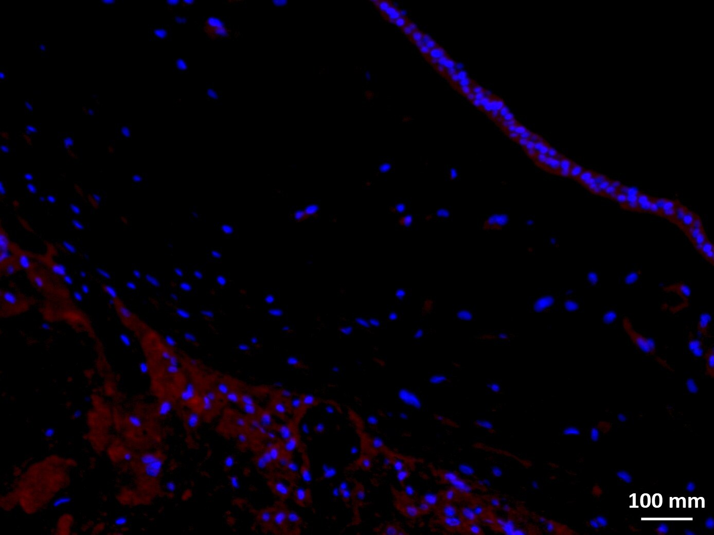
Immunocytochemistry/ Immunofluorescence: 15-PGDH/HPGD Antibody [NB200-179]
15-PGDH-HPGD-Antibody-Immunocytochemistry-Immunofluorescence-NB200-179-img0017.jpg![Immunocytochemistry/ Immunofluorescence: 15-PGDH/HPGD Antibody [NB200-179]](https://resources.rndsystems.com/images/products/15-PGDH-HPGD-Antibody-Immunocytochemistry-Immunofluorescence-NB200-179-img0004.jpg "Immunocytochemistry/ Immunofluorescence: 15-PGDH/HPGD Antibody [NB200-179]")
Immunocytochemistry/ Immunofluorescence: 15-PGDH/HPGD Antibody [NB200-179]
Immunocytochemistry/Immunofluorescence: 15-PGDH/HPGD Antibody [NB200-179] - 15-PGDH antibody was tested in HeLa cells with Dylight 488 (green). Nuclei and alpha-tubulin were counterstained with DAPI (blue) and Dylight 550 (red).![Immunohistochemistry-Paraffin: 15-PGDH/HPGD Antibody [NB200-179]](https://resources.rndsystems.com/images/products/15-PGDH-HPGD-Antibody-Immunohistochemistry-Paraffin-NB200-179-img0003.jpg "Immunohistochemistry-Paraffin: 15-PGDH/HPGD Antibody [NB200-179]")
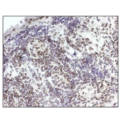
Immunohistochemistry-Paraffin: 15-PGDH/HPGD Antibody [NB200-179]
Immunohistochemistry-Paraffin: 15-PGDH/HPGD Antibody [NB200-179] - IHC analysis of 15-PGDH in human synovial tissue from patient with rheumatoid arthritis. Image courtesy of an anonymous customer review.![Immunohistochemistry: 15-PGDH/HPGD Antibody [NB200-179]](https://resources.rndsystems.com/images/products/15-PGDH-HPGD-Antibody-Immunohistochemistry-NB200-179-img0009.jpg "Immunohistochemistry: 15-PGDH/HPGD Antibody [NB200-179]")
Immunohistochemistry: 15-PGDH/HPGD Antibody [NB200-179]
Immunohistochemistry: 15-PGDH/HPGD Antibody [NB200-179] - Staining in human fetal membrane tissue. Image from verified customer review.![Knockdown Validated: 15-PGDH/HPGD Antibody [NB200-179]](https://resources.rndsystems.com/images/products/15-PGDH-HPGD%20Antibody-Knockdown%20Validated-NB200-179-img0018.jpg "Western Blot: 15-PGDH/HPGD Antibody [NB200-179]")

Immunohistochemistry: 15-PGDH/HPGD Antibody [NB200-179] -
Immunohistochemistry: 15-PGDH/HPGD Antibody [NB200-179] - Expression of 15-prostaglandin dehydrogenase (15-PGDH) in healthy & inflamed synovial tissue. Immunohistochemical staining reveals positive (brown) staining for 15-PGDH (hematoxilin counterstained) in synovial tissue from healthy individuals (a) & patients with rheumatoid arthritis (b), osteoarthritis (c), or psoriatic arthritis (d). Original magnifications: ×250 (a, b), ×100 (c, d). Image collected & cropped by CiteAb from the following publication (https://pubmed.ncbi.nlm.nih.gov/22616846), licensed under a CC-BY license. Not internally tested by Novus Biologicals.
Immunocytochemistry/ Immunofluorescence: 15-PGDH/HPGD Antibody [NB200-179] -
Immunocytochemistry/ Immunofluorescence: 15-PGDH/HPGD Antibody [NB200-179] - Cellular localization of 15-prostaglandin dehydrogenase (15-PGDH) in rheumatoid arthritis synovial tissue & the effects of anti-rheumatic treatment on its expression. (a) Double-immunofluorescence images show staining for 15-PGDH-positive (red) cells & cell marker staining (green) for macrophage CD163, fibroblast prolyl-4-hydroxylase, & endothelial cell CD31. Merged images display double-stained cells in yellow. Original magnification: ×400. Light microscopy pictures of representative synovial biopsy sections show immunohistochemical positive (brown) staining for 15-PGDH before & after intra-articular treatment with glucocorticoids (b) & before & 8 weeks after initiation of methotrexate therapy (c) (hematoxilin counterstained). Graphs display the comparative 15-PGDH expression in rheumatoid arthritis synovial tissue before & after treatment with glucocorticoids or methotrexate as a percentage of the positive stained area versus the total tissue area. GC, glucocorticoids; Mtx, methotrexate. Image collected & cropped by CiteAb from the following publication (https://pubmed.ncbi.nlm.nih.gov/22616846), licensed under a CC-BY license. Not internally tested by Novus Biologicals.
Western Blot: 15-PGDH/HPGD Antibody [NB200-179] -
Western Blot: 15-PGDH/HPGD Antibody [NB200-179] - Effect of LTC4 on 15-PGDH expression in the colon cancer cell line Caco-2(A) Quantification by qPCR of the mRNA expression of 15-PGDH following treatment with 40 nM LTC4 for 48 h in the presence or absence of AP100984 (a CysLT2 receptor antagonist, 1 μM) & JNK inhibitor I (10 μM). (B) Western blot showing 15-PGDH expression (antibody dilution, 1:5000) & densitometric analysis of LTC4-induced 15-PGDH up-regulation before or after the cells were stimulated with 40 nM LTC4 in the presence or absence of AP100984 & JNK inhibitor I for 48 h. (C) Confocal microscopy immunofluorescence images showing the expression of 15-PGDH (in green; antibody dilution, 1:200; DAPI in blue) after 24 h of stimulation with LTC4 in Caco-2 cells. The objective used was 63x. The data are presented as the percent of untreated control cells & represent the mean ± SEM of at least three separate experiments. Statistical analysis was conducted using an unpaired t-test; *P≤0.05, **P<0.01, ***P<0.001. Image collected & cropped by CiteAb from the following publication (https://pubmed.ncbi.nlm.nih.gov/28402256), licensed under a CC-BY license. Not internally tested by Novus Biologicals.
Western Blot: 15-PGDH/HPGD Antibody [NB200-179] -
Western Blot: 15-PGDH/HPGD Antibody [NB200-179] - Effect of 15-PGDH down-regulation on the cell differentiation markers SI & Mucin-2(A) A representative western blot & densitometric analysis of LTC4-induced SI protein expression after transfection with a scrambled control siRNA or 15-PGDH siRNA in HT-29 cells are shown. The cells were treated with or without 40 nM LTC4 for 48 h, & the change in the SI protein level was detected using an SI-specific antibody (1:1000 dilution) & 15-PGDH was detected using a 15-PGDH antibody (1:5000 dilution). The membrane was re-probed with an antibody against beta –actin to ensure equal loading. (B) A representative western blot & densitometric analysis performed as in (A) shown here for Caco-2 cells. (C, D) Representative confocal microscopy immunofluorescence images from cells transfected with a scrambled control siRNA or 15-PGDH siRNA with or without stimulation with 40 nM LTC4 for 48 h. The expression of Mucin-2 (in red; antibody dilution, 1:500; DAPI in blue, dilution 1:1000) (C) in HT-29 cells & (D) Caco-2 cells is shown. The objective used was 63x, & the scale bar is 50 μm. The data are presented as the percent of control cells & represent the mean ± SEM of at least three separate experiments. Statistical analysis was conducted using an unpaired t-test; *P≤0.05, **P<0.01. Image collected & cropped by CiteAb from the following publication (https://pubmed.ncbi.nlm.nih.gov/28402256), licensed under a CC-BY license. Not internally tested by Novus Biologicals.
Immunohistochemistry: 15-PGDH/HPGD Antibody [NB200-179] -
Immunohistochemistry: 15-PGDH/HPGD Antibody [NB200-179] - Cellular localization of 15-prostaglandin dehydrogenase (15-PGDH) in rheumatoid arthritis synovial tissue & the effects of anti-rheumatic treatment on its expression. (a) Double-immunofluorescence images show staining for 15-PGDH-positive (red) cells & cell marker staining (green) for macrophage CD163, fibroblast prolyl-4-hydroxylase, & endothelial cell CD31. Merged images display double-stained cells in yellow. Original magnification: ×400. Light microscopy pictures of representative synovial biopsy sections show immunohistochemical positive (brown) staining for 15-PGDH before & after intra-articular treatment with glucocorticoids (b) & before & 8 weeks after initiation of methotrexate therapy (c) (hematoxilin counterstained). Graphs display the comparative 15-PGDH expression in rheumatoid arthritis synovial tissue before & after treatment with glucocorticoids or methotrexate as a percentage of the positive stained area versus the total tissue area. GC, glucocorticoids; Mtx, methotrexate. Image collected & cropped by CiteAb from the following publication (https://pubmed.ncbi.nlm.nih.gov/22616846), licensed under a CC-BY license. Not internally tested by Novus Biologicals.Applications for 15-PGDH/HPGD Antibody - BSA Free
Application
Recommended Usage
Immunohistochemistry
1:200
Immunohistochemistry-Paraffin
1:200
Simple Western
1:500
Western Blot
1:5000-1:6000
Reviewed Applications
Read 3 reviews rated 4.7 using NB200-179 in the following applications:
Formulation, Preparation, and Storage
Purification
Protein A purified
Formulation
PBS
Format
BSA Free
Preservative
0.05% Sodium Azide
Concentration
2 mg/ml
Shipping
The product is shipped with polar packs. Upon receipt, store it immediately at the temperature recommended below.
Stability & Storage
Store at 4C short term. Aliquot and store at -20C long term. Avoid freeze-thaw cycles.
Background: 15-PGDH/HPGD
Long Name
15-Hydroxyprostaglandin Dehydrogenase
Alternate Names
15PGDH, HPGD, PGDH1, SDR36C1
Gene Symbol
HPGD
Additional 15-PGDH/HPGD Products
Product Documents for 15-PGDH/HPGD Antibody - BSA Free
Certificate of Analysis
To download a Certificate of Analysis, please enter a lot or batch number in the search box below.
Product Specific Notices for 15-PGDH/HPGD Antibody - BSA Free
This product is for research use only and is not approved for use in humans or in clinical diagnosis. Primary Antibodies are guaranteed for 1 year from date of receipt.
Related Research Areas
Citations for 15-PGDH/HPGD Antibody - BSA Free
 Powered by Bioz
Customer Reviews for 15-PGDH/HPGD Antibody - BSA Free (3)
4.7 out of 5
3 Customer Ratings
Have you used 15-PGDH/HPGD Antibody - BSA Free?
Submit a review and receive an Amazon gift card!
$25/€18/£15/$25CAN/¥2500 Yen for a review with an image
$10/€7/£6/$10CAN/¥1110 Yen for a review without an image
Submit a review
Customer Images
-(01-ml)_NB200-179_7871.jpg)
Showing
1
-
3 of
3 reviews
Showing All
Filter By:
-
Application: ImmunofluorescenceSample Tested: Fetal membranesSpecies: HumanVerified Customer | Posted 06/07/2018

-
Application: Western BlotSample Tested: Human ling tissue lysatesSpecies: HumanVerified Customer | Posted 05/28/201415-PGDH expression in human lung tissue
-
Application: ImmunocytochemistrySample Tested: synovial tissue from patient with rheumatoid arthritisSpecies: HumanVerified Customer | Posted 08/17/2012
There are no reviews that match your criteria.
Protocols
View specific protocols for 15-PGDH/HPGD Antibody - BSA Free (NB200-179):
Culture cells to appropriate density in 35 mm culture dishes or 6-well plates.
1. Remove culture medium and add 10% formalin to the dish. Fix at room temperature for 30 minutes.
2. Remove the formalin and add ice cold methanol. Incubate for 5-10 minutes.
3. Remove methanol and add washing solution (i.e. PBS). Be sure to not let the specimen dry out. Wash three times for 10 minutes.
4. To block nonspecific antibody binding incubate in 10% normal goat serum from 1 hour to overnight at room temperature.
5. Add primary antibody at appropriate dilution and incubate at room temperature from 2 hours to overnight at room temperature.
6. Remove primary antibody and replace with washing solution. Wash three times for 10 minutes.
7. Add secondary antibody at appropriate dilution. Incubate for 1 hour at room temperature.
8. Remove antibody and replace with wash solution, then wash for 10 minutes. Add Hoechst 33258 to wash solution at 1:25,0000 and incubate for 10 minutes. Wash a third time for 10 minutes.
9. Cells can be viewed directly after washing. The plates can also be stored in PBS containing Azide covered in Parafilm (TM). Cells can also be cover-slipped using Fluoromount, with appropriate sealing.
*The above information is only intended as a guide. The researcher should determine what protocol best meets their needs. Please follow safe laboratory procedures.
Immunohistochemistry
1. 5 uM-thick formalin-fixed paraffin-embedded tissue sections were baked at 60C for 75 minutes, deparaffinized, and rehydrated.
2. Antigen retrieval was performed by steaming the sections at 96C for 5 minutes in 10 mM citrate buffer (pH 6.0), plus a cool-down period of 20 minutes.
3. Reduction of peroxidases was accomplished by incubating in 3% H2O2 in water for 30 minutes at room temperature.
4. Avidin ??biotin blocking was performed for 15 minutes each, followed by nonspecific protein blocking (Serum-Free Protein Block, Dako, Carpenteria, CA) performed for 60 minutes.
5. Primary antibody was diluted in 1% BSA and incubated overnight at 4C in humidified chambers.
6. The slides were washed thoroughly, and Protein Block was added again for 30 minutes.
7. LSAB+ anti-rabbit kit (Dako) was used for development, applying the secondary antibody and HRP-conjugated streptavidin per the manufacturer's instructions.
8. Diaminobenzidine (Dako) was added to the slides for 10 minutes.
9. The sections were then counterstained by using Harris modified hematoxylin stain (Fisher Scientific) for 1 minute.
**Note: All washes were done with TBS (50 mM Tris-HCl/150 mM NaCl, pH 7.6) diluted in deionized water.
Western Blot I (LoVo lysates)
1. Perform SDS-PAGE (4-12%) on samples to be analyzed, loading 20ug of total protein per lane.
2. Transfer proteins to Nitrocellulose according to the instructions provided by the manufacturer of the transferapparatus.
3. Stain the blot using ponceau S for 1-2 minutes to access the transfer of proteins onto the nitrocellulose membrane. Rinse the blot in water to remove excess stain and mark the lane locations and locations of molecular weight markers using a pencil.
4. Rinse the blot in TBS for approximately 5 minutes.
5. Block the membrane using 5% non-fat dry milk in TBS for 1 hour.
6. Dilute the rabbit anti-15-PGDH primary antibody (NB 200-179) in blocking buffer and incubate overnight at 4C.
7. Wash the membrane in water for 5 minutes and apply the diluted rabbit-IgG HRP-conjugated secondary antibody in blocking buffer (as per manufacturers instructions) and incubate 1 hour at room temperature.
8. Wash the blot in TBS containing 0.05-0.1% Tween-20 for 10-20 minutes.
9. Wash the blot in type I water for an additional 10-20 minutes (this step can be repeated as required to reduce background).
10. Apply the detection reagent of choice in accordance with the manufacturers instructions (Amersham's ECL is the standard reagent used at Novus Biologicals).
**Note: Tween-20 can be added to the blocking buffer at a final concentration of 0.05-0.2%, provided it does not interfere with antibody-antigen binding.
Western Blot II (A549 lysates)
1. Cell lysates are prepared in RIPA buffer (50 mM 50 mM Tris-HCL/1% Nonidet P-40/0.25% Na-deoxycholate/150 mM NaCl/1 mM EDTA/1 mM PMSF) supplemented with a protease inhibitor mixture (Roche Applied Sciences).
2. They are then separated on 10% or 12% SDS/PAGE (30-150 ug per lane) and transferred to a Immobilon PVDF membrane (Millipore).
3. The membrane is blocked with 5% NFDM in TBS-T (TBS + 0.1% Tween-20).
4. The membrane is then probed with the diluted anti-PGDH antibody (NB 200-179), diluted in blocking buffer at RT for 1 hour.
5. The membrane is washed 3 times with TBS-T.
6. The membrane is then incubated with a biotinylated goat anti-rabbit IgG (diluted as per manufacturer's guidelines in blocking buffer) at RT for 1 hour.
7. The membrane was washed extensively.
8. The membrane is then incubated with an HRP-conjugated streptavidin (1:2,000) complex at RT for 1 hour.
9. The membrane is washed extensively.
10. The membrane is developed using and ECL detection system.
Find general support by application which include: protocols, troubleshooting, illustrated assays, videos and webinars.
- Antigen Retrieval Protocol (PIER)
- Antigen Retrieval for Frozen Sections Protocol
- Appropriate Fixation of IHC/ICC Samples
- Cellular Response to Hypoxia Protocols
- Chromogenic IHC Staining of Formalin-Fixed Paraffin-Embedded (FFPE) Tissue Protocol
- Chromogenic Immunohistochemistry Staining of Frozen Tissue
- ClariTSA™ Fluorophore Kits
- Detection & Visualization of Antibody Binding
- Fluorescent IHC Staining of Frozen Tissue Protocol
- Graphic Protocol for Heat-induced Epitope Retrieval
- Graphic Protocol for the Preparation and Fluorescent IHC Staining of Frozen Tissue Sections
- Graphic Protocol for the Preparation and Fluorescent IHC Staining of Paraffin-embedded Tissue Sections
- Graphic Protocol for the Preparation of Gelatin-coated Slides for Histological Tissue Sections
- ICC Cell Smear Protocol for Suspension Cells
- ICC Immunocytochemistry Protocol Videos
- ICC for Adherent Cells
- IHC Sample Preparation (Frozen sections vs Paraffin)
- Immunocytochemistry (ICC) Protocol
- Immunocytochemistry Troubleshooting
- Immunofluorescence of Organoids Embedded in Cultrex Basement Membrane Extract
- Immunofluorescent IHC Staining of Formalin-Fixed Paraffin-Embedded (FFPE) Tissue Protocol
- Immunohistochemistry (IHC) and Immunocytochemistry (ICC) Protocols
- Immunohistochemistry Frozen Troubleshooting
- Immunohistochemistry Paraffin Troubleshooting
- Preparing Samples for IHC/ICC Experiments
- Preventing Non-Specific Staining (Non-Specific Binding)
- Primary Antibody Selection & Optimization
- Protocol for Heat-Induced Epitope Retrieval (HIER)
- Protocol for Making a 4% Formaldehyde Solution in PBS
- Protocol for VisUCyte™ HRP Polymer Detection Reagent
- Protocol for the Fluorescent ICC Staining of Cell Smears - Graphic
- Protocol for the Fluorescent ICC Staining of Cultured Cells on Coverslips - Graphic
- Protocol for the Preparation & Fixation of Cells on Coverslips
- Protocol for the Preparation and Chromogenic IHC Staining of Frozen Tissue Sections
- Protocol for the Preparation and Chromogenic IHC Staining of Frozen Tissue Sections - Graphic
- Protocol for the Preparation and Chromogenic IHC Staining of Paraffin-embedded Tissue Sections
- Protocol for the Preparation and Chromogenic IHC Staining of Paraffin-embedded Tissue Sections - Graphic
- Protocol for the Preparation and Fluorescent ICC Staining of Cells on Coverslips
- Protocol for the Preparation and Fluorescent ICC Staining of Non-adherent Cells
- Protocol for the Preparation and Fluorescent ICC Staining of Stem Cells on Coverslips
- Protocol for the Preparation and Fluorescent IHC Staining of Frozen Tissue Sections
- Protocol for the Preparation and Fluorescent IHC Staining of Paraffin-embedded Tissue Sections
- Protocol for the Preparation of Gelatin-coated Slides for Histological Tissue Sections
- Protocol for the Preparation of a Cell Smear for Non-adherent Cell ICC - Graphic
- R&D Systems Quality Control Western Blot Protocol
- TUNEL and Active Caspase-3 Detection by IHC/ICC Protocol
- The Importance of IHC/ICC Controls
- Troubleshooting Guide: Immunohistochemistry
- Troubleshooting Guide: Western Blot Figures
- Western Blot Conditions
- Western Blot Protocol
- Western Blot Protocol for Cell Lysates
- Western Blot Troubleshooting
- Western Blot Troubleshooting Guide
- View all Protocols, Troubleshooting, Illustrated assays and Webinars
Loading...