4-Hydroxynonenal Antibody (198960)
R&D Systems | Catalog # MAB3249

Key Product Details
Validated by
Species Reactivity
Validated:
Cited:
Applications
Validated:
Cited:
Label
Antibody Source
Product Specifications
Immunogen
Specificity
Clonality
Host
Isotype
Scientific Data Images for 4-Hydroxynonenal Antibody (198960)
Detection of Human 4‑Hydroxynonenal by Western Blot.
Western blot shows lysates of HepG2 human hepatocellular carcinoma cell line untreated (-) or treated (+) with 4-Hydroxynonenal (4-HNE). PVDF membrane was probed with 1 µg/mL of Mouse Anti-4-Hydroxynonenal Monoclonal Antibody (Catalog # MAB3249) followed by HRP-conjugated Anti-Mouse IgG Secondary Antibody (Catalog # HAF007). Specific bands were detected for 4-Hydroxynonenal at adducts of histidine residues (as indicated). This experiment was conducted under reducing conditions and using Immunoblot Buffer Group 1.
Detection of Human 4‑Hydroxynonenal by Simple WesternTM.
Simple Western lane view shows lysates of HepG2 human hepatocellular carcinoma cell line untreated (-) or treated (+) with 4-Hydroxynonenal (4-HNE), loaded at 0.2 mg/mL. Specific bands were detected for 4‑Hydroxynonenal at adducts of histidine residues kDa (as indicated) using 5 µg/mL of Mouse Anti-4‑Hydroxynonenal Monoclonal Antibody (Catalog # MAB3249). This experiment was conducted under reducing conditions and using the 12-230 kDa separation system.
4‑Hydroxynonenal in Human Prostate.
4-Hydroxynonenal was detected in immersion fixed paraffin-embedded sections of human prostate using Mouse Anti-4-Hydroxynonenal Monoclonal Antibody (Catalog # MAB3249) at 0.1 µg/mL for 1 hour at room temperature followed by incubation with the Anti-Mouse IgG VisUCyte™ HRP Polymer Antibody (Catalog # VC001). Before incubation with the primary antibody, tissue was subjected to heat-induced epitope retrieval using Antigen Retrieval Reagent-Basic (Catalog # CTS013). Tissue was stained using DAB (brown) and counterstained with hematoxylin (blue). Specific staining was localized to cytoplasm. View our protocol for IHC Staining with VisUCyte HRP Polymer Detection Reagents.
4‑Hydroxynonenal in Human Prostate Cancer Tissue.
4-Hydroxynonenal was detected in immersion fixed paraffin-embedded sections of human prostate cancer tissue using Mouse Anti-4-Hydroxynonenal Monoclonal Antibody (Catalog # MAB3249) at 0.5 µg/mL for 1 hour at room temperature followed by incubation with the Anti-Mouse IgG VisUCyte™ HRP Polymer Antibody (Catalog # VC001). Before incubation with the primary antibody, tissue was subjected to heat-induced epitope retrieval using Antigen Retrieval Reagent-Basic (Catalog # CTS013). Tissue was stained using DAB (brown) and counterstained with hematoxylin (blue). Specific staining was localized to cytoplasm. View our protocol for IHC Staining with VisUCyte HRP Polymer Detection Reagents.
Detection of Mouse 4-Hydroxynonenal by Immunocytochemistry/Immunofluorescence
Diapocynin inhibits the formation of 3-nitrotyrosine (3-NT) and 4-hydroxynonenal (4-HNE) in the substantia nigra (SN) of MPTP-treated mice. (A) Representative Western blots illustrating the expression of 3-NT in SN. (B) Bar graph showing mean Western blot 3-NT/ beta -actin ratios ± SEM in SN of 6 mice per group. (C) Double labeling of tyrosine hydroxylase (TH) and 3-NT in SN region of ventral midbrain. (D) Representative Western blots illustrating the expression of 4-HNE in SN. (E) Bar graph showing mean Western blot 4-HNE/ beta -actin ratios ± SEM in SN of 6 mice per group. (F) Double labeling of TH and 4-HNE in SN region of ventral midbrain. Images were captured at 60× magnification. The SN zone is outlined in white dots. Inset pictures demonstrated colocalization of TH and 3-NT/4-HNE. ***P <0.001 vs the control group; **P <0.01 vs the control group; *P <0.05 vs the MPTP group; #P <0.001 vs the MPTP group. Image collected and cropped by CiteAb from the following publication (https://pubmed.ncbi.nlm.nih.gov/23092448), licensed under a CC-BY license. Not internally tested by R&D Systems.
Detection of Mouse 4-Hydroxynonenal by Western Blot
Analysis of inflammatory response and oxidative stress levels of atherosclerotic VSMCs in mice: (A–E) representative images (A) and data of the protein levels of 4-HNE (B), Nrf2 (C), TNF-alpha (D), and IL-1 beta (E). (F–H) The expression of Nrf2, TNF-alpha, and IL-1 beta was quantified by qPCR and standardized to GAPDH. (I,O) ROS fluorescence detection (red) and quantification. Scale bar 100 μm. (J–N) Representative images (J) and data of the protein levels of P-PI3K (K), PI3K (L), P-Akt (M), and Akt (N). The bar graphs depict means ± SEM; statistical significance * p < 0.05, ** p < 0.01, and *** p < 0.001 and ns by the one-way ANOVA test. Image collected and cropped by CiteAb from the following open publication (https://pubmed.ncbi.nlm.nih.gov/37047626), licensed under a CC-BY license. Not internally tested by R&D Systems.
Detection of 4-Hydroxynonenal by Immunohistochemistry
CYP1B1 expression attenuates the effect of anti-PD-1 therapy. A Western blot analysis of MC38 cells expressing ctrl shRNA or CYP1B1 shRNAs. B C57BL/6 J mice carrying the indicated MC38 cell formed tumors at the right flank were treated with anti-mPD-1 antibody or isotype control IgG for a total of five treatments. C, D Tumor images C and tumor growth curve D in mice bearing indicated MC38 cell tumors treated with anti-mPD-1 or isotype control IgG antibodies. E IHC staining of 4-HNE in MC38 tumors expressing ctrl or CYP1B1 shRNA. F Quantification of 4-HNE IHC staining. n = 5 per group. p value was determined by Student’s two-sided t test. *p < 0.05, **p < 0.01, ***p < 0.001. Image collected and cropped by CiteAb from the following open publication (https://pubmed.ncbi.nlm.nih.gov/37059712), licensed under a CC-BY license. Not internally tested by R&D Systems.
Detection of 4-Hydroxynonenal by Immunohistochemistry
B cell-derived antibodies mediate HHcy-aggravated kidney lipid peroxidation in hypertensive mice. a Immunohistochemical staining of 4-HNE (brown) from kidney sections in sham or 2K1C mice with or without Hcy (1.8 g/L) in drinking water for 4 weeks, indicative of lipid peroxidation. Rituximab was administered to HHcy 2K1C mice at the start of modeling (i.p. 75 μg/20 g body weights every other day for 4 weeks). The immunohistochemical staining was calculated as the percentage of brown signals over the total area. n = 3. b, c Effects of 2K1C and HHcy on lipid peroxidation in the kidney were measured using ELISA for LPO (b) and MDA (c). n = 6. d Quantitative PCR analysis of redox enzyme expression in kidney tissues, including acsl4, lox15, slc7a11 and gpx4. n = 6. e Western blot analysis of LOX15, GPX4 and SLC7A11 protein expression and quantification. beta -actin was used as an internal control. n = 3. f, g The redox balance in the kidney was assayed for the ratio of GSSG/GSH and NADP+/NADPH using ELISA. n = 6. h Quantitative PCR analysis of redox enzyme expression in the renal CD31-positive GECs from frozen section by laser capture microdissection, including acsl4, lox15, slc7a11 and gpx4. n = 3. LPO lipid peroxide, MDA malondialdehyde, 4-HNE 4-hydroxynonenal, lox15 lipoxygenase 15, acsl4 acyl-CoA synthetase long chain family member 4, slc7a11 cystine/glutamate transporter, gpx4 glutathione peroxidase 4, GSH glutathione, GSSG glutathione disulfide. All data are expressed as the means ± SEM. *P < 0.05, **P < 0.01 Image collected and cropped by CiteAb from the following open publication (https://pubmed.ncbi.nlm.nih.gov/36907919), licensed under a CC-BY license. Not internally tested by R&D Systems.
Detection of 4-Hydroxynonenal by Immunohistochemistry
CYP1B1 expression attenuates the effect of anti-PD-1 therapy. A Western blot analysis of MC38 cells expressing ctrl shRNA or CYP1B1 shRNAs. B C57BL/6 J mice carrying the indicated MC38 cell formed tumors at the right flank were treated with anti-mPD-1 antibody or isotype control IgG for a total of five treatments. C, D Tumor images C and tumor growth curve D in mice bearing indicated MC38 cell tumors treated with anti-mPD-1 or isotype control IgG antibodies. E IHC staining of 4-HNE in MC38 tumors expressing ctrl or CYP1B1 shRNA. F Quantification of 4-HNE IHC staining. n = 5 per group. p value was determined by Student’s two-sided t test. *p < 0.05, **p < 0.01, ***p < 0.001. Image collected and cropped by CiteAb from the following open publication (https://pubmed.ncbi.nlm.nih.gov/37059712), licensed under a CC-BY license. Not internally tested by R&D Systems.
Detection of 4-Hydroxynonenal by Immunohistochemistry
B cell-derived antibodies mediate HHcy-aggravated kidney lipid peroxidation in hypertensive mice. a Immunohistochemical staining of 4-HNE (brown) from kidney sections in sham or 2K1C mice with or without Hcy (1.8 g/L) in drinking water for 4 weeks, indicative of lipid peroxidation. Rituximab was administered to HHcy 2K1C mice at the start of modeling (i.p. 75 μg/20 g body weights every other day for 4 weeks). The immunohistochemical staining was calculated as the percentage of brown signals over the total area. n = 3. b, c Effects of 2K1C and HHcy on lipid peroxidation in the kidney were measured using ELISA for LPO (b) and MDA (c). n = 6. d Quantitative PCR analysis of redox enzyme expression in kidney tissues, including acsl4, lox15, slc7a11 and gpx4. n = 6. e Western blot analysis of LOX15, GPX4 and SLC7A11 protein expression and quantification. beta -actin was used as an internal control. n = 3. f, g The redox balance in the kidney was assayed for the ratio of GSSG/GSH and NADP+/NADPH using ELISA. n = 6. h Quantitative PCR analysis of redox enzyme expression in the renal CD31-positive GECs from frozen section by laser capture microdissection, including acsl4, lox15, slc7a11 and gpx4. n = 3. LPO lipid peroxide, MDA malondialdehyde, 4-HNE 4-hydroxynonenal, lox15 lipoxygenase 15, acsl4 acyl-CoA synthetase long chain family member 4, slc7a11 cystine/glutamate transporter, gpx4 glutathione peroxidase 4, GSH glutathione, GSSG glutathione disulfide. All data are expressed as the means ± SEM. *P < 0.05, **P < 0.01 Image collected and cropped by CiteAb from the following open publication (https://pubmed.ncbi.nlm.nih.gov/36907919), licensed under a CC-BY license. Not internally tested by R&D Systems.
Detection of Mouse 4-Hydroxynonenal by Immunohistochemistry
RMZL suppressed oxidative stress and inflammation in HIRI mice. Mice were subjected to ischemia for 1 h and then reperfusion for 6 h through liver surgery, and followed by RMZL treatment. (A) SOD and MDA levels in the left hepatic tissues were determined using commercial kits. (C) 4-HNE expression in the left hepatic tissues was detected by IHC. (D) The levels of TNF-alpha, IL-1 beta, IL-6 and IL-10 in serum from mice were evaluated using ELISA. All data were indicated as mean ± SD (n = 6 per group). *P < 0.05, **P < 0.01, ***P < 0.001 Image collected and cropped by CiteAb from the following open publication (https://pubmed.ncbi.nlm.nih.gov/40263992), licensed under a CC-BY license. Not internally tested by R&D Systems.
Detection of 4-Hydroxynonenal by Western Blot
S. aureus and E. coli infections induced lipid peroxides accumulation in infected bone microenvironment and BMSCs. A) Lipid peroxidation levels in bone marrow on Day 3 post‐infection in murine osteomyelitis model induced by S. aureus and E. coli were determined by WB analysis of 4‐HNE and MDA protein modifications. Each group contained four mice. B) Representative tissue immunofluorescence images of LepR, MDA, 4‐HNE, ACSL4, GPX4 in the uninfected murine femur and implant‐associated bone infection model induced by S. aureus and E. coli. Scale bar = 50 µm. C) The schematic route of animal experiments and gating strategy were depicted. D) Lipid peroxide regulation by S. aureus and E. coli infections and Fer‐1 in CD45−CD31−Ter119−LepR+ BMSCs were analyzed in murine osteomyelitis model by Liperfluo staining and analyzed by flow cytometry. E,F) Quantification of median fluorescent intensity in flow cytometry analysis in (D). Values are means ± SDs. Each group contained six mice. Multiple comparison was performed by one‐way analysis of variance (ANOVA) with Tukey's post‐hoc analysis. Image collected and cropped by CiteAb from the following open publication (https://pubmed.ncbi.nlm.nih.gov/39166412), licensed under a CC-BY license. Not internally tested by R&D Systems.
Detection of Mouse 4-Hydroxynonenal by Western Blot
Inhibition of H3K14la improved functional outcomes after ICH in mice. Immunostaining of H3K14la and NeuN was performed at 24 h after ICH. “H” indicates hematoma. The representative images (A) and quantifications (B) are shown. n = 5 mice. C The expression of 4-HNE was measured at 24 h after ICH by western blot analysis. Tubulin serves as the loading control. n = 4 mice. D FJC staining was performed at 24 h after ICH. The representative images and quantification are shown. n = 4 mice. E–G 1 μl of 500 μM GSK or 10 mM Caloxin 2A1 was injected at 30 min after ICH. Neurological function and motor function were determined at indicated time points after ICH. n = 8 mice. FJC staining was performed at 24 h after ICH. The representative images (H) and quantification (I) are shown. n = 4 mice. J–L AAV virus of NC or shP300 virus was injected at 1 week before ICH and Neurological function and motor function were examined at indicated time points after ICH. Results are shown as scatter plots (Mean ± SD). n = 8 mice. One-way (B–D and I) or two-way (E–G and J–L) ANOVA followed by Tukey’s multiple comparisons tests was performed, *p < 0.05, **p < 0.01, ***p < 0.001, ****p < 0.0001vs Sham; #p < 0.05 vs ICH + NC/Veh; NS, not significant. Image collected and cropped by CiteAb from the following open publication (https://pubmed.ncbi.nlm.nih.gov/40701963), licensed under a CC-BY license. Not internally tested by R&D Systems.
Detection of Mouse 4-Hydroxynonenal by Immunohistochemistry
RMZL suppressed oxidative stress and inflammation in HIRI mice. Mice were subjected to ischemia for 1 h and then reperfusion for 6 h through liver surgery, and followed by RMZL treatment. (A) SOD and MDA levels in the left hepatic tissues were determined using commercial kits. (C) 4-HNE expression in the left hepatic tissues was detected by IHC. (D) The levels of TNF-alpha, IL-1 beta, IL-6 and IL-10 in serum from mice were evaluated using ELISA. All data were indicated as mean ± SD (n = 6 per group). *P < 0.05, **P < 0.01, ***P < 0.001 Image collected and cropped by CiteAb from the following open publication (https://pubmed.ncbi.nlm.nih.gov/40263992), licensed under a CC-BY license. Not internally tested by R&D Systems.Applications for 4-Hydroxynonenal Antibody (198960)
Immunohistochemistry
Sample: Immersion fixed paraffin-embedded sections of human normal prostate and prostate cancer tissue
Simple Western
Sample: HepG2 human hepatocellular carcinoma cell line treated with 4-Hydroxynonenal (4-HNE)
Western Blot
Sample: HepG2 human hepatocellular carcinoma cell line treated with 4-Hydroxynonenal (4-HNE)
Reviewed Applications
Read 3 reviews rated 4.3 using MAB3249 in the following applications:
Formulation, Preparation, and Storage
Purification
Reconstitution
Reconstitute at 0.5 mg/mL in sterile PBS. For liquid material, refer to CoA for concentration.
Formulation
Shipping
Stability & Storage
- 12 months from date of receipt, -20 to -70 °C as supplied.
- 1 month, 2 to 8 °C under sterile conditions after reconstitution.
- 6 months, -20 to -70 °C under sterile conditions after reconstitution.
Calculators
Background: 4-Hydroxynonenal
Alternate Names
Additional 4-Hydroxynonenal Products
Product Documents for 4-Hydroxynonenal Antibody (198960)
Certificate of Analysis
To download a Certificate of Analysis, please enter a lot or batch number in the search box below.
Note: Certificate of Analysis not available for kit components.
Product Specific Notices for 4-Hydroxynonenal Antibody (198960)
For research use only
Citations for 4-Hydroxynonenal Antibody (198960)
 Powered by Bioz
Customer Reviews for 4-Hydroxynonenal Antibody (198960) (3)
Have you used 4-Hydroxynonenal Antibody (198960)?
Submit a review and receive an Amazon gift card!
$25/€18/£15/$25CAN/¥2500 Yen for a review with an image
$10/€7/£6/$10CAN/¥1110 Yen for a review without an image
Submit a review
Customer Images
-
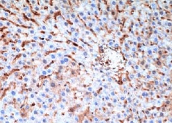
Application: ImmunohistochemistrySample Tested: Liver tissueSpecies: MouseVerified Customer | Posted 08/11/2021

-
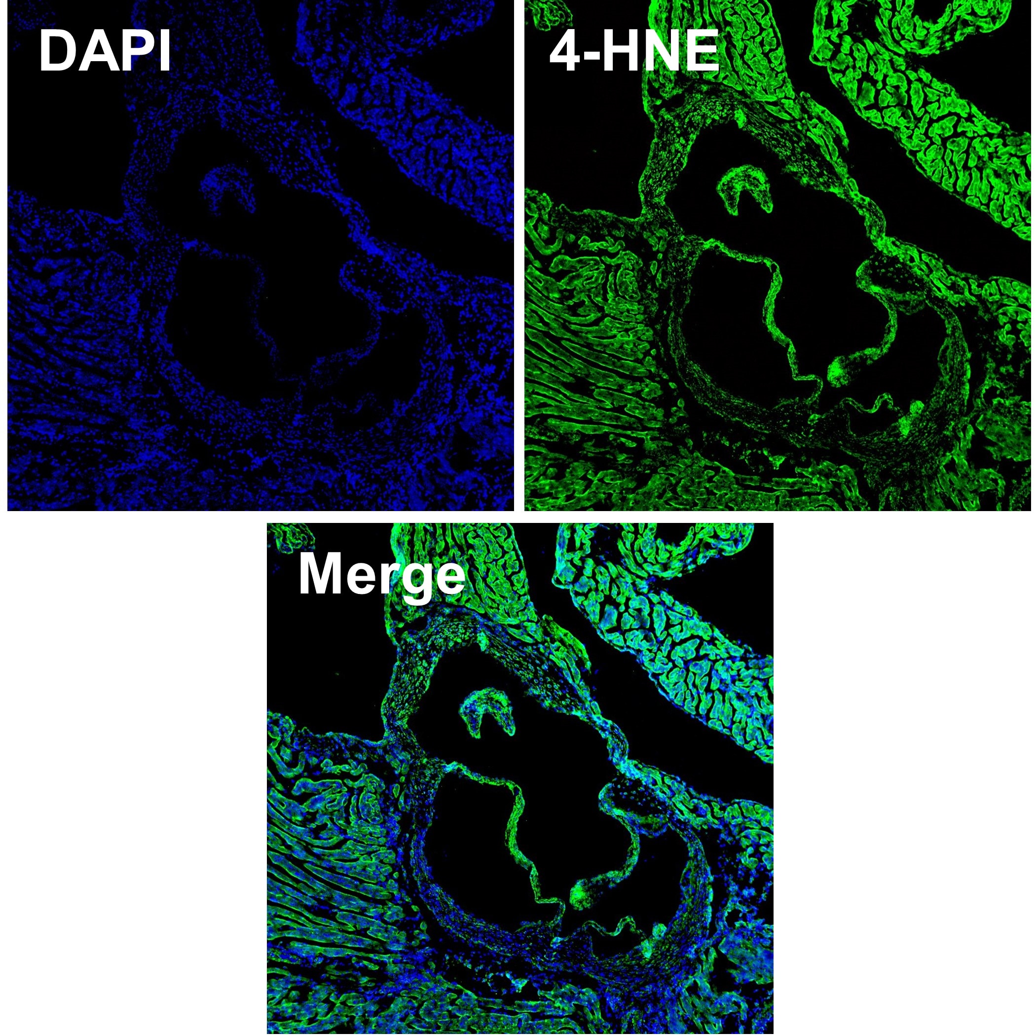
Application: Immunocytochemistry/ImmunofluorescenceSample Tested: Aortic valvesSpecies: MouseVerified Customer | Posted 03/29/2021-Fixed in 10% neutral buffered formalin followed by dehydration process -10 um thick cryosection - Blocking: Normal Goat Serum (NGS) - Primary Ab (4-HNE): diluted in NGS at 20 ug/mL; 4C overnight - Secondary (AF488): 1:100 dilution in NGS; room temp 1hr - Vector anti-fade mounting media with DAPI
-
Application: Western BlotSample Tested: See PMID 21429624Species: MouseVerified Customer | Posted 02/19/2015
There are no reviews that match your criteria.
Protocols
Find general support by application which include: protocols, troubleshooting, illustrated assays, videos and webinars.
- Antigen Retrieval Protocol (PIER)
- Antigen Retrieval for Frozen Sections Protocol
- Appropriate Fixation of IHC/ICC Samples
- Cellular Response to Hypoxia Protocols
- Chromogenic IHC Staining of Formalin-Fixed Paraffin-Embedded (FFPE) Tissue Protocol
- Chromogenic Immunohistochemistry Staining of Frozen Tissue
- ClariTSA™ Fluorophore Kits
- Detection & Visualization of Antibody Binding
- Fluorescent IHC Staining of Frozen Tissue Protocol
- Graphic Protocol for Heat-induced Epitope Retrieval
- Graphic Protocol for the Preparation and Fluorescent IHC Staining of Frozen Tissue Sections
- Graphic Protocol for the Preparation and Fluorescent IHC Staining of Paraffin-embedded Tissue Sections
- Graphic Protocol for the Preparation of Gelatin-coated Slides for Histological Tissue Sections
- IHC Sample Preparation (Frozen sections vs Paraffin)
- Immunofluorescent IHC Staining of Formalin-Fixed Paraffin-Embedded (FFPE) Tissue Protocol
- Immunohistochemistry (IHC) and Immunocytochemistry (ICC) Protocols
- Immunohistochemistry Frozen Troubleshooting
- Immunohistochemistry Paraffin Troubleshooting
- Preparing Samples for IHC/ICC Experiments
- Preventing Non-Specific Staining (Non-Specific Binding)
- Primary Antibody Selection & Optimization
- Protocol for Heat-Induced Epitope Retrieval (HIER)
- Protocol for Making a 4% Formaldehyde Solution in PBS
- Protocol for VisUCyte™ HRP Polymer Detection Reagent
- Protocol for the Preparation & Fixation of Cells on Coverslips
- Protocol for the Preparation and Chromogenic IHC Staining of Frozen Tissue Sections
- Protocol for the Preparation and Chromogenic IHC Staining of Frozen Tissue Sections - Graphic
- Protocol for the Preparation and Chromogenic IHC Staining of Paraffin-embedded Tissue Sections
- Protocol for the Preparation and Chromogenic IHC Staining of Paraffin-embedded Tissue Sections - Graphic
- Protocol for the Preparation and Fluorescent IHC Staining of Frozen Tissue Sections
- Protocol for the Preparation and Fluorescent IHC Staining of Paraffin-embedded Tissue Sections
- Protocol for the Preparation of Gelatin-coated Slides for Histological Tissue Sections
- R&D Systems Quality Control Western Blot Protocol
- TUNEL and Active Caspase-3 Detection by IHC/ICC Protocol
- The Importance of IHC/ICC Controls
- Troubleshooting Guide: Immunohistochemistry
- Troubleshooting Guide: Western Blot Figures
- Western Blot Conditions
- Western Blot Protocol
- Western Blot Protocol for Cell Lysates
- Western Blot Troubleshooting
- Western Blot Troubleshooting Guide
- View all Protocols, Troubleshooting, Illustrated assays and Webinars
FAQs for 4-Hydroxynonenal Antibody (198960)
-
Q: For the Western Blot data image for Catalog # MAB3249, how long were the HepG2 cells treated with 4-HNE prior to analysis?
A: The HepG2 cells were treated with 100 µM 4-HNE for 1 hour before lysing the cells.