Ago2/eIF2C2 Antibody (2E12-1C9) - Azide and BSA Free
Novus Biologicals | Catalog # H00027161-M01
![Knockdown Validated: Ago2/eIF2C2 Antibody (2E12-1C9) [H00027161-M01]](https://resources.rndsystems.com/images/products/Ago2-eIF2C2-Antibody-2E12-1C9-Western-Blot-H00027161-M01-img0018.jpg "Western Blot: Ago2/eIF2C2 Antibody (2E12-1C9) [H00027161-M01]")
Key Product Details
Validated by
Species Reactivity
Validated:
Cited:
Applications
Validated:
Cited:
Label
Antibody Source
Format
Product Specifications
Immunogen
Reactivity Notes
Specificity
Clonality
Host
Isotype
Scientific Data Images for Ago2/eIF2C2 Antibody (2E12-1C9) - Azide and BSA Free
![Western Blot: Ago2/eIF2C2 Antibody (2E12-1C9) [H00027161-M01]](https://resources.rndsystems.com/images/products/Ago2-eIF2C2-Antibody-2E12-1C9-Western-Blot-H00027161-M01-img0017.jpg "Western Blot: Ago2/eIF2C2 Antibody (2E12-1C9) [H00027161-M01]")
Western Blot: Ago2/eIF2C2 Antibody (2E12-1C9) [H00027161-M01]
Ago2-eIF2C2-Antibody-2E12-1C9-Western-Blot-H00027161-M01-img0017.jpg![Immunocytochemistry/ Immunofluorescence: Ago2/eIF2C2 Antibody (2E12-1C9) [H00027161-M01]](https://resources.rndsystems.com/images/products/Ago2-eIF2C2-Antibody-2E12-1C9-Immunocytochemistry-Immunofluorescence-H00027161-M01-img0008.jpg "Immunocytochemistry/ Immunofluorescence: Ago2/eIF2C2 Antibody (2E12-1C9) [H00027161-M01]")
Immunocytochemistry/ Immunofluorescence: Ago2/eIF2C2 Antibody (2E12-1C9) [H00027161-M01]
Immunocytochemistry/Immunofluorescence: Ago2/eIF2C2 Antibody (2E12-1C9) [H00027161-M01] - Analysis of monoclonal antibody to EIF2C2 on HeLa cell. Antibody concentration 10 ug/ml.![Immunohistochemistry-Paraffin: Ago2/eIF2C2 Antibody (2E12-1C9) [H00027161-M01]](https://resources.rndsystems.com/images/products/Ago2-eIF2C2-Antibody-2E12-1C9-Immunohistochemistry-Paraffin-H00027161-M01-img0010.jpg "Immunohistochemistry-Paraffin: Ago2/eIF2C2 Antibody (2E12-1C9) [H00027161-M01]")
Immunohistochemistry-Paraffin: Ago2/eIF2C2 Antibody (2E12-1C9) [H00027161-M01]
Immunohistochemistry-Paraffin: Ago2/eIF2C2 Antibody (2E12-1C9) [H00027161-M01] - Analysis of monoclonal antibody to EIF2C2 on formalin-fixed paraffin-embedded human stomach. Antibody concentration 3 ug/ml.![Western Blot: Ago2/eIF2C2 Antibody (2E12-1C9) [H00027161-M01]](https://resources.rndsystems.com/images/products/Ago2-eIF2C2-Antibody-2E12-1C9-Western-Blot-H00027161-M01-img0011.jpg "Western Blot: Ago2/eIF2C2 Antibody (2E12-1C9) [H00027161-M01]")
Western Blot: Ago2/eIF2C2 Antibody (2E12-1C9) [H00027161-M01]
Western Blot: Ago2/eIF2C2 Antibody (2E12-1C9) [H00027161-M01] - Analysis of EIF2C2 over-expressed 293 cell line, cotransfected with EIF2C2 Validated Chimera RNAi ( Cat # H00027161-R01V ) (Lane 2) or non-transfected control (Lane 1). Blot probed with EIF2C2 monoclonal antibody (M01), clone 2E12-1C9 (Cat # H00027161-M01 ). GAPDH ( 36.1 kDa ) used as specificity and loading control.![ELISA: Ago2/eIF2C2 Antibody (2E12-1C9) [H00027161-M01]](https://resources.rndsystems.com/images/products/Ago2-eIF2C2-Antibody-2E12-1C9-ELISA-H00027161-M01-img0015.jpg "ELISA: Ago2/eIF2C2 Antibody (2E12-1C9) [H00027161-M01]")
ELISA: Ago2/eIF2C2 Antibody (2E12-1C9) [H00027161-M01]
ELISA: Ago2/eIF2C2 Antibody (2E12-1C9) [H00027161-M01] - Detection limit for recombinant GST tagged EIF2C2 is 0.3 ng/ml as a capture antibody. [H00027161-M01] -")
Western Blot: Ago2/eIF2C2 Antibody (2E12-1C9) [H00027161-M01] -
Western Blot: Ago2/eIF2C2 Antibody (2E12-1C9) [H00027161-M01] - The impact of different HAd infections on RNAi/miRNA-pathway proteins.(A) Efficiency of different HAd infections. HeLa cells were infected with the indicated viruses, followed by a 35S-methionine pulse labeling after 24 & 48 hpi. Total protein lysates were separated on an SDS-PAGE & protein synthesis visualized by autoradiography. Accumulation of late viral hexon protein is indicated by an arrow. (B) HAd infections do not affect RNAi/miRNA-pathway protein levels. Western blot analysis on the same protein samples as in panel A was used to monitor the levels of RNAi/miRNA-pathway proteins Exportin 5, Dicer, TRBP & Ago2. Detection of the Lamin B protein served as a loading control. Letter “M” denotes mock, non-infected samples. The different panels were repeated at least two times. Image collected & cropped by CiteAb from the following publication (https://pubmed.ncbi.nlm.nih.gov/25144466), licensed under a CC-BY license. Not internally tested by Novus Biologicals. [H00027161-M01] -")
Western Blot: Ago2/eIF2C2 Antibody (2E12-1C9) [H00027161-M01] -
Western Blot: Ago2/eIF2C2 Antibody (2E12-1C9) [H00027161-M01] - AGO2 contributed predominately to silencing activity in both regions.(A) The relative AGO mRNA levels were measured by quantitative RT-PCR 24 hours post siAgo transfection. Results were average values of assays in triplicates, & all experiments were repeated three times. (B) The relative AGO protein levels were detected by western blot 56 hours post siAgo transfection, at the time point when AGO ablated cells were harvested to evaluate luciferase activities. GAPDH was included as loading control. The intensity of protein bands was quantified by ImageJ software (NIH, USA). All experiments were performed at least twice. (C) The normalized silencing efficacies of siR-04 on perfectly matched target in CDS versus 3′-UTR after AGOs ablation. Silencing of AGO expression was carried out by gene-specific siRNA assessed in the previous study [9], & subsequently, influence of the gene silencing on perfectly-match tolerance was evaluated by reporter system. All data were normalized to mock. (D) The normalized silencing efficacies of siR-04 on single-nucleotide mismatched target sites in CDS versus 3′-UTR at the indicated positions (4C, 10U, 12G, 17A) after AGOs ablation. The target site location & siRNA:mRNA match pattern were given under the x-axis. Error bars represented SD. Data were average values of assays in triplicates, & all experiments were repeated at least twice. Image collected & cropped by CiteAb from the following publication (https://pubmed.ncbi.nlm.nih.gov/23145149), licensed under a CC-BY license. Not internally tested by Novus Biologicals. [H00027161-M01] -")
Immunoprecipitation: Ago2/eIF2C2 Antibody (2E12-1C9) [H00027161-M01] -
Immunoprecipitation: Ago2/eIF2C2 Antibody (2E12-1C9) [H00027161-M01] - Snail1 is a target of miR‐30c. (A) Sequence alignment between miR‐30c & the 3′‐UTR of Snail1 among several species. (B) Ago2 protein levels in co‐immunoprecipitated products detected by Western blot. IgGHC, IgG heavy chain; IgGLC, IgG light chain. (C) Relative expression of Snail1 in the whole RNA (left) & RNA of the nonspecific IgG or anti‐Ago2 co‐IP (right) from the HG‐treated HK2 cell lysates. #P < 0.05 vs. miR‐con + input, *P < 0.05 vs. miR‐con + IgG IP. (D) Schematic diagram of the luciferase reporter plasmids of pMIR‐Snail1 3′‐UTR & pMIR‐Snail1 3′‐UTR mut, & the potential target site of miR‐30c on the 3′‐UTR of Snail1. (E) Regulation of miR‐30c on 3′‐UTR of Snail1 in HEK293 cells by luciferase reporter assay. *P < 0.05 vs. Snail1 3′‐UTR + miR‐con. (F) Snail1 protein levels of HK2 cells with different treatments detected by Western blot. *P < 0.05 vs. NG, #P < 0.05 vs. HG + miR‐con, &P < 0.05 vs. HG + inhibitor‐con. (G) Snail1 protein levels of renal cortex detected by Western blot. *P < 0.05 vs. C57BL/Ks. #P < 0.05 vs. db/db control. (H) Stability curves of Snail1 mRNA in HG‐treated HK2 cells after transfection of miR‐30c mimics (left) or inhibitor (right). (I) The relative abundance of individual mRNA in each fraction was presented as the percentage of the total fraction following miR‐con (left) or miR‐30c (right) transfection. (J) The association of the Snail1 mRNA with putative polysome fractions (fraction 12 & fraction 13) after miR‐30c mimics transfection. Data are representative of three experiments. Data are expressed as mean ± SEM, n ≥ 3. Image collected & cropped by CiteAb from the following publication (https://pubmed.ncbi.nlm.nih.gov/28127848), licensed under a CC-BY license. Not internally tested by Novus Biologicals.Applications for Ago2/eIF2C2 Antibody (2E12-1C9) - Azide and BSA Free
Chromatin Immunoprecipitation (ChIP)
ELISA
Immunocytochemistry/ Immunofluorescence
Immunohistochemistry
Immunohistochemistry-Paraffin
Western Blot
Reviewed Applications
Read 2 reviews rated 5 using H00027161-M01 in the following applications:
Formulation, Preparation, and Storage
Purification
Formulation
Format
Preservative
Concentration
Shipping
Stability & Storage
Background: Ago2/eIF2C2
Alternate Names
Entrez Gene IDs
Gene Symbol
OMIM
UniProt
Additional Ago2/eIF2C2 Products
Product Documents for Ago2/eIF2C2 Antibody (2E12-1C9) - Azide and BSA Free
Certificate of Analysis
To download a Certificate of Analysis, please enter a lot or batch number in the search box below.
Product Specific Notices for Ago2/eIF2C2 Antibody (2E12-1C9) - Azide and BSA Free
This product is produced by and distributed for Abnova, a company based in Taiwan.
This product is for research use only and is not approved for use in humans or in clinical diagnosis. Primary Antibodies are guaranteed for 1 year from date of receipt.
Citations for Ago2/eIF2C2 Antibody (2E12-1C9) - Azide and BSA Free
 Powered by Bioz
Customer Reviews for Ago2/eIF2C2 Antibody (2E12-1C9) - Azide and BSA Free (2)
Have you used Ago2/eIF2C2 Antibody (2E12-1C9) - Azide and BSA Free?
Submit a review and receive an Amazon gift card!
$25/€18/£15/$25CAN/¥2500 Yen for a review with an image
$10/€7/£6/$10CAN/¥1110 Yen for a review without an image
Submit a review
Customer Images
-
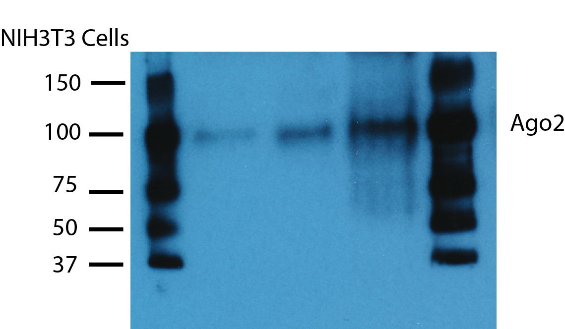
Application: Western BlotSample Tested: mouse cellSpecies: MouseVerified Customer | Posted 04/27/2018FACS Sorted NIH3T3 cells-Lysed in RIPA Buffer-Resolved on 10% SDS PAGE.NIH3T3 cells were FACS sorted based on marker gene expression.Cell lysates were compared for Ago2 expression levels

-
Application: Western BlotSample Tested: HeLa 4Species: OtherVerified Customer | Posted 04/26/2010Image Caption 1
There are no reviews that match your criteria.
Protocols
Find general support by application which include: protocols, troubleshooting, illustrated assays, videos and webinars.
- Antigen Retrieval Protocol (PIER)
- Antigen Retrieval for Frozen Sections Protocol
- Appropriate Fixation of IHC/ICC Samples
- Cellular Response to Hypoxia Protocols
- ChIP Protocol Video
- Chromatin Immunoprecipitation (ChIP) Protocol
- Chromatin Immunoprecipitation Protocol
- Chromogenic IHC Staining of Formalin-Fixed Paraffin-Embedded (FFPE) Tissue Protocol
- Chromogenic Immunohistochemistry Staining of Frozen Tissue
- ClariTSA™ Fluorophore Kits
- Detection & Visualization of Antibody Binding
- ELISA Sample Preparation & Collection Guide
- ELISA Troubleshooting Guide
- Fluorescent IHC Staining of Frozen Tissue Protocol
- Graphic Protocol for Heat-induced Epitope Retrieval
- Graphic Protocol for the Preparation and Fluorescent IHC Staining of Frozen Tissue Sections
- Graphic Protocol for the Preparation and Fluorescent IHC Staining of Paraffin-embedded Tissue Sections
- Graphic Protocol for the Preparation of Gelatin-coated Slides for Histological Tissue Sections
- How to Run an R&D Systems DuoSet ELISA
- How to Run an R&D Systems Quantikine ELISA
- How to Run an R&D Systems Quantikine™ QuicKit™ ELISA
- ICC Cell Smear Protocol for Suspension Cells
- ICC Immunocytochemistry Protocol Videos
- ICC for Adherent Cells
- IHC Sample Preparation (Frozen sections vs Paraffin)
- Immunocytochemistry (ICC) Protocol
- Immunocytochemistry Troubleshooting
- Immunofluorescence of Organoids Embedded in Cultrex Basement Membrane Extract
- Immunofluorescent IHC Staining of Formalin-Fixed Paraffin-Embedded (FFPE) Tissue Protocol
- Immunohistochemistry (IHC) and Immunocytochemistry (ICC) Protocols
- Immunohistochemistry Frozen Troubleshooting
- Immunohistochemistry Paraffin Troubleshooting
- Immunoprecipitation Protocol
- Preparing Samples for IHC/ICC Experiments
- Preventing Non-Specific Staining (Non-Specific Binding)
- Primary Antibody Selection & Optimization
- Protocol for Heat-Induced Epitope Retrieval (HIER)
- Protocol for Making a 4% Formaldehyde Solution in PBS
- Protocol for VisUCyte™ HRP Polymer Detection Reagent
- Protocol for the Fluorescent ICC Staining of Cell Smears - Graphic
- Protocol for the Fluorescent ICC Staining of Cultured Cells on Coverslips - Graphic
- Protocol for the Preparation & Fixation of Cells on Coverslips
- Protocol for the Preparation and Chromogenic IHC Staining of Frozen Tissue Sections
- Protocol for the Preparation and Chromogenic IHC Staining of Frozen Tissue Sections - Graphic
- Protocol for the Preparation and Chromogenic IHC Staining of Paraffin-embedded Tissue Sections
- Protocol for the Preparation and Chromogenic IHC Staining of Paraffin-embedded Tissue Sections - Graphic
- Protocol for the Preparation and Fluorescent ICC Staining of Cells on Coverslips
- Protocol for the Preparation and Fluorescent ICC Staining of Non-adherent Cells
- Protocol for the Preparation and Fluorescent ICC Staining of Stem Cells on Coverslips
- Protocol for the Preparation and Fluorescent IHC Staining of Frozen Tissue Sections
- Protocol for the Preparation and Fluorescent IHC Staining of Paraffin-embedded Tissue Sections
- Protocol for the Preparation of Gelatin-coated Slides for Histological Tissue Sections
- Protocol for the Preparation of a Cell Smear for Non-adherent Cell ICC - Graphic
- Quantikine HS ELISA Kit Assay Principle, Alkaline Phosphatase
- Quantikine HS ELISA Kit Principle, Streptavidin-HRP Polymer
- R&D Systems Quality Control Western Blot Protocol
- Sandwich ELISA (Colorimetric) – Biotin/Streptavidin Detection Protocol
- Sandwich ELISA (Colorimetric) – Direct Detection Protocol
- TUNEL and Active Caspase-3 Detection by IHC/ICC Protocol
- The Importance of IHC/ICC Controls
- Troubleshooting Guide: ELISA
- Troubleshooting Guide: Immunohistochemistry
- Troubleshooting Guide: Western Blot Figures
- Western Blot Conditions
- Western Blot Protocol
- Western Blot Protocol for Cell Lysates
- Western Blot Troubleshooting
- Western Blot Troubleshooting Guide
- View all Protocols, Troubleshooting, Illustrated assays and Webinars
FAQs for Ago2/eIF2C2 Antibody (2E12-1C9) - Azide and BSA Free
-
Q: I ordered the H00027161-M01 antibody for detecting AGO2. I am using it for both IPs and westerns. The product sheet says the AGO2 should run at ~42 kDa, but online, in HL-60 cells, AGO2 runs at ~80-90 kDa, which is more of what I would expect. Can you please explain this discrepancy?
A: Ago2 should run at around 80-90 kDa. Abnova generated their antibody against a 42 kDa partial recombinant protein. The image you see showing Ago2 at 42 kDa is from them probing the partial recombinant protein with H00027161-M01.