Alkaline Phosphatase/ALPP Antibody (8B6) - BSA Free
Novus Biologicals | Catalog # NB110-3638
Key Product Details
Species Reactivity
Validated:
Cited:
Applications
Validated:
Cited:
Label
Antibody Source
Format
Product Specifications
Immunogen
Reactivity Notes
Localization
Specificity
Clonality
Host
Isotype
Theoretical MW
Disclaimer note: The observed molecular weight of the protein may vary from the listed predicted molecular weight due to post translational modifications, post translation cleavages, relative charges, and other experimental factors.
Scientific Data Images for Alkaline Phosphatase/ALPP Antibody (8B6) - BSA Free
![Immunocytochemistry/ Immunofluorescence: Alkaline Phosphatase/ALPP Antibody (8B6) - BSA Free [NB110-3638]](https://resources.rndsystems.com/images/products/Alkaline-Phosphatase-ALPP-Antibody-8B6-Immunocytochemistry-Immunofluorescence-NB110-3638-img0005.jpg "Immunocytochemistry/ Immunofluorescence: Alkaline Phosphatase/ALPP Antibody (8B6) - BSA Free [NB110-3638]")
Immunocytochemistry/ Immunofluorescence: Alkaline Phosphatase/ALPP Antibody (8B6) - BSA Free [NB110-3638]
Alkaline-Phosphatase-ALPP-Antibody-8B6-Immunocytochemistry-Immunofluorescence-NB110-3638-img0005.jpg![Immunocytochemistry/ Immunofluorescence: Alkaline Phosphatase/ALPP Antibody (8B6) - BSA Free [NB110-3638]](https://resources.rndsystems.com/images/products/Alkaline-Phosphatase-ALPP-Antibody-8B6-Immunocytochemistry-Immunofluorescence-NB110-3638-img0003.jpg "Immunocytochemistry/ Immunofluorescence: Alkaline Phosphatase/ALPP Antibody (8B6) - BSA Free [NB110-3638]")
Immunocytochemistry/ Immunofluorescence: Alkaline Phosphatase/ALPP Antibody (8B6) - BSA Free [NB110-3638]
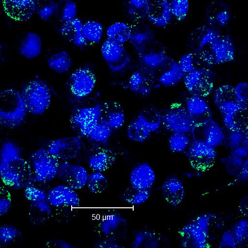
Immunocytochemistry/Immunofluorescence: Alkaline Phosphatase/ALPP Antibody (8B6) [NB110-3638] - analysis of ALPP in MDA-MB-231 cells using an anti-ALPP antibody (blue - cell membrane, green - ALPP). Image from verified customer review.![Western Blot: Alkaline Phosphatase/ALPP Antibody (8B6)BSA Free [NB110-3638]](https://resources.rndsystems.com/images/products/Alkaline-Phosphatase-ALPP-Antibody-8B6-Western-Blot-NB110-3638-img0002.jpg "Western Blot: Alkaline Phosphatase/ALPP Antibody (8B6)BSA Free [NB110-3638]")
Western Blot: Alkaline Phosphatase/ALPP Antibody (8B6)BSA Free [NB110-3638]
Western Blot: Alkaline Phosphatase/ALPP Antibody (8B6) [NB110-3638] - Analysis of Alkaline Phosphatase (Placental) expression in JAR whole cell lysate.![Immunohistochemistry-Paraffin: Alkaline Phosphatase/ALPP Antibody (8B6) - BSA Free [NB110-3638]](https://resources.rndsystems.com/images/products/Alkaline-Phosphatase-ALPP-Antibody-8B6-Immunohistochemistry-Paraffin-NB110-3638-img0001.jpg "Immunohistochemistry-Paraffin: Alkaline Phosphatase/ALPP Antibody (8B6) - BSA Free [NB110-3638]")
Immunohistochemistry-Paraffin: Alkaline Phosphatase/ALPP Antibody (8B6) - BSA Free [NB110-3638]
Immunohistochemistry-Paraffin: Alkaline Phosphatase/ALPP Antibody (8B6) [NB110-3638] - Alkaline Phosphatase, Placental Antibody (8B6) [NB110-3638] - IHC staining of Alkaline Phosphatase (Placental) in human placenta using DAB with hematoxylin counterstain. Proteolytic Induced Epitope Retrieval (PIER) was used.![Flow Cytometry: Alkaline Phosphatase/ALPP Antibody (8B6) - BSA Free [NB110-3638]](https://resources.rndsystems.com/images/products/Alkaline-Phosphatase-ALPP-Antibody-8B6-Flow-Cytometry-NB110-3638-img0004.jpg "Flow Cytometry: Alkaline Phosphatase/ALPP Antibody (8B6) - BSA Free [NB110-3638]")
Flow Cytometry: Alkaline Phosphatase/ALPP Antibody (8B6) - BSA Free [NB110-3638]
Flow Cytometry: Alkaline Phosphatase/ALPP Antibody (8B6) [NB110-3638] - Detection of Alkaline Phosphatase/ALPP in Human HeLa Cell Line by Flow Cytometry. Human HeLa cell line was stained with Mouse Anti- Alkaline Phosphatase/ALPP Monoclonal Antibody (Catalog # NB110-3638, filled histogram), or Mouse IgG2A isotype control (Catalog # MAB003, open histogram) followed by APC-conjugated Anti-Mouse IgG Secondary Antibody (Catalog # F0101B). To facilitate intracellular staining, cells were fixed with Flow Cytometry Fixation Buffer (Catalog # FC004) and permeabilized with Flow Cytometry Permeabilization/Wash Buffer I (Catalog # FC005).Images may not be copied, printed or otherwise disseminated without express written permission of Novus Biologicals a bio-techne brand.
 - BSA Free [NB110-3638] -")
Immunocytochemistry/ Immunofluorescence: Alkaline Phosphatase/ALPP Antibody (8B6) - BSA Free [NB110-3638] -
Immunocytochemistry/ Immunofluorescence: Alkaline Phosphatase/ALPP Antibody (8B6) - BSA Free [NB110-3638] - GRPs enhance axonal growth in DRGs in vitro. A–C, Representative montage images are shown for control dissociated DRG culture (A), dissociated DRGs cocultured with rat GRPs (B), & dissociated DRGs cultures exposed to conditioned medium from alkaline phosphatase-expressing rat GRPs (C). beta III-tubulin (red) immunofluorescence highlights the neurons; GRPs are visualized by immunostaining for alkaline phosphatase (green). D shows quantification for the average length of the longest axon per neuron ± SEM (n ≥ 30 neurons in three separate experiments; **p ≤ 0.01 by Student’s t test). Scale bar, 250 μm. Image collected & cropped by CiteAb from the following publication (https://www.eneuro.org/lookup/doi/10.1523/ENEURO.0171-16.2017), licensed under a CC-BY license. Not internally tested by Novus Biologicals. - BSA Free [NB110-3638] -")
Immunocytochemistry/ Immunofluorescence: Alkaline Phosphatase/ALPP Antibody (8B6) - BSA Free [NB110-3638] -
Immunocytochemistry/ Immunofluorescence: Alkaline Phosphatase/ALPP Antibody (8B6) - BSA Free [NB110-3638] - Coculture with neural & glial progenitor cells increases axonal outgrowth from adult DRG neurons. A–C, Representative montage images of control-dissociated DRG cultures (A), dissociated DRGs cocultured with rat GRPs/NRPs (B), & dissociated DRG cultures exposed to conditioned medium from parallel rat GRP/NRP cultures (C) are shown. Immunofluorescence for beta III-tubulin (red) & nestin (green) highlight DRG neurons & GRP/NRP nuclei, respectively. D, Quantitation of the average lengths of the longest axon per neuron (±SEM) for the above conditions is shown. Coculture with GRPs/NRPs significantly increases in axon length compared with the standard DRG culture; exposure to conditioned medium from GRP/NRP cultures showed a further increase in axon length (n ≥ 30 neurons in three separate experiments; *p ≤ 0.05 & ***p ≤ 0.001 by Student’s t test). Scale bar, 250 μm. E, Quantitation of axon growth parameters for 7 d injury-conditioned DRG neurons cultured on coverslips laid over a bed of GRP/NRP cells (coculture) or control conditions is shown (n ≥ 200 neurons analyzed in three separate experiments; p values represent ANOVA with Tukey post hoc analyses). Image collected & cropped by CiteAb from the following publication (https://www.eneuro.org/lookup/doi/10.1523/ENEURO.0171-16.2017), licensed under a CC-BY license. Not internally tested by Novus Biologicals. - BSA Free [NB110-3638] -")
Immunocytochemistry/ Immunofluorescence: Alkaline Phosphatase/ALPP Antibody (8B6) - BSA Free [NB110-3638] -
Immunocytochemistry/ Immunofluorescence: Alkaline Phosphatase/ALPP Antibody (8B6) - BSA Free [NB110-3638] - Coculture with neural & glial progenitor cells increases axonal outgrowth from adult DRG neurons. A–C, Representative montage images of control-dissociated DRG cultures (A), dissociated DRGs cocultured with rat GRPs/NRPs (B), & dissociated DRG cultures exposed to conditioned medium from parallel rat GRP/NRP cultures (C) are shown. Immunofluorescence for beta III-tubulin (red) & nestin (green) highlight DRG neurons & GRP/NRP nuclei, respectively. D, Quantitation of the average lengths of the longest axon per neuron (±SEM) for the above conditions is shown. Coculture with GRPs/NRPs significantly increases in axon length compared with the standard DRG culture; exposure to conditioned medium from GRP/NRP cultures showed a further increase in axon length (n ≥ 30 neurons in three separate experiments; *p ≤ 0.05 & ***p ≤ 0.001 by Student’s t test). Scale bar, 250 μm. E, Quantitation of axon growth parameters for 7 d injury-conditioned DRG neurons cultured on coverslips laid over a bed of GRP/NRP cells (coculture) or control conditions is shown (n ≥ 200 neurons analyzed in three separate experiments; p values represent ANOVA with Tukey post hoc analyses). Image collected & cropped by CiteAb from the following publication (https://www.eneuro.org/lookup/doi/10.1523/ENEURO.0171-16.2017), licensed under a CC-BY license. Not internally tested by Novus Biologicals.Applications for Alkaline Phosphatase/ALPP Antibody (8B6) - BSA Free
ELISA
Flow Cytometry
Immunocytochemistry/ Immunofluorescence
Immunohistochemistry
Immunohistochemistry-Frozen
Immunohistochemistry-Paraffin
Western Blot
Reviewed Applications
Read 1 review rated 5 using NB110-3638 in the following applications:
Flow Cytometry Panel Builder
Bio-Techne Knows Flow Cytometry
Save time and reduce costly mistakes by quickly finding compatible reagents using the Panel Builder Tool.
Advanced Features
- Spectra Viewer - Custom analysis of spectra from multiple fluorochromes
- Spillover Popups - Visualize the spectra of individual fluorochromes
- Antigen Density Selector - Match fluorochrome brightness with antigen density

Formulation, Preparation, and Storage
Purification
Formulation
Format
Preservative
Concentration
Shipping
Stability & Storage
Background: Alkaline Phosphatase/ALPP
Long Name
Alternate Names
Entrez Gene IDs
Gene Symbol
UniProt
Additional Alkaline Phosphatase/ALPP Products
Product Documents for Alkaline Phosphatase/ALPP Antibody (8B6) - BSA Free
Certificate of Analysis
To download a Certificate of Analysis, please enter a lot or batch number in the search box below.
Product Specific Notices for Alkaline Phosphatase/ALPP Antibody (8B6) - BSA Free
This product is for research use only and is not approved for use in humans or in clinical diagnosis. Primary Antibodies are guaranteed for 1 year from date of receipt.
Related Research Areas
Citations for Alkaline Phosphatase/ALPP Antibody (8B6) - BSA Free
 Powered by Bioz
Customer Reviews for Alkaline Phosphatase/ALPP Antibody (8B6) - BSA Free (1)
Have you used Alkaline Phosphatase/ALPP Antibody (8B6) - BSA Free?
Submit a review and receive an Amazon gift card!
$25/€18/£15/$25CAN/¥2500 Yen for a review with an image
$10/€7/£6/$10CAN/¥1110 Yen for a review without an image
Submit a review
Customer Images
-
Application: ImmunofluorescenceSample Tested: MDA-MB-231 cellSpecies: HumanVerified Customer | Posted 11/24/2014MDA-MB-231 cells having internalized PLAP (blue-cell membrane, green-PLAP)

There are no reviews that match your criteria.
Protocols
View specific protocols for Alkaline Phosphatase/ALPP Antibody (8B6) - BSA Free (NB110-3638):
Antigen Unmasking - Proteolytic Induced Epitope Retrieval (PIER):
Trypsin Working Solution (0.05%):
Trypsin stock solution (0.5%) -1 ml
Calcium chloride stock solution 1% - 1 ml Distilled Water - 8 ml Adjust pH to 7.8 with 1N NaOH.
Cover sections with trypsin working solution and incubate for 10-20 minutes at 37 degrees Celsius in humidified chamber (optimal incubation time may vary depending on tissue type and degree of fixation, and should be determined by user). Allow sections to cool at room temperature for 10 minutes.
Staining:
1. Wash sections in deionized water three times for 5 minutes each.
2. Wash sections in wash buffer for 5 minutes.
3. Block each section with 100-400 ul blocking solution for 1 hour at room temperature.
4. Remove blocking solution and add 100-400 ul diluted primary antibody. Incubate overnight at 4 C.
5. Remove antibody solution and wash sections in wash buffer three times for 5 minutes each.
6. Add 100-400 ul biotinylated diluted secondary antibody. Incubate 30 minutes at room temperature.
7. Remove secondary antibody solution and wash sections three times with wash buffer for 5 minutes each.
8. Add 100-400 ul Streptavidin-HRP reagent to each section and incubate for 30 minutes at room temperature.
9. Wash sections three times in wash buffer for 5 minutes each.
10. Add 100-400 ul DAB substrate to each section and monitor staining closely.
11. As soon as the sections develop, immerse slides in deionized water.
12. Counterstain sections in hematoxylin.
13. Wash sections in deionized water two times for 5 minutes each.
14. Dehydrate sections.
15. Mount coverslips.
Find general support by application which include: protocols, troubleshooting, illustrated assays, videos and webinars.
- 7-Amino Actinomycin D (7-AAD) Cell Viability Flow Cytometry Protocol
- Antigen Retrieval Protocol (PIER)
- Antigen Retrieval for Frozen Sections Protocol
- Appropriate Fixation of IHC/ICC Samples
- Cellular Response to Hypoxia Protocols
- Chromogenic IHC Staining of Formalin-Fixed Paraffin-Embedded (FFPE) Tissue Protocol
- Chromogenic Immunohistochemistry Staining of Frozen Tissue
- ClariTSA™ Fluorophore Kits
- Detection & Visualization of Antibody Binding
- ELISA Sample Preparation & Collection Guide
- ELISA Troubleshooting Guide
- Extracellular Membrane Flow Cytometry Protocol
- Flow Cytometry Protocol for Cell Surface Markers
- Flow Cytometry Protocol for Staining Membrane Associated Proteins
- Flow Cytometry Staining Protocols
- Flow Cytometry Troubleshooting Guide
- Fluorescent IHC Staining of Frozen Tissue Protocol
- Graphic Protocol for Heat-induced Epitope Retrieval
- Graphic Protocol for the Preparation and Fluorescent IHC Staining of Frozen Tissue Sections
- Graphic Protocol for the Preparation and Fluorescent IHC Staining of Paraffin-embedded Tissue Sections
- Graphic Protocol for the Preparation of Gelatin-coated Slides for Histological Tissue Sections
- How to Run an R&D Systems DuoSet ELISA
- How to Run an R&D Systems Quantikine ELISA
- How to Run an R&D Systems Quantikine™ QuicKit™ ELISA
- ICC Cell Smear Protocol for Suspension Cells
- ICC Immunocytochemistry Protocol Videos
- ICC for Adherent Cells
- IHC Sample Preparation (Frozen sections vs Paraffin)
- Immunocytochemistry (ICC) Protocol
- Immunocytochemistry Troubleshooting
- Immunofluorescence of Organoids Embedded in Cultrex Basement Membrane Extract
- Immunofluorescent IHC Staining of Formalin-Fixed Paraffin-Embedded (FFPE) Tissue Protocol
- Immunohistochemistry (IHC) and Immunocytochemistry (ICC) Protocols
- Immunohistochemistry Frozen Troubleshooting
- Immunohistochemistry Paraffin Troubleshooting
- Intracellular Flow Cytometry Protocol Using Alcohol (Methanol)
- Intracellular Flow Cytometry Protocol Using Detergents
- Intracellular Nuclear Staining Flow Cytometry Protocol Using Detergents
- Intracellular Staining Flow Cytometry Protocol Using Alcohol Permeabilization
- Intracellular Staining Flow Cytometry Protocol Using Detergents to Permeabilize Cells
- Preparing Samples for IHC/ICC Experiments
- Preventing Non-Specific Staining (Non-Specific Binding)
- Primary Antibody Selection & Optimization
- Propidium Iodide Cell Viability Flow Cytometry Protocol
- Protocol for Heat-Induced Epitope Retrieval (HIER)
- Protocol for Liperfluo
- Protocol for Making a 4% Formaldehyde Solution in PBS
- Protocol for VisUCyte™ HRP Polymer Detection Reagent
- Protocol for the Characterization of Human Th22 Cells
- Protocol for the Characterization of Human Th9 Cells
- Protocol for the Fluorescent ICC Staining of Cell Smears - Graphic
- Protocol for the Fluorescent ICC Staining of Cultured Cells on Coverslips - Graphic
- Protocol for the Preparation & Fixation of Cells on Coverslips
- Protocol for the Preparation and Chromogenic IHC Staining of Frozen Tissue Sections
- Protocol for the Preparation and Chromogenic IHC Staining of Frozen Tissue Sections - Graphic
- Protocol for the Preparation and Chromogenic IHC Staining of Paraffin-embedded Tissue Sections
- Protocol for the Preparation and Chromogenic IHC Staining of Paraffin-embedded Tissue Sections - Graphic
- Protocol for the Preparation and Fluorescent ICC Staining of Cells on Coverslips
- Protocol for the Preparation and Fluorescent ICC Staining of Non-adherent Cells
- Protocol for the Preparation and Fluorescent ICC Staining of Stem Cells on Coverslips
- Protocol for the Preparation and Fluorescent IHC Staining of Frozen Tissue Sections
- Protocol for the Preparation and Fluorescent IHC Staining of Paraffin-embedded Tissue Sections
- Protocol for the Preparation of Gelatin-coated Slides for Histological Tissue Sections
- Protocol for the Preparation of a Cell Smear for Non-adherent Cell ICC - Graphic
- Protocol: Annexin V and PI Staining by Flow Cytometry
- Protocol: Annexin V and PI Staining for Apoptosis by Flow Cytometry
- Quantikine HS ELISA Kit Assay Principle, Alkaline Phosphatase
- Quantikine HS ELISA Kit Principle, Streptavidin-HRP Polymer
- R&D Systems Quality Control Western Blot Protocol
- Sandwich ELISA (Colorimetric) – Biotin/Streptavidin Detection Protocol
- Sandwich ELISA (Colorimetric) – Direct Detection Protocol
- TUNEL and Active Caspase-3 Detection by IHC/ICC Protocol
- The Importance of IHC/ICC Controls
- Troubleshooting Guide: ELISA
- Troubleshooting Guide: Fluorokine Flow Cytometry Kits
- Troubleshooting Guide: Immunohistochemistry
- Troubleshooting Guide: Western Blot Figures
- Western Blot Conditions
- Western Blot Protocol
- Western Blot Protocol for Cell Lysates
- Western Blot Troubleshooting
- Western Blot Troubleshooting Guide
- View all Protocols, Troubleshooting, Illustrated assays and Webinars
FAQs for Alkaline Phosphatase/ALPP Antibody (8B6) - BSA Free
-
Q: I am looking to use a placental alkaline phosphatase antibody for sorting syncytiotrophoblast material from female tissue. It looks like most people who use these antibodies use in-house versions and references for commercially available antibodies I've found are from the '80's. I noticed you have several different monoclonal antibodies and was hoping you could give me a little more information on them and potential applications they've been tested for. Also, do you have a biotinylated or fluorophore-conjugated version?
A:
None of our placental alkaline phosphatase antibodies are biotinylated or fluorchrome conjugated but we offer a number of kits for this application and our lab can do a custom conjugation as well, for an additional fee. NB600-750 is useful in FACS with human cells, so I would recommend this antibody for your experiment. We back the use of this antibody as stated on the datasheet with the 100% Novus Guarantee. If you have questions about another antibody, I will be happy to answer them.
-
Q: Our customer asked us to submit the inquiry to confirm if it is true that catalog # NB110-3638 has not been tested for WB. The customer seems to believe NB110-3638 shall be able to be used for WB.
A: NB110-3638 has not been tested in Western blot and we cannot guarantee that application. Please advise your customer that the applications listed on the datasheet are the ones we have tested and will guarantee.
-
Q: I am looking to use a placental alkaline phosphatase antibody for sorting syncytiotrophoblast material from female tissue. It looks like most people who use these antibodies use in-house versions and references for commercially available antibodies I've found are from the '80's. I noticed you have several different monoclonal antibodies and was hoping you could give me a little more information on them and potential applications they've been tested for. Also, do you have a biotinylated or fluorophore-conjugated version?
A:
None of our placental alkaline phosphatase antibodies are biotinylated or fluorchrome conjugated but we offer a number of kits for this application and our lab can do a custom conjugation as well, for an additional fee. NB600-750 is useful in FACS with human cells, so I would recommend this antibody for your experiment. We back the use of this antibody as stated on the datasheet with the 100% Novus Guarantee. If you have questions about another antibody, I will be happy to answer them.
-
Q: Our customer asked us to submit the inquiry to confirm if it is true that catalog # NB110-3638 has not been tested for WB. The customer seems to believe NB110-3638 shall be able to be used for WB.
A: NB110-3638 has not been tested in Western blot and we cannot guarantee that application. Please advise your customer that the applications listed on the datasheet are the ones we have tested and will guarantee.