alpha Tubulin Antibody (YL1/2) - BSA Free
Novus Biologicals | Catalog # NB600-506
Key Product Details
Species Reactivity
Validated:
Human, Mouse, Rat, Avian, C. elegans, Drosophila, Invertebrate, Mammal, Monkey, Primate, Yeast
Cited:
Human, Mouse, Rat, Insect - Drosophila, Invertebrate, Nematode - Caenorhabditis elegans
Applications
Validated:
Immunohistochemistry, Immunohistochemistry-Paraffin, Immunohistochemistry-Frozen, Western Blot, ELISA, Flow Cytometry, Immunocytochemistry/ Immunofluorescence, Immunoprecipitation, Functional, Radioimmunoassay
Cited:
Western Blot, Immunocytochemistry/ Immunofluorescence, Functional Assay, IF/IHC
Label
Unconjugated
Antibody Source
Monoclonal Rat IgG2A Clone # YL1/2
Format
BSA Free
Loading...
Product Specifications
Immunogen
This alpha Tubulin Antibody (YL1/2) was developed against full length native protein (purified) (S. cerevisiae).
Epitope
A linear sequence requiring an aromatic residue at the C terminus, with the two adjacent amino acids being negatively charged (represented by Gly-Gly-Tyr in Tyr-Tubulin).
Reactivity Notes
S. cerevisiae, S. pombe, Slime molds, Allium. Other species have not been tested. Expected to react with most eukaryotes due to sequence identity. Drosophila reactivity reported in scientific literature (PMID: 24019759). C. elegans reactivity reported in scientific literature (PMID: 29118344). Toxoplasma gondii reactivity reported by customer review.
Marker
Microtubule Marker
Clonality
Monoclonal
Host
Rat
Isotype
IgG2A
Theoretical MW
50 kDa.
Disclaimer note: The observed molecular weight of the protein may vary from the listed predicted molecular weight due to post translational modifications, post translation cleavages, relative charges, and other experimental factors.
Disclaimer note: The observed molecular weight of the protein may vary from the listed predicted molecular weight due to post translational modifications, post translation cleavages, relative charges, and other experimental factors.
Scientific Data Images for alpha Tubulin Antibody (YL1/2) - BSA Free
![Immunocytochemistry/ Immunofluorescence: alpha Tubulin Antibody (YL1/2) - BSA Free [NB600-506]](https://resources.rndsystems.com/images/products/alpha-Tubulin-Antibody-YL1-2-Immunocytochemistry-Immunofluorescence-NB600-506-img0021.jpg "Immunocytochemistry/ Immunofluorescence: alpha Tubulin Antibody (YL1/2) - BSA Free [NB600-506]")
Immunocytochemistry/ Immunofluorescence: alpha Tubulin Antibody (YL1/2) - BSA Free [NB600-506]
Immunocytochemistry/Immunofluorescence: alpha Tubulin Antibody (YL1/2) [NB600-506] - HeLa cells were fixed and permeabilized for 10 minutes with -20C MeOH. The cells were incubated with alpha Tubulin Antibody [YL1/2] (NB600-506) at 1ug/ml overnight at 4C and detected with an anti-rat DyLight 488 (Green) at a 1:1000 dilution for 60 minutes. Nuclei were counterstained with DAPI (Blue). Cells were imaged using a 100X objective and digitally deconvolved.![Flow Cytometry: alpha Tubulin Antibody (YL1/2) - BSA Free [NB600-506]](https://resources.rndsystems.com/images/products/alpha-Tubulin-Antibody-YL1-2-Flow-Cytometry-NB600-506-img0018.jpg "Flow Cytometry: alpha Tubulin Antibody (YL1/2) - BSA Free [NB600-506]")
Flow Cytometry: alpha Tubulin Antibody (YL1/2) - BSA Free [NB600-506]
Flow Cytometry: alpha Tubulin Antibody (YL1/2) [NB600-506] - An intracellular stain was performed on SH-SY5Y cells with alpha Tubulin (YL1/2) Antibody NB600-506G (blue) and a matched isotype control (orange). Cells were fixed with 4% PFA and then permeabilized with 0.1% saponin. Cells were incubated in an antibody dilution of 5 ug/mL for 30 minutes at room temperature. Both antibodies were conjugated to DyLight 488.![Immunocytochemistry/ Immunofluorescence: alpha Tubulin Antibody (YL1/2) - BSA Free [NB600-506]](https://resources.rndsystems.com/images/products/alpha-Tubulin-Antibody-YL1-2-NB600-506-img0022.jpg "Immunocytochemistry/ Immunofluorescence: alpha Tubulin Antibody (YL1/2) - BSA Free [NB600-506]")
Immunocytochemistry/ Immunofluorescence: alpha Tubulin Antibody (YL1/2) - BSA Free [NB600-506]
Immunocytochemistry/Immunofluorescence: alpha Tubulin Antibody (YL1/2) [NB600-506] - NIH3T3 cells were fixed and permeabilized for 10 minutes with -20C MeOH. The cells were incubated with alpha Tubulin Antibody [YL1/2] (NB600-506) at 1ug/ml overnight at 4C and detected with an anti-rat DyLight 488 (Green) at a 1:1000 dilution for 60 minutes. Nuclei were counterstained with DAPI (Blue). Cells were imaged using a 100X objective and digitally deconvolved.![Immunocytochemistry/ Immunofluorescence: alpha Tubulin Antibody (YL1/2) - BSA Free [NB600-506]](https://resources.rndsystems.com/images/products/alpha-Tubulin-Antibody-YL1-2-Immunocytochemistry-Immunofluorescence-NB600-506-img0023.jpg "Immunocytochemistry/ Immunofluorescence: alpha Tubulin Antibody (YL1/2) - BSA Free [NB600-506]")
Immunocytochemistry/ Immunofluorescence: alpha Tubulin Antibody (YL1/2) - BSA Free [NB600-506]
Immunocytochemistry/Immunofluorescence: alpha Tubulin Antibody (YL1/2) [NB600-506] - Rat FR cells were fixed and permeabilized for 10 minutes with -20C MeOH. The cells were incubated with alpha Tubulin Antibody [YL1/2] (NB600-506) at 1ug/ml overnight at 4C and detected with an anti-rat DyLight 488 (Green) at a 1:1000 dilution for 60 minutes. Nuclei were counterstained with DAPI (Blue). Cells were imaged using a 100X objective and digitally deconvolved.![Western Blot: alpha Tubulin Antibody (YL1/2)BSA Free [NB600-506]](https://resources.rndsystems.com/images/products/alpha-Tubulin-Antibody-YL1-2-Western-Blot-NB600-506-img0015.jpg "Western Blot: alpha Tubulin Antibody (YL1/2)BSA Free [NB600-506]")
Western Blot: alpha Tubulin Antibody (YL1/2)BSA Free [NB600-506]
Western Blot: alpha Tubulin Antibody (YL1/2) [NB600-506] - Total protein from human HeLa, mouse 3T3, rat PC12 and African green monkey Cos7 cell lines was separated on a 12% gel by SDS-PAGE, transferred to PVDF membrane and blocked in 5% non-fat milk in TBST. The membrane was probed with a 1:6000 dilution of anti-Tubulin (YL 1/2) in 1% non-fat milk in TBST and detected with an anti-rat HRP secondary antibody using chemiluminescence. Alpha tubulin molecular weight: 50 kDa.![Immunohistochemistry-Paraffin: alpha Tubulin Antibody (YL1/2) - BSA Free [NB600-506]](https://resources.rndsystems.com/images/products/alpha-Tubulin-Antibody-YL1-2-Immunohistochemistry-Paraffin-NB600-506-img0005.jpg "Immunohistochemistry-Paraffin: alpha Tubulin Antibody (YL1/2) - BSA Free [NB600-506]")
Immunohistochemistry-Paraffin: alpha Tubulin Antibody (YL1/2) - BSA Free [NB600-506]
Immunohistochemistry-Paraffin: alpha Tubulin Antibody (YL1/2) [NB600-506] - Tubulin antibody was tested in human breast cancer xenograft using DAB with hematoxylin counterstain.![Flow Cytometry: alpha Tubulin Antibody (YL1/2) - BSA Free [NB600-506]](https://resources.rndsystems.com/images/products/alpha-Tubulin-Antibody-YL1-2-Flow-Cytometry-NB600-506-img0017.jpg "Flow Cytometry: alpha Tubulin Antibody (YL1/2) - BSA Free [NB600-506]")
Flow Cytometry: alpha Tubulin Antibody (YL1/2) - BSA Free [NB600-506]
Flow Cytometry: alpha Tubulin Antibody (YL1/2) [NB600-506] - An intracellular stain was performed on PC12 cells with Tubulin [YL1/2] Antibody NB600-506AF488 (blue) and a matched isotype control (orange). Cells were fixed with 4% PFA and then permeabilized with 0.1% saponin. Cells were incubated in an antibody dilution of 5 ug/mL for 30 minutes at room temperature. Both antibodies were conjugated to Alexa Fluor 488.![Western Blot: alpha Tubulin Antibody (YL1/2)BSA Free [NB600-506]](https://resources.rndsystems.com/images/products/alpha-Tubulin-Antibody-YL1-2-Western-Blot-NB600-506-img0019.jpg "Western Blot: alpha Tubulin Antibody (YL1/2)BSA Free [NB600-506]")
Western Blot: alpha Tubulin Antibody (YL1/2)BSA Free [NB600-506]
alpha-Tubulin-Antibody-YL1-2-Western-Blot-NB600-506-img0019.jpg![Western Blot: alpha Tubulin Antibody (YL1/2)BSA Free [NB600-506]](https://resources.rndsystems.com/images/products/alpha-Tubulin-Antibody-YL1-2-Western-Blot-NB600-506-img0002.jpg "Western Blot: alpha Tubulin Antibody (YL1/2)BSA Free [NB600-506]")
Western Blot: alpha Tubulin Antibody (YL1/2)BSA Free [NB600-506]
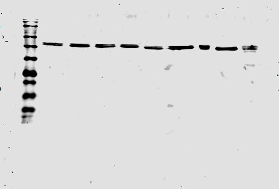
Western Blot: alpha Tubulin Antibody (YL1/2) [NB600-506] - Western blot analysis of alpha Tubulin (molecular weight: 50 kDa) expression in 1) HeLa, 2) NTERA-2, 3) A431, 4) HepG2, 5) MCF7, 6) NIH-3T3, 7) PC-12 and 8) Cos 7 whole cell lysates using NB600-506.![Western Blot: alpha Tubulin Antibody (YL1/2)BSA Free [NB600-506]](https://resources.rndsystems.com/images/products/alpha-Tubulin-Antibody-YL1-2-Western-Blot-NB600-506-img0011.jpg "Western Blot: alpha Tubulin Antibody (YL1/2)BSA Free [NB600-506]")
Western Blot: alpha Tubulin Antibody (YL1/2)BSA Free [NB600-506]
Western Blot: alpha Tubulin Antibody (YL1/2) [NB600-506] - Western Blot analysis of whole cell lysates of HeLa, NIH-3T3 and PC-12 cell lines using Tubulin antibody (clone YL1/2). Alpha tubulin molecular weight: 50 kDa.![Immunocytochemistry/ Immunofluorescence: alpha Tubulin Antibody (YL1/2) - BSA Free [NB600-506]](https://resources.rndsystems.com/images/products/alpha-Tubulin-Antibody-YL1-2-Immunocytochemistry-Immunofluorescence-NB600-506-img0004.jpg "Immunocytochemistry/ Immunofluorescence: alpha Tubulin Antibody (YL1/2) - BSA Free [NB600-506]")
Immunocytochemistry/ Immunofluorescence: alpha Tubulin Antibody (YL1/2) - BSA Free [NB600-506]
Immunocytochemistry/Immunofluorescence: alpha Tubulin Antibody (YL1/2) [NB600-506] - Tubulin YL1/2 antibody was tested in HeLa cells with Dylight 550 (red). Nuclei were counterstained with DAPI (blue).![Immunocytochemistry/ Immunofluorescence: alpha Tubulin Antibody (YL1/2) - BSA Free [NB600-506]](https://resources.rndsystems.com/images/products/alpha-Tubulin-Antibody-YL1-2-Immunocytochemistry-Immunofluorescence-NB600-506-img0008.jpg "Immunocytochemistry/ Immunofluorescence: alpha Tubulin Antibody (YL1/2) - BSA Free [NB600-506]")
Immunocytochemistry/ Immunofluorescence: alpha Tubulin Antibody (YL1/2) - BSA Free [NB600-506]
Immunocytochemistry/Immunofluorescence: alpha Tubulin Antibody (YL1/2) [NB600-506] - B-Tubulin staining (488) and Pericentrin (594) in Mitotic Cell. Image from verified customer review.![Immunocytochemistry/ Immunofluorescence: alpha Tubulin Antibody (YL1/2) - BSA Free [NB600-506]](https://resources.rndsystems.com/images/products/alpha-Tubulin-Antibody-YL1-2-Immunocytochemistry-Immunofluorescence-NB600-506-img0010.jpg "Immunocytochemistry/ Immunofluorescence: alpha Tubulin Antibody (YL1/2) - BSA Free [NB600-506]")
Immunocytochemistry/ Immunofluorescence: alpha Tubulin Antibody (YL1/2) - BSA Free [NB600-506]
Immunocytochemistry/Immunofluorescence: alpha Tubulin Antibody (YL1/2) [NB600-506] - IF Confocal analysis of NIH/3T3 cells using Tubulin antibody (NB600-506, 1:5). An Alexa Fluor 488-conjugated Goat to rat IgG was used as secondary antibody (green). Actin filaments were labeled with Alexa Fluor 568 phalloidin (red). DAPI was used to stain the cell nuclei (blue).![Immunocytochemistry/ Immunofluorescence: alpha Tubulin Antibody (YL1/2) - BSA Free [NB600-506]](https://resources.rndsystems.com/images/products/alpha-Tubulin-Antibody-YL1-2-Immunocytochemistry-Immunofluorescence-NB600-506-img0013.jpg "Immunocytochemistry/ Immunofluorescence: alpha Tubulin Antibody (YL1/2) - BSA Free [NB600-506]")
Immunocytochemistry/ Immunofluorescence: alpha Tubulin Antibody (YL1/2) - BSA Free [NB600-506]
Immunocytochemistry/Immunofluorescence: alpha Tubulin Antibody (YL1/2) [NB600-506] - HeLa cells were fixed and permeabilized for 10 minutes using -20C MeOH. The cells were incubated with anti-Tubulin (YL1/2) at a 1:200 dilution overnight at 4C and detected with an anti-rat Dylight 488 (Green) at a 1:500 dilution. Nuclei were counterstained with DAPI (Blue). Cells were imaged using a 40X objective.![Flow Cytometry: alpha Tubulin Antibody (YL1/2) - BSA Free [NB600-506]](https://resources.rndsystems.com/images/products/alpha-Tubulin-Antibody-YL1-2-Flow-Cytometry-NB600-506-img0012.jpg "Flow Cytometry: alpha Tubulin Antibody (YL1/2) - BSA Free [NB600-506]")
Flow Cytometry: alpha Tubulin Antibody (YL1/2) - BSA Free [NB600-506]
Flow Cytometry: alpha Tubulin Antibody (YL1/2) [NB600-506] - Analysis of Alexa Fluor (R) 647 conjugate of NB600-506. An intracellular stain was performed on HeLa cells with Tubulin antibody (YL1/2) NB600-506AF647 (blue) and a matched isotype control (orange). Cells were fixed with 4% PFA and then permeablized with 0.1% saponin. Cells were incubated in an antibody dilution of 2.5 ug/mL for 30 minutes at room temperature. Both antibodies were conjugated to Alexa Fluor 647.![Flow Cytometry: alpha Tubulin Antibody (YL1/2) - BSA Free [NB600-506]](https://resources.rndsystems.com/images/products/alpha-Tubulin-Antibody-YL1-2-Flow-Cytometry-NB600-506-img0016.jpg "Flow Cytometry: alpha Tubulin Antibody (YL1/2) - BSA Free [NB600-506]")
Flow Cytometry: alpha Tubulin Antibody (YL1/2) - BSA Free [NB600-506]
Flow Cytometry: alpha Tubulin Antibody (YL1/2) [NB600-506] - An intracellular stain was performed on NIH3T3 cells with Tubulin [YL1/2] Antibody NB600-506AF488 (blue) and a matched isotype control (orange). Cells were fixed with 4% PFA and then permeabilized with 0.1% saponin. Cells were incubated in an antibody dilution of 5 ug/mL for 30 minutes at room temperature. Both antibodies were conjugated to Alexa Fluor 488. - BSA Free [NB600-506] -")
Western Blot: alpha Tubulin Antibody (YL1/2) - BSA Free [NB600-506] -
Western Blot: alpha Tubulin Antibody (YL1/2) - BSA Free [NB600-506] - Tissue levels of VEGF & PCNA.VEGF levels were estimated by (A) western blot & (B) ELISA. (C) PCNA levels estimated by western blot. Data were normalized with tubulin. (D) Representative images of PCNA immunohistochemistry staining in the nucleus is shown for CS-exposed mice (CSE) & CS-exposed & LGF-treated mice (CSE+LGF). Scale bars = 50 µm. LGF promoted an increase of PCNA+ cells as showed in the bar graph. ** P<0.01 vs. air-exposed mice; ¥ P<0.05 vs. CS-exposed mice (n = 5 per group). Data are presented as mean ± SEM. Image collected & cropped by CiteAb from the following publication (https://pubmed.ncbi.nlm.nih.gov/25401951), licensed under a CC-BY license. Not internally tested by Novus Biologicals. - BSA Free [NB600-506] -")
Western Blot: alpha Tubulin Antibody (YL1/2) - BSA Free [NB600-506] -
Western Blot: alpha Tubulin Antibody (YL1/2) - BSA Free [NB600-506] - LGF ameliorates oxidative stress.Bars represent (A) 3NT & (B) Nrf2 levels estimated by western blot. Data were normalized with tubulin. * P<0.05 vs. air-exposed mice; ¥ P<0.05 vs. CS-exposed mice (n = 5 per group). Data are presented as mean ± SEM. Image collected & cropped by CiteAb from the following publication (https://pubmed.ncbi.nlm.nih.gov/25401951), licensed under a CC-BY license. Not internally tested by Novus Biologicals. - BSA Free [NB600-506] -")
Western Blot: alpha Tubulin Antibody (YL1/2) - BSA Free [NB600-506] -
Western Blot: alpha Tubulin Antibody (YL1/2) - BSA Free [NB600-506] - Role of cell-type-specific cyclooxygenase-2 (Cox-2) expression in Ras/p53-mediated tumor formation. a Experimental scheme. b Microscopic phenotype & histology of three-dimensional (3D) organoids with/without celecoxib treatment. c Relative 3D organoid formation was determined by the number of organoids larger than 100 µm, n = 3 independent experiments, 25 fields at high power field (HPF) per sample. Veh, vehicle control; C25, 25 µM; C50, 50 µM celecoxib. d Experimental scheme. e Ras/p53-mediated tumor incidence with/without condition Cox-2 knockout. Ptgs2wt/wt, n = 15 animals; Ptgs2flox/flox, n = 14 animals. f, g Histological phenotypes. h–k Immunoblotting of Keratin5 (Krt5), Krt6 & Loricrin (Lor) demonstrated decreased tumor susceptibility but increased differentiation status, n = 4 independent experiments. l Summary of the contribution of physiological stress factors in the susceptibility of tumor development from tumor-competent Krt15+ progenitors. Data with bar graphs are represented as mean ± SEM. Statistical significance was determined by pair-wise comparison using t test; ns = not significant, *p < 0.05, **p < 0.005. Scale bars, 100 µm. Ptgs2 prostaglandin-endoperoxide synthase 2 Image collected & cropped by CiteAb from the following publication (https://pubmed.ncbi.nlm.nih.gov/31110179), licensed under a CC-BY license. Not internally tested by Novus Biologicals. - BSA Free [NB600-506] -")
Western Blot: alpha Tubulin Antibody (YL1/2) - BSA Free [NB600-506] -
Western Blot: alpha Tubulin Antibody (YL1/2) - BSA Free [NB600-506] - Microenvironmental acidic stressors & tumor susceptibility. a Experimental scheme. Epithelial cells isolated from the regions adjacent or distant from squamocolumnar junction (SCJ). b Microscopic phenotype of three-dimensional (3D) organoids from control & Krt15-CrePR; LSL-KrasG12D; p53flox/flox mice. Histology of 3D organoids demonstrated that oncogenic Ras/p53 increased abnormal growth features. c–e Quantification of relative organoid formation (n = 3 independent experiments, 25 fields at high power field (HPF) per sample) & size distribution (n ≥ 150) from control & experimental mice expressing oncogenic Ras/p53 combination. f Experimental scheme. Phosphate-buffered saline (PBS) (vehicle) or PPI were daily treated by intraperitoneal (i.p.) injections (5 consecutive days per week). g Tumor incidence from Krt15-CrePR; LSL-KrasG12D; p53flox/flox mice, with/without PPI treatment, n = 9 animals per each. Statistical significance was determined by Fisher’s exact test; *p < 0.05. h–j Immunoblotting of Keratin5 (Krt5) & Krt6 demonstrated relatively less tumor formation in the PPI treatment group, n = 5 independent experiments. k–m Immunostaining of Krt5, Krt6, & phospho-histone H3 (ph-H3), & histology demonstrated suppressed tumor formation by daily PPI treatment in Krt15-CrePR; LSL-KrasG12D; p53flox/flox mice. PPI, proton pump inhibitor. Data with bar graphs are represented as mean ± SEM. Statistical significance was determined by pair-wise comparison using t test; *p < 0.05, **p < 0.005. Scale bars, 100 µm Image collected & cropped by CiteAb from the following publication (https://pubmed.ncbi.nlm.nih.gov/31110179), licensed under a CC-BY license. Not internally tested by Novus Biologicals. in U-2 OS Human Cell Line.")
Alpha Tubulin (YL1/2) in U-2 OS Human Cell Line.
Alpha Tubulin (YL1/2) was detected in immersion fixed U-2 OS human osteosarcoma cell line using Rat anti-Alpha Tubulin (YL1/2) Protein G Purified Monoclonal Antibody conjugated to DyLight 488 (Catalog # NB600-506G) (green) at 5 µg/mL overnight at 4C. Cells were counterstained with DAPI (dark blue). Cells were imaged using a 100X objective and digitally deconvolved.Applications for alpha Tubulin Antibody (YL1/2) - BSA Free
Application
Recommended Usage
ELISA
1:100-1:1000
Flow Cytometry
2-5 ug/0.1x10^6 cells
Functional
reported in scientific literature (PMID 31358662)
Immunocytochemistry/ Immunofluorescence
1:1000-1:10000. Use reported in scientific literature (PMID 28001364)
Immunohistochemistry
1:200
Immunohistochemistry-Frozen
1:200
Immunohistochemistry-Paraffin
1:200
Immunoprecipitation
1:10-1:500
Western Blot
1:5000-1:10000
Application Notes
NB600-506 is ideal for use as a Western blot loading control, where a band can be seen around 50-55 kDa and as a cytoskeletal marker in Immunocytochemistry.
Reviewed Applications
Read 4 reviews rated 5 using NB600-506 in the following applications:
Flow Cytometry Panel Builder
Bio-Techne Knows Flow Cytometry
Save time and reduce costly mistakes by quickly finding compatible reagents using the Panel Builder Tool.
Advanced Features
- Spectra Viewer - Custom analysis of spectra from multiple fluorochromes
- Spillover Popups - Visualize the spectra of individual fluorochromes
- Antigen Density Selector - Match fluorochrome brightness with antigen density

Formulation, Preparation, and Storage
Purification
Protein G purified
Formulation
PBS
Format
BSA Free
Preservative
0.02% Sodium Azide
Concentration
1.0 mg/ml
Shipping
The product is shipped with polar packs. Upon receipt, store it immediately at the temperature recommended below.
Stability & Storage
Store at 4C short term. Aliquot and store at -20C long term. Avoid freeze-thaw cycles.
Background: alpha Tubulin
Tyrosine ligase adds a C-terminal tyrosine to monomeric alpha tubulin. Assembled microtubules can again be detyrosinated by a cytoskeleton associated carboxypeptidase. Detyrosinated alpha tubulin is referred to as Glu-tubulin. Another post-translational modification of detyrosinated alpha tubulin is C-terminal polyglutamylation which is characteristic for microtubules in neuronal cells and the mitotic spindle.
Like GAPDH and beta-actin, alpha/beta tubulin is often used as a loading control in immunoblot applications (1). Alpha/beta tubulin is also good for counterstaining microtubules in immunofluorescence (2).
References
1. Hannen, R., Selmansberger, M., Hauswald, M., Pagenstecher, A., Nist, A., Stiewe, T.,... Bartsch, J. W. (2019). Comparative Transcriptomic Analysis of Temozolomide Resistant Primary GBM Stem-Like Cells and Recurrent GBM Identifies Up-Regulation of the Carbonic Anhydrase CA2 Gene as Resistance Factor. Cancers (Basel), 11(7). doi:10.3390/cancers11070921
2. Nel, M., Joubert, A. M., Dohle, W., Potter, B. V., & Theron, A. E. (2018). Modes of cell death induced by tetrahydroisoquinoline-based analogs in MDA-MB-231 breast and A549 lung cancer cell lines. Drug Des Devel Ther, 12, 1881-1904. doi:10.2147/dddt.S152718
Long Name
Tubulin Alpha 1a
Alternate Names
Alpha-Tubulin 3, B-ALPHA-1, LIS3, TUBA1A, TUBA3, Tubulin B-Alpha-1
Gene Symbol
TUBA1A
Additional alpha Tubulin Products
Product Documents for alpha Tubulin Antibody (YL1/2) - BSA Free
Certificate of Analysis
To download a Certificate of Analysis, please enter a lot or batch number in the search box below.
Product Specific Notices for alpha Tubulin Antibody (YL1/2) - BSA Free
This product is for research use only and is not approved for use in humans or in clinical diagnosis. Primary Antibodies are guaranteed for 1 year from date of receipt.
Related Research Areas
Citations for alpha Tubulin Antibody (YL1/2) - BSA Free
 Powered by Bioz
Customer Reviews for alpha Tubulin Antibody (YL1/2) - BSA Free (4)
5 out of 5
4 Customer Ratings
Have you used alpha Tubulin Antibody (YL1/2) - BSA Free?
Submit a review and receive an Amazon gift card!
$25/€18/£15/$25CAN/¥2500 Yen for a review with an image
$10/€7/£6/$10CAN/¥1110 Yen for a review without an image
Submit a review
Customer Images
-(01-ml)_NB600-506_8211.bmp)
-(01-ml)_NB600-506_8206.jpg)
-(01-ml)_NB600-506_7126.jpg)
Showing
1
-
4 of
4 reviews
Showing All
Filter By:
-
Application: Western BlotSample Tested: Whole cell lysatesSpecies: Toxoplasma gondiiVerified Customer | Posted 05/22/2019Expected molecular weight is 50kDa. When compared to the protein ladder, a clear band between molecular weight of 63 and 48 kDa can be observed.Western Blot analysis from T. gondii tachyzoites whole cell lysates. This antibody was used at 1:10000 dilution.

-
Application: ImmunocytochemistrySample Tested:Species: HumanVerified Customer | Posted 06/11/2014IF Confocal analysis of NIH/3T3 cells using Tubulin antibody (NB600-506, 1:5).
-
Application: Western BlotSample Tested:Species: HumanVerified Customer | Posted 06/11/2014Western blot analysis of extracts from HeLa, COS and NIH/3T3 cells using Tubulin antibody (NB600-506, 1:100).
-
Application: ImmunofluorescenceVerified Customer | Posted 04/28/2014B-Tubulin staining (488) and Pericentrin (594) in Mitotic Cell
There are no reviews that match your criteria.
Protocols
View specific protocols for alpha Tubulin Antibody (YL1/2) - BSA Free (NB600-506):
Protocol for Flow Cytometry Intracellular Staining
Sample Preparation.
1. Grow cells to 60-85% confluency. Flow cytometry requires between 2 x 10⁵ and 1 x 10⁶ cells for optimal performance.
2. If cells are adherent, harvest gently by washing once with staining buffer and then scraping. Avoid using trypsin as this can disrupt certain epitopes of interest. If enzymatic harvest is required, use Accutase, Collagenase, or TrypLE Express for a less damaging option.
3. Reserve 100 uL for counting, then transfer cell volume into a 50 mL conical tube and centrifuge for 8 minutes at 400 RCF.
a. Count cells using a hemocytometer and a 1:1 trypan blue exclusion stain to determine cell viability before starting the flow protocol. If cells appear blue, do not proceed.
4. Re-suspend cells to a concentration of 1 x 10⁶ cells/mL in staining buffer (NBP2-26247).
5. Aliquot out 100 uL samples in accordance with your experimental samples.
Tip: When cell surface and intracellular staining are required in the same sample, it is advisable that the cell surface staining be performed first since the fixation and permeabilization steps might reduce the availability of surface antigens.
Intracellular Staining.
Tip: When performing intracellular staining, it is important to use appropriate fixation and permeabilization reagents based upon the target and its subcellular location. Generally, our Intracellular Flow Assay Kit (NBP2-29450) is a good place to start as it contains an optimized combination of reagents for intracellular staining as well as an inhibitor of intracellular protein transport (necessary if staining secreted proteins). Certain targets may require more gentle or transient permeabilization protocols such as the commonly employed methanol or saponin-based methods.
Protocol for Cytoplasmic Targets:
1. Fix the cells by adding 100 uL fixation solution (such as 4% PFA) to each sample for 10-15 minutes.
2. Permeabilize cells by adding 100 uL of a permeabilization buffer to every 1 x 10⁶ cells present in the sample. Mix well and incubate at room temperature for 15 minutes.
a. For cytoplasmic targets, use a gentle permeabilization solution such as 1X PBS + 0.5% Saponin or 1X PBS + 0.5% Tween-20.
b. To maintain the permeabilized state throughout your experiment, use staining buffer + 0.1% of the permeabilization reagent (i.e. 0.1% Tween-20 or 0.1% Saponin).
3. Following the 15 minute incubation, add 2 mL of the staining buffer + 0.1% permeabilizer to each sample.
4. Centrifuge for 1 minute at 400 RCF.
5. Discard supernatant and re-suspend in 100 uL of staining buffer + 0.1% permeabilizer.
6. Add appropriate amount of each antibody (eg. 1 test or 1 ug per sample, as experimentally determined).
7. Mix well and incubate at room temperature for 30 minutes- 1 hour. Gently mix samples every 10-15 minutes.
8. Following the primary/conjugate incubation, add 1-2 mL/sample of staining buffer +0.1% permeabilizer and centrifuge for 1 minute at 400 RCF.
9. Wash twice by re-suspending cells in staining buffer (2 mL for tubes or 200 uL for wells) and centrifuging at 400 RCF for 5 minutes. Discard supernatant.
10. Add appropriate amount of secondary antibody (as experimentally determined) to each sample.
11. Incubate at room temperature in dark for 20 minutes.
12. Add 1-2 mL of staining buffer and centrifuge at 400 RCF for 1 minute and discard supernatant.
13. Wash twice by re-suspending cells in staining buffer (2 mL for tubes or 200 uL for wells) and centrifuging at 400 RCF for 5 minutes. Discard supernatant.
14. Resuspend in an appropriate volume of staining buffer (usually 500 uL per sample) and proceed with analysis on your flow cytometer.
Immunocytochemistry Protocol
Culture cells to appropriate density in 35 mm culture dishes or 6-well plates.
1. Remove culture medium and wash the cells briefly in PBS. Add 10% formalin to the dish and fix at room temperature for 10 minutes.
2. Remove the formalin and wash the cells in PBS.
3. Permeablize the cells with 0.1% Triton X100 or other suitable detergent for 10 min.
4. Remove the permeablization buffer and wash three times for 10 minutes each in PBS. Be sure to not let the specimen dry out.
5. To block nonspecific antibody binding, incubate in 10% normal goat serum from 1 hour to overnight at room temperature.
6. Add primary antibody at appropriate dilution and incubate overnight at 4C.
7. Remove primary antibody and replace with PBS. Wash three times for 10 minutes each.
8. Add secondary antibody at appropriate dilution. Incubate for 1 hour at room temperature.
9. Remove secondary antibody and replace with PBS. Wash three times for 10 minutes each.
10. Counter stain DNA with DAPi if required.
Immunohistochemistry-Paraffin Embedded Sections
Antigen Unmasking:
Bring slides to a boil in 10 mM sodium citrate buffer (pH 6.0) then maintain at a sub-boiling temperature for 10 minutes. Cool slides on bench-top for 30 minutes (keep slides in the sodium citrate buffer at all times).
Staining:
1. Wash sections in deionized water three times for 5 minutes each.
2. Wash sections in PBS for 5 minutes.
3. Block each section with 100-400 ul blocking solution (1% BSA in PBS) for 1 hour at room temperature.
4. Remove blocking solution and add 100-400 ul diluted primary antibody. Incubate overnight at 4 C.
5. Remove antibody solution and wash sections in wash buffer three times for 5 minutes each.
6. Add 100-400 ul HRP polymer conjugated secondary antibody. Incubate 30 minutes at room temperature.
7. Wash sections three times in wash buffer for 5 minutes each.
8. Add 100-400 ul DAB substrate to each section and monitor staining closely.
9. As soon as the sections develop, immerse slides in deionized water.
10. Counterstain sections in hematoxylin.
11. Wash sections in deionized water two times for 5 minutes each.
12. Dehydrate sections.
13. Mount coverslips.
Western Blot Protocol
1. Perform SDS-PAGE on samples to be analyzed, loading 10-25 ug of total protein per lane.
2. Transfer proteins to PVDF membrane according to the instructions provided by the manufacturer of the membrane and transfer apparatus.
3. Stain the membrane with Ponceau S (or similar product) to assess transfer success, and mark molecular weight standards where appropriate.
4. Rinse the blot TBS -0.05% Tween 20 (TBST).
5. Block the membrane in 5% Non-fat milk in TBST (blocking buffer) for at least 1 hour.
6. Wash the membrane in TBST three times for 10 minutes each.
7. Dilute primary antibody in blocking buffer and incubate overnight at 4C with gentle rocking.
8. Wash the membrane in TBST three times for 10 minutes each.
9. Incubate the membrane in diluted HRP conjugated secondary antibody in blocking buffer (as per manufacturer's instructions) for 1 hour at room temperature.
10. Wash the blot in TBST three times for 10 minutes each (this step can be repeated as required to reduce background).
11. Apply the detection reagent of choice in accordance with the manufacturer's instructions.
Find general support by application which include: protocols, troubleshooting, illustrated assays, videos and webinars.
- 7-Amino Actinomycin D (7-AAD) Cell Viability Flow Cytometry Protocol
- Antigen Retrieval Protocol (PIER)
- Antigen Retrieval for Frozen Sections Protocol
- Appropriate Fixation of IHC/ICC Samples
- Cellular Response to Hypoxia Protocols
- Chromogenic IHC Staining of Formalin-Fixed Paraffin-Embedded (FFPE) Tissue Protocol
- Chromogenic Immunohistochemistry Staining of Frozen Tissue
- ClariTSA™ Fluorophore Kits
- Detection & Visualization of Antibody Binding
- ELISA Sample Preparation & Collection Guide
- ELISA Troubleshooting Guide
- Extracellular Membrane Flow Cytometry Protocol
- Flow Cytometry Protocol for Cell Surface Markers
- Flow Cytometry Protocol for Staining Membrane Associated Proteins
- Flow Cytometry Staining Protocols
- Flow Cytometry Troubleshooting Guide
- Fluorescent IHC Staining of Frozen Tissue Protocol
- Graphic Protocol for Heat-induced Epitope Retrieval
- Graphic Protocol for the Preparation and Fluorescent IHC Staining of Frozen Tissue Sections
- Graphic Protocol for the Preparation and Fluorescent IHC Staining of Paraffin-embedded Tissue Sections
- Graphic Protocol for the Preparation of Gelatin-coated Slides for Histological Tissue Sections
- How to Run an R&D Systems DuoSet ELISA
- How to Run an R&D Systems Quantikine ELISA
- How to Run an R&D Systems Quantikine™ QuicKit™ ELISA
- ICC Cell Smear Protocol for Suspension Cells
- ICC Immunocytochemistry Protocol Videos
- ICC for Adherent Cells
- IHC Sample Preparation (Frozen sections vs Paraffin)
- Immunocytochemistry (ICC) Protocol
- Immunocytochemistry Troubleshooting
- Immunofluorescence of Organoids Embedded in Cultrex Basement Membrane Extract
- Immunofluorescent IHC Staining of Formalin-Fixed Paraffin-Embedded (FFPE) Tissue Protocol
- Immunohistochemistry (IHC) and Immunocytochemistry (ICC) Protocols
- Immunohistochemistry Frozen Troubleshooting
- Immunohistochemistry Paraffin Troubleshooting
- Immunoprecipitation Protocol
- Intracellular Flow Cytometry Protocol Using Alcohol (Methanol)
- Intracellular Flow Cytometry Protocol Using Detergents
- Intracellular Nuclear Staining Flow Cytometry Protocol Using Detergents
- Intracellular Staining Flow Cytometry Protocol Using Alcohol Permeabilization
- Intracellular Staining Flow Cytometry Protocol Using Detergents to Permeabilize Cells
- Preparing Samples for IHC/ICC Experiments
- Preventing Non-Specific Staining (Non-Specific Binding)
- Primary Antibody Selection & Optimization
- Propidium Iodide Cell Viability Flow Cytometry Protocol
- Protocol for Heat-Induced Epitope Retrieval (HIER)
- Protocol for Liperfluo
- Protocol for Making a 4% Formaldehyde Solution in PBS
- Protocol for VisUCyte™ HRP Polymer Detection Reagent
- Protocol for the Characterization of Human Th22 Cells
- Protocol for the Characterization of Human Th9 Cells
- Protocol for the Fluorescent ICC Staining of Cell Smears - Graphic
- Protocol for the Fluorescent ICC Staining of Cultured Cells on Coverslips - Graphic
- Protocol for the Preparation & Fixation of Cells on Coverslips
- Protocol for the Preparation and Chromogenic IHC Staining of Frozen Tissue Sections
- Protocol for the Preparation and Chromogenic IHC Staining of Frozen Tissue Sections - Graphic
- Protocol for the Preparation and Chromogenic IHC Staining of Paraffin-embedded Tissue Sections
- Protocol for the Preparation and Chromogenic IHC Staining of Paraffin-embedded Tissue Sections - Graphic
- Protocol for the Preparation and Fluorescent ICC Staining of Cells on Coverslips
- Protocol for the Preparation and Fluorescent ICC Staining of Non-adherent Cells
- Protocol for the Preparation and Fluorescent ICC Staining of Stem Cells on Coverslips
- Protocol for the Preparation and Fluorescent IHC Staining of Frozen Tissue Sections
- Protocol for the Preparation and Fluorescent IHC Staining of Paraffin-embedded Tissue Sections
- Protocol for the Preparation of Gelatin-coated Slides for Histological Tissue Sections
- Protocol for the Preparation of a Cell Smear for Non-adherent Cell ICC - Graphic
- Protocol: Annexin V and PI Staining by Flow Cytometry
- Protocol: Annexin V and PI Staining for Apoptosis by Flow Cytometry
- Quantikine HS ELISA Kit Assay Principle, Alkaline Phosphatase
- Quantikine HS ELISA Kit Principle, Streptavidin-HRP Polymer
- R&D Systems Quality Control Western Blot Protocol
- Sandwich ELISA (Colorimetric) – Biotin/Streptavidin Detection Protocol
- Sandwich ELISA (Colorimetric) – Direct Detection Protocol
- TUNEL and Active Caspase-3 Detection by IHC/ICC Protocol
- The Importance of IHC/ICC Controls
- Troubleshooting Guide: ELISA
- Troubleshooting Guide: Fluorokine Flow Cytometry Kits
- Troubleshooting Guide: Immunohistochemistry
- Troubleshooting Guide: Western Blot Figures
- Western Blot Conditions
- Western Blot Protocol
- Western Blot Protocol for Cell Lysates
- Western Blot Troubleshooting
- Western Blot Troubleshooting Guide
- View all Protocols, Troubleshooting, Illustrated assays and Webinars
FAQs for alpha Tubulin Antibody (YL1/2) - BSA Free
Showing
1
-
2 of
2 FAQs
Showing All
-
Q: We would like to stain cilia with an acetylated alpha tubulin antibody in our cells, but I am unsure if this antibody will be able to conclusively differentiate cilia from other structures such as spindle pole bodies. Does anyone know what acetylated alpha tubulin antibodies might bind to apart from cilia?
A: Acetylated alpha tubulin is found in relatively stable microtubules. It is best practice to use this marker together with a centrosome/centriole marker, which will stain the basal bodies at the base of the cilium. After that, it is relatively straightforward to identify the acetylated alpha tubulin signal that corresponds to the cilium.
-
Q: Will this alpha tubulin antibody recognize both isoforms of alpha tubulin?
A: The epitope for this alpha tubulin antibody lies on the C-terminus of the protein and the difference between the two major isoforms is within the first 35 aa of the N-terminus so this alpha tubulin antibody will recognize both isoforms.
-
Q: We would like to stain cilia with an acetylated alpha tubulin antibody in our cells, but I am unsure if this antibody will be able to conclusively differentiate cilia from other structures such as spindle pole bodies. Does anyone know what acetylated alpha tubulin antibodies might bind to apart from cilia?
A: Acetylated alpha tubulin is found in relatively stable microtubules. It is best practice to use this marker together with a centrosome/centriole marker, which will stain the basal bodies at the base of the cilium. After that, it is relatively straightforward to identify the acetylated alpha tubulin signal that corresponds to the cilium.
-
Q: Will this alpha tubulin antibody recognize both isoforms of alpha tubulin?
A: The epitope for this alpha tubulin antibody lies on the C-terminus of the protein and the difference between the two major isoforms is within the first 35 aa of the N-terminus so this alpha tubulin antibody will recognize both isoforms.
Loading...