APE Antibody (13B8E5C2) - Azide and BSA Free
Novus Biologicals | Catalog # NBP2-80578
Key Product Details
Validated by
Knockout/Knockdown, Biological Validation
Species Reactivity
Human, Mouse, Rat, Primate
Applications
Immunohistochemistry, Immunohistochemistry-Paraffin, Immunohistochemistry-Frozen, Western Blot, Immunoblotting, Immunocytochemistry/ Immunofluorescence, Simple Western, Immunoprecipitation, Chromatin Immunoprecipitation (ChIP), Proximity Ligation Assay, Gel Super Shift Assays, Knockdown Validated
Label
Unconjugated
Antibody Source
Monoclonal Mouse IgG2B Clone # 13B8E5C2
Format
Azide and BSA Free
Loading...
Product Specifications
Immunogen
Purified human APE1 [Uniprot: P27695]
Epitope
The epitope is within amino acids 80-100.
Localization
In immunohistochemical experiments punctate nuclear staining is observed in most cell types. Cytoplasmic staining may be seen in hepatocytes, large neurons and granule cells.
Clonality
Monoclonal
Host
Mouse
Isotype
IgG2B
Theoretical MW
37 kDa.
Disclaimer note: The observed molecular weight of the protein may vary from the listed predicted molecular weight due to post translational modifications, post translation cleavages, relative charges, and other experimental factors.
Disclaimer note: The observed molecular weight of the protein may vary from the listed predicted molecular weight due to post translational modifications, post translation cleavages, relative charges, and other experimental factors.
Scientific Data Images for APE Antibody (13B8E5C2) - Azide and BSA Free
![Western Blot: APE Antibody (13B8E5C2)Azide and BSA Free [NBP2-80578]](https://resources.rndsystems.com/images/products/APE-Antibody-13B8E5C2-Azide-and-BSA-Free-Western-Blot-NBP2-80578-img0002.jpg "Western Blot: APE Antibody (13B8E5C2)Azide and BSA Free [NBP2-80578]")
Western Blot: APE Antibody (13B8E5C2)Azide and BSA Free [NBP2-80578]
Western Blot: APE Antibody (13B8E5C2) - Azide and BSA Free [NBP2-80578] - Whole cell protein from human HeLa, A431, mouse Neuro2A and rat PC12 cells was separated on a 12% gel by SDS-PAGE, transferred to PVDF membrane and blocked in 5% non-fat milk in TBST. The membrane was probed with 2.0 ug/mL anti-APE-1 in block buffer and detected with an anti-mouse HRP secondary antibody using chemiluminescence. Image from the standard format of this antibody.![Immunocytochemistry: APE Antibody (13B8E5C2) - Azide and BSA Free [NBP2-80578]](https://resources.rndsystems.com/images/products/APE-Antibody-13B8E5C2-Azide-and-BSA-Free-Immunocytochemistry-NBP2-80578-img0006.jpg "Immunocytochemistry: APE Antibody (13B8E5C2) - Azide and BSA Free [NBP2-80578]")
Immunocytochemistry: APE Antibody (13B8E5C2) - Azide and BSA Free [NBP2-80578]
Immunocytochemistry: APE Antibody (13B8E5C2) - Azide and BSA Free [NBP2-80578] - Immunohistochemical staining of APE1. A. Nuclear staining in the luminal epithelium of normal breast ducts and lobules. B. Low, and C. high nuclear APE1 expression in invasive breast cancer. (magnification x400). Image collected and cropped by CiteAb from![Immunohistochemistry: APE Antibody (13B8E5C2) - Azide and BSA Free [NBP2-80578]](https://resources.rndsystems.com/images/products/APE-Antibody-13B8E5C2-Azide-and-BSA-Free-Immunohistochemistry-NBP2-80578-img0007.jpg "Immunohistochemistry: APE Antibody (13B8E5C2) - Azide and BSA Free [NBP2-80578]")
Immunohistochemistry: APE Antibody (13B8E5C2) - Azide and BSA Free [NBP2-80578]
Immunohistochemistry: APE Antibody (13B8E5C2) - Azide and BSA Free [NBP2-80578] - APE1 antibody was tested in human breast cancer xenograft using DAB with hematoxylin counterstain. Image from the standard format of this antibody.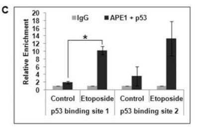
Chromatin Immunoprecipitation: APE Antibody (13B8E5C2) - Azide and BSA Free [NBP2-80578] - Association of p53 and APE1 on p53-binding sites in p21 promoter. Re-ChIP analysis (first IP with alpha-APE1 and the second IP with alpha-p53 antibody) showing simultaneous recruitment of APE1 and p53 in control vs. EPE treated cells; *: p value <0.05 (n=2) calculated based on APE1/p53 enriched DNA from control vs. etoposide treated cells. Image collected and cropped by CiteAb from the following publication (https://dx.plos.org/10.1371/journal.pone.0068467) licensed under a CC-BY license. Image from the standard format of this antibody.
![Western Blot: APE Antibody (13B8E5C2)Azide and BSA Free [NBP2-80578]](https://resources.rndsystems.com/images/products/APE-Antibody-13B8E5C2-Azide-and-BSA-Free-Western-Blot-NBP2-80578-img0001.jpg "Western Blot: APE Antibody (13B8E5C2)Azide and BSA Free [NBP2-80578]")
Western Blot: APE Antibody (13B8E5C2)Azide and BSA Free [NBP2-80578]
Western Blot: APE Antibody (13B8E5C2) - Azide and BSA Free [NBP2-80578] - Ovarian Cancer cell lines. Image from the standard format of this antibody.![Immunocytochemistry/ Immunofluorescence: APE Antibody (13B8E5C2) - Azide and BSA Free [NBP2-80578]](https://resources.rndsystems.com/images/products/APE-Antibody-13B8E5C2-Azide-and-BSA-Free-Immunocytochemistry-Immunofluorescence-NBP2-80578-img0003.jpg "Immunocytochemistry/ Immunofluorescence: APE Antibody (13B8E5C2) - Azide and BSA Free [NBP2-80578]")
Immunocytochemistry/ Immunofluorescence: APE Antibody (13B8E5C2) - Azide and BSA Free [NBP2-80578]
Immunocytochemistry/Immunofluorescence: APE Antibody (13B8E5C2) - Azide and BSA Free [NBP2-80578] - HeLa cells stained NB100-116 (Green) detected with DyLight Fluor 488 conjugated anti-rabbit IgG secondary antibody. Nuclei are counterstained with Hoechst 33258 (Blue). Image from the standard format of this antibody.![Immunocytochemistry/ Immunofluorescence: APE Antibody (13B8E5C2) - Azide and BSA Free [NBP2-80578]](https://resources.rndsystems.com/images/products/APE-Antibody-13B8E5C2-Azide-and-BSA-Free-Immunocytochemistry-Immunofluorescence-NBP2-80578-img0004.jpg "Immunocytochemistry/ Immunofluorescence: APE Antibody (13B8E5C2) - Azide and BSA Free [NBP2-80578]")
Immunocytochemistry/ Immunofluorescence: APE Antibody (13B8E5C2) - Azide and BSA Free [NBP2-80578]
Immunocytochemistry/Immunofluorescence: APE Antibody (13B8E5C2) - Azide and BSA Free [NBP2-80578] - HeLa cells were fixed for 10 minutes using 10% formalin and then permeabilized for 5 minutes using 1X PBS + 0.05% Triton X-100. The cells were incubated with anti-APE (13B8E5C2) at 5 ug/mL overnight at 4C and detected with an anti-mouse DyLight 488 (Green) at a 1:500 dilution. Nuclei were counterstained with DAPI (Blue). Cells were imaged using a 40X objective. Image from the standard format of this antibody.![Immunocytochemistry: APE Antibody (13B8E5C2) - Azide and BSA Free [NBP2-80578]](https://resources.rndsystems.com/images/products/APE-Antibody-13B8E5C2-Azide-and-BSA-Free-Immunocytochemistry-NBP2-80578-img0005.jpg "Immunocytochemistry: APE Antibody (13B8E5C2) - Azide and BSA Free [NBP2-80578]")
Immunocytochemistry: APE Antibody (13B8E5C2) - Azide and BSA Free [NBP2-80578]
Immunocytochemistry: APE Antibody (13B8E5C2) - Azide and BSA Free [NBP2-80578] - Immunocytochemical detection of APE-ref-1 in breast cancer cell line MDA MB 231. Image from the standard format of this antibody.![Simple Western: APE Antibody (13B8E5C2)Azide and BSA Free [NBP2-80578]](https://resources.rndsystems.com/images/products/APE-Antibody-13B8E5C2-Azide-and-BSA-Free-Simple-Western-NBP2-80578-img0010.jpg "Simple Western: APE Antibody (13B8E5C2)Azide and BSA Free [NBP2-80578]")
Simple Western: APE Antibody (13B8E5C2)Azide and BSA Free [NBP2-80578]
Simple Western: APE Antibody (13B8E5C2) - Azide and BSA Free [NBP2-80578] - Image shows a specific band for APE1 in 0.1 mg/mL of HeLa lysate. This experiment was performed under reducing conditions using the 12-230kDa separation system. * Non-specific interaction with the 230 kDa Simple Western standard may be seen with this antibody. Image from the standard format of this antibody.![Knockdown Validated: APE Antibody (13B8E5C2) - Azide and BSA Free [NBP2-80578]](https://resources.rndsystems.com/images/products/APE-Antibody-13B8E5C2-Azide-and-BSA-Free-Knockdown-Validated-NBP2-80578-img0009.jpg "Western Blot: APE Antibody (13B8E5C2) - Azide and BSA Free [NBP2-80578]")
Western Blot: APE Antibody (13B8E5C2) - Azide and BSA Free [NBP2-80578]
Western Blot: APE Antibody (13B8E5C2) - Azide and BSA Free [NBP2-80578] - Repression of p21 by APE1 in p53-null cells and effect of ectopic p53 in this repression. Representative Western analysis of p53, APE1, p21 and alpha-Tubulin levels in the same HCT116p53null cells. Image collected and cropped by CiteAb from the following publication (https://dx.plos.org/10.1371/journal.pone.0068467) licensed under a CC-BY license. Image from the standard format of this antibody.Applications for APE Antibody (13B8E5C2) - Azide and BSA Free
Application
Recommended Usage
Chromatin Immunoprecipitation (ChIP)
1:10-1:500
Gel Super Shift Assays
reported in scientific literature
Immunoblotting
reported in scientific literature (PMID 27608656)
Immunocytochemistry/ Immunofluorescence
1:50 - 1:200
Immunohistochemistry
1:100
Immunohistochemistry-Frozen
1:10 - 1:500
Immunohistochemistry-Paraffin
1:100
Immunoprecipitation
1:10 - 1:500
Proximity Ligation Assay
reported in scientific literature (PMID 27808278)
Simple Western
1:25
Western Blot
1:100 - 1:2000
Application Notes
In Western blot, this antibody detects a single band at 37 kDa. In IHC, it can be competitively inhibited from recognizing the APE1 antigen in tissues using APE1 protein. It can also be used on frozen and fixed-paraffin sections and cytospin preps. In IHC-P, staining was observed in the nucleus of a human breast cancer xenograft. Prior to immunostaining paraffin tissues, antigen retrieval with sodium citrate buffer (pH 6.0) is recommended. In Simple Western only 10 - 15 uL of the recommended dilution is used per data point.
See Simple Western Antibody Database for Simple Western validation: antibody dilution of 1:25. Separated by Size-Wes, Sally Sue/Peggy Sue.
See Simple Western Antibody Database for Simple Western validation: antibody dilution of 1:25. Separated by Size-Wes, Sally Sue/Peggy Sue.
Formulation, Preparation, and Storage
Purification
Protein G purified
Formulation
PBS
Format
Azide and BSA Free
Preservative
No Preservative
Concentration
1 mg/ml
Shipping
The product is shipped with polar packs. Upon receipt, store it immediately at the temperature recommended below.
Stability & Storage
Store at 4C short term. Aliquot and store at -20C long term. Avoid freeze-thaw cycles.
Background: APE
Long Name
Apurinic/Apyrimidinic Endonuclease
Alternate Names
APEX1
Gene Symbol
APEX1
Additional APE Products
Product Documents for APE Antibody (13B8E5C2) - Azide and BSA Free
Certificate of Analysis
To download a Certificate of Analysis, please enter a lot or batch number in the search box below.
Product Specific Notices for APE Antibody (13B8E5C2) - Azide and BSA Free
This product is for research use only and is not approved for use in humans or in clinical diagnosis. Primary Antibodies are guaranteed for 1 year from date of receipt.
Related Research Areas
Customer Reviews for APE Antibody (13B8E5C2) - Azide and BSA Free
There are currently no reviews for this product. Be the first to review APE Antibody (13B8E5C2) - Azide and BSA Free and earn rewards!
Have you used APE Antibody (13B8E5C2) - Azide and BSA Free?
Submit a review and receive an Amazon gift card!
$25/€18/£15/$25CAN/¥2500 Yen for a review with an image
$10/€7/£6/$10CAN/¥1110 Yen for a review without an image
Submit a review
Protocols
Find general support by application which include: protocols, troubleshooting, illustrated assays, videos and webinars.
- Antigen Retrieval Protocol (PIER)
- Antigen Retrieval for Frozen Sections Protocol
- Appropriate Fixation of IHC/ICC Samples
- Cellular Response to Hypoxia Protocols
- ChIP Protocol Video
- Chromatin Immunoprecipitation (ChIP) Protocol
- Chromatin Immunoprecipitation Protocol
- Chromogenic IHC Staining of Formalin-Fixed Paraffin-Embedded (FFPE) Tissue Protocol
- Chromogenic Immunohistochemistry Staining of Frozen Tissue
- ClariTSA™ Fluorophore Kits
- Detection & Visualization of Antibody Binding
- Fluorescent IHC Staining of Frozen Tissue Protocol
- Graphic Protocol for Heat-induced Epitope Retrieval
- Graphic Protocol for the Preparation and Fluorescent IHC Staining of Frozen Tissue Sections
- Graphic Protocol for the Preparation and Fluorescent IHC Staining of Paraffin-embedded Tissue Sections
- Graphic Protocol for the Preparation of Gelatin-coated Slides for Histological Tissue Sections
- ICC Cell Smear Protocol for Suspension Cells
- ICC Immunocytochemistry Protocol Videos
- ICC for Adherent Cells
- IHC Sample Preparation (Frozen sections vs Paraffin)
- Immunocytochemistry (ICC) Protocol
- Immunocytochemistry Troubleshooting
- Immunofluorescence of Organoids Embedded in Cultrex Basement Membrane Extract
- Immunofluorescent IHC Staining of Formalin-Fixed Paraffin-Embedded (FFPE) Tissue Protocol
- Immunohistochemistry (IHC) and Immunocytochemistry (ICC) Protocols
- Immunohistochemistry Frozen Troubleshooting
- Immunohistochemistry Paraffin Troubleshooting
- Immunoprecipitation Protocol
- Preparing Samples for IHC/ICC Experiments
- Preventing Non-Specific Staining (Non-Specific Binding)
- Primary Antibody Selection & Optimization
- Protocol for Heat-Induced Epitope Retrieval (HIER)
- Protocol for Making a 4% Formaldehyde Solution in PBS
- Protocol for VisUCyte™ HRP Polymer Detection Reagent
- Protocol for the Fluorescent ICC Staining of Cell Smears - Graphic
- Protocol for the Fluorescent ICC Staining of Cultured Cells on Coverslips - Graphic
- Protocol for the Preparation & Fixation of Cells on Coverslips
- Protocol for the Preparation and Chromogenic IHC Staining of Frozen Tissue Sections
- Protocol for the Preparation and Chromogenic IHC Staining of Frozen Tissue Sections - Graphic
- Protocol for the Preparation and Chromogenic IHC Staining of Paraffin-embedded Tissue Sections
- Protocol for the Preparation and Chromogenic IHC Staining of Paraffin-embedded Tissue Sections - Graphic
- Protocol for the Preparation and Fluorescent ICC Staining of Cells on Coverslips
- Protocol for the Preparation and Fluorescent ICC Staining of Non-adherent Cells
- Protocol for the Preparation and Fluorescent ICC Staining of Stem Cells on Coverslips
- Protocol for the Preparation and Fluorescent IHC Staining of Frozen Tissue Sections
- Protocol for the Preparation and Fluorescent IHC Staining of Paraffin-embedded Tissue Sections
- Protocol for the Preparation of Gelatin-coated Slides for Histological Tissue Sections
- Protocol for the Preparation of a Cell Smear for Non-adherent Cell ICC - Graphic
- R&D Systems Quality Control Western Blot Protocol
- TUNEL and Active Caspase-3 Detection by IHC/ICC Protocol
- The Importance of IHC/ICC Controls
- Troubleshooting Guide: Immunohistochemistry
- Troubleshooting Guide: Western Blot Figures
- Western Blot Conditions
- Western Blot Protocol
- Western Blot Protocol for Cell Lysates
- Western Blot Troubleshooting
- Western Blot Troubleshooting Guide
- View all Protocols, Troubleshooting, Illustrated assays and Webinars
Loading...