ATM Antibody (2C1) - Azide and BSA Free
Novus Biologicals | Catalog # NB100-309
![Western Blot: ATM Antibody (2C1) [NB100-309]](https://resources.rndsystems.com/images/products/ATM-Antibody-2C1-Western-Blot-NB100-309-img0018.jpg "Western Blot: ATM Antibody (2C1) [NB100-309]")
Loading...
Key Product Details
Validated by
Knockout/Knockdown, Biological Validation
Species Reactivity
Validated:
Human, Mouse, Rat, Monkey
Cited:
Human, Mouse, Hamster - Cricetulus (Chinese Hamster)
Applications
Validated:
Immunohistochemistry, Immunohistochemistry-Paraffin, Western Blot, ELISA, Flow Cytometry, Immunocytochemistry/ Immunofluorescence, Immunoprecipitation, Chromatin Immunoprecipitation (ChIP), Knockdown Validated, SDS-Page, Single Cell Western
Cited:
Immunohistochemistry-Paraffin, Western Blot, ELISA, Flow Cytometry, Immunocytochemistry/ Immunofluorescence, Immunoprecipitation, SDS-Page
Label
Unconjugated
Antibody Source
Monoclonal Mouse IgG1 Clone # 2C1
Format
Azide and BSA Free
Loading...
Product Specifications
Immunogen
Recombinant protein expressed in E. coli corresponding to amino acids 2577-3056.
Reactivity Notes
Use in Human reported in scientific literature (PMID:33743824).
Localization
Nucleus. Cytoplasmic vesicle.
Specificity
ATM Antibody (2C1) recognizes full-length ATM, a 350-kDa nuclear phosphoprotein.
Clonality
Monoclonal
Host
Mouse
Isotype
IgG1
Theoretical MW
351 kDa.
Disclaimer note: The observed molecular weight of the protein may vary from the listed predicted molecular weight due to post translational modifications, post translation cleavages, relative charges, and other experimental factors.
Disclaimer note: The observed molecular weight of the protein may vary from the listed predicted molecular weight due to post translational modifications, post translation cleavages, relative charges, and other experimental factors.
Scientific Data Images for ATM Antibody (2C1) - Azide and BSA Free
Western Blot: ATM Antibody (2C1) [NB100-309]
Western Blot: ATM Antibody (2C1) [NB100-309] - Detection of human ATM protein using ATM antibody (2C1) [NB100-309] by western blot or immunoprecipitation. Theoretical molecular weight 351 kDa.![Immunohistochemistry-Paraffin: ATM Antibody (2C1) [NB100-309]](https://resources.rndsystems.com/images/products/ATM-Antibody-2C1-Immunohistochemistry-Paraffin-NB100-309-img0021.jpg "Immunohistochemistry-Paraffin: ATM Antibody (2C1) [NB100-309]")
Immunohistochemistry-Paraffin: ATM Antibody (2C1) [NB100-309]
Immunohistochemistry-Paraffin: ATM Antibody (2C1) [NB100-309] - Human breast carcinoma. ATM stained by ATM antibody [2C1] diluted at 1:100.Antigen Retrieval: Citrate buffer, pH 6.0, 15 min.![Western Blot: ATM Antibody (2C1) [NB100-309]](https://resources.rndsystems.com/images/products/ATM-Antibody-2C1-Western-Blot-NB100-309-img0010.jpg "Western Blot: ATM Antibody (2C1) [NB100-309]")
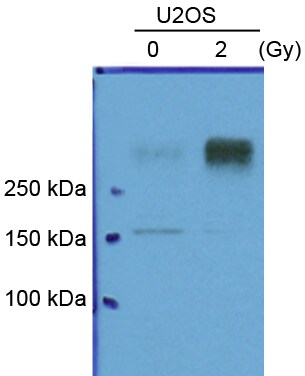
Western Blot: ATM Antibody (2C1) [NB100-309]
Western Blot: ATM Antibody (2C1) [NB100-309] - Analysis of ATM in U2OS sarcoma cells using ATM antibody (2C1) [NB100-309]. Theoretical molecular weight 351 kDa. Western blot image submitted by a verifeid customer review.![Immunohistochemistry-Paraffin: ATM Antibody (2C1) [NB100-309]](https://resources.rndsystems.com/images/products/ATM-Antibody-2C1-Immunohistochemistry-Paraffin-NB100-309-img0012.jpg "Immunohistochemistry-Paraffin: ATM Antibody (2C1) [NB100-309]")
Immunohistochemistry-Paraffin: ATM Antibody (2C1) [NB100-309]
Immunohistochemistry-Paraffin: ATM Antibody (2C1) [NB100-309] - Human Kidney (formalin-fixed, paraffin-embedded) stained with ATM antibody (2C1) [NB100-309] at 5 ug/ml followed by biotinylated anti-mouse IgG secondary antibody, alkaline phosphatase-streptavidin and chromogen.![Knockdown Validated: ATM Antibody (2C1) [NB100-309]](https://resources.rndsystems.com/images/products/ATM-Antibody-2C1-Knockdown-Validated-NB100-309-img0017.jpg "Western Blot: ATM Antibody (2C1) [NB100-309]")
![Knockdown Validated: ATM Antibody (2C1) [NB100-309]](https://resources.rndsystems.com/images/products/ATM-Antibody-2C1-Western-Blot-NB100-309-img0019.jpg "Western Blot: ATM Antibody (2C1) [NB100-309]")
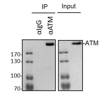
![Immunoprecipitation: ATM Antibody (2C1) [NB100-309]](https://resources.rndsystems.com/images/products/ATM-Antibody-2C1-Western-Blot-NB100-309-img0020.jpg "Immunoprecipitation: ATM Antibody (2C1) [NB100-309]")
Immunoprecipitation: ATM Antibody (2C1) [NB100-309]
Immunoprecipitation: ATM Antibody (2C1) [NB100-309] - MDA-MB-231 whole cell lysates were immunoprecipitated with 2 ug ATM Antibody (NB100-309), followed by Western blot (primary antibody: NB100-309 at 1:1000, 4C overnight. Western blot image submitted by a verified customer review. [NB100-309] -")
 [NB100-309] -")
Western Blot: ATM Antibody (2C1) [NB100-309] -
Western Blot: ATM Antibody (2C1) [NB100-309] - Western blot analysis of the extent of knockdown MRE11 & ATM, & the lack of effect of Mirin activation of ATM. The extent of shRNA-mediated knockdown of MRE11 & ATM are shown for (A) clones GFP-7F1 & GFP-6D1 & (B) clones EDS-7F2 & EDS-6J8. The extent of knockdown was determined by analyzing the intensity of the ATM & MRE11 bands relative to the intensity of the loading control GAPDH bands (see Table 1). (C) The effect of Mirin on activation of ATM was determined by analysis of phosphorylation of ATM in response to ionizing radiation. Cultures treated with DMSO alone, 20 μM Mirin, or knockdown of ATM were analyzed by western blot 30 min after exposure to 10 Gy of ionizing radiation. Image collected & cropped by CiteAb from the following publication (https://pubmed.ncbi.nlm.nih.gov/26209132), licensed under a CC-BY license. Not internally tested by Novus Biologicals. [NB100-309] -")
Western Blot: ATM Antibody (2C1) [NB100-309] -
Western Blot: ATM Antibody (2C1) [NB100-309] - Rac1 inhibition abolishes IR-induced activation of both ATM & ATR signaling. (A) MCF-7 cells were treated with/without 20-Gy IR in the presence or absence of 100 μM NSC23766 & incubated for 1 hour at 37°C before analysis. To assess the ATM kinase activity, ATM was immunoprecipitated from cell lysate by using anti-ATM antibody (2C1) & assayed for ATM activity by using p53 recombinant protein as substrate. To measure the Chk2 activity, Chk2 was immunoprecipitated from cell lysate by using B-4 anti-Chk2 antibody & assayed for Chk2 activity by using Cdc25C recombinant protein as substrate. As controls, ATM & Chk2 protein levels in the immunoprecipitates (IP-WB) as well as in cell lysates (WB) were assessed with immunoblotting. (B) ATR & Chk1 were immunoprecipitated from cell lysates by using N-19 anti-ATR & G-4 anti-Chk1 antibody, respectively. ATR activity was assayed by using p53 recombinant protein substrate, & Chk1 activity assayed by using Cdc25C recombinant protein substrate. As controls, ATR & Chk1 protein levels in the immunoprecipitates (IP-WB), as well as in cell lysates (WB) were assessed with immunoblotting. (C) MCF-7 cells were exposed to IR at the indicated doses in the presence or absence of NSC23766, incubated for 1 hour, & assessed for Chk1 & Chk2 activities. *Kinase assay does not contain Cdc25C substrate. (D) T47D & ZR-75-1 cells were exposed to 10-Gy IR in the presence or absence of 100 μM NSC23766, incubated for 1 hour, & analyzed for Chk1 & Chk2 activities. Image collected & cropped by CiteAb from the following publication (http://breast-cancer-research.biomedcentral.com/articles/10.1186/bcr3164), licensed under a CC-BY license. Not internally tested by Novus Biologicals. [NB100-309] -")
Western Blot: ATM Antibody (2C1) [NB100-309] -
Western Blot: ATM Antibody (2C1) [NB100-309] - Inhibition of Rac1 by N17Rac1 mutant or Rac1 siRNA diminishes IR-induced G2/M checkpoint activation. (A) MCF-7 cells were infected with Ad.N17Rac1 or Ad.Control for 24 hours & exposed to 15-Gy IR. Left panel: the cells were analyzed for DNA content 24 hours after IR. The result depicts the percentage of cells with 4N-DNA content & is shown as mean ± SD of quadruplicate samples. *P < 0.001 (n = 4), significant difference from the irradiated Ad.Control-infected cells. Right panel: Inset: at 15 minutes after IR, the infected cells were analyzed for Rac1 activities (Rac1-GTP) & protein levels (total Rac1). Bar graph: mitotic cells in the cell samples were analyzed 2 hours after IR. The result depicts the percentage of mitotic cells & is shown as mean ± SD of triplicate samples. **P = 0.002 (n = 3), significant difference from the irradiated Ad.Control-infected cells. (B) Upper panel: MCF-7 cells transfected with Rac1 siRNA (Rac1) or control siRNA (Control) were incubated for the indicated times & analyzed for protein levels of Rac1 & Actin. Lower panel: After 2-day incubation, the siRNA-transfected cells were exposed to IR, incubated for 24 hours, & assessed for DNA content. Results depict the percentage of cells with 4N-DNA content & represent the mean ± SD of three separate experiments in duplicate samples. *P < 0.001 (n = 6), significant difference from the irradiated Control-siRNA transfected cells. (C) After 2-day incubation, siRNA-transfected cells were treated with/without 20-Gy IR, incubated for 1 hour, & analyzed for ATM, ATR, Chk1, & Chk2 activities. Image collected & cropped by CiteAb from the following publication (http://breast-cancer-research.biomedcentral.com/articles/10.1186/bcr3164), licensed under a CC-BY license. Not internally tested by Novus Biologicals. [NB100-309] -")
Western Blot: ATM Antibody (2C1) [NB100-309] -
Western Blot: ATM Antibody (2C1) [NB100-309] - HeLa whole cell extract and nuclear extracts (30 ug) were separated by 5% SDS-PAGE, and the membrane was blotted with ATM antibody [2C1] (NB100-309) diluted at 1:500. The HRP-conjugated anti-mouse IgG antibody was used to detect the primary antibody. [NB100-309] -")
Western Blot: ATM Antibody (2C1) [NB100-309] -
Western Blot: ATM Antibody (2C1) [NB100-309] - Whole cell extract (30 ug) was separated by 5% SDS-PAGE, and the membrane was blotted with ATM antibody [2C1] (NB100-309) diluted at 1:1000. [NB100-309] -")
Immunohistochemistry: ATM Antibody (2C1) [NB100-309] -
Human Testis (formalin-fixed, paraffin-embedded) stained with ATM antibody at 5 ug/ml followed by biotinylated anti-mouse IgG secondary antibody, alkaline phosphatase-streptavidin and chromogen.Applications for ATM Antibody (2C1) - Azide and BSA Free
Application
Recommended Usage
Chromatin Immunoprecipitation (ChIP)
1:10-1:500
ELISA
1:100 - 1:2000
Immunocytochemistry/ Immunofluorescence
1:10 - 1:500
Immunohistochemistry
5 ug/mL
Immunohistochemistry-Paraffin
5 ug/mL
Immunoprecipitation
1 - 10 ug/mL
Single Cell Western
100 ug/mL
Western Blot
1:500 - 1:3000
Application Notes
Use in SDS-PAGE reported in scientific literature (PMID:34210973). Use in IHC-P reported in scientific literature (PMID: 25895060). Use in FLOW reported in scientific literature (PMID: 15197179). ATM antibody is validated for WB, IP from a verified customer reviews.
Reviewed Applications
Read 3 reviews rated 4.7 using NB100-309 in the following applications:
Flow Cytometry Panel Builder
Bio-Techne Knows Flow Cytometry
Save time and reduce costly mistakes by quickly finding compatible reagents using the Panel Builder Tool.
Advanced Features
- Spectra Viewer - Custom analysis of spectra from multiple fluorochromes
- Spillover Popups - Visualize the spectra of individual fluorochromes
- Antigen Density Selector - Match fluorochrome brightness with antigen density

Formulation, Preparation, and Storage
Purification
Antigen Affinity-purified
Formulation
PBS
Format
Azide and BSA Free
Preservative
No Preservative
Concentration
Concentrations vary lot to lot. See vial label for concentration. If unlisted please contact technical services.
Shipping
The product is shipped with polar packs. Upon receipt, store it immediately at the temperature recommended below.
Stability & Storage
Store at 4C short term. Aliquot and store at -20C long term. Avoid freeze-thaw cycles.
Background: ATM
The theoretical molecular weight of ATM is 350 kDa and it has 3 main domains: a FAT (focal adhesion targeting) domain (aa 1960-2566), a PI-3/PI-4 kinase catalytic domain (aa 2712-2962), and a C-terminal FAT domain (aa 3024-3056). ATM exists as a dimer or tetramer in its inactive state. Upon sensing DNA damage, the MRE11-RAD50-NBS1 (MRN) complex recruits ATM. The intricate process of ATM activation involves acetylation by KAT5/TIP60, autophosphorylation at Ser-1981, and dissociation into catalytically active monomers (5). Following activation, ATM phosphorylates multiple substrates such as p53/TP53 and Chk2 involved in DNA repair, checkpoint signaling, and the apoptosis pathway.
References
1. Paull TT. (2015) Mechanisms of ATM Activation. Annu Rev Biochem. 84:711-38. PMID: 25580527
2. Chaudhary MW and Al-Baradie RS. (2014) Ataxia-telangiectasia: future prospects. Appl Clin Genet. 7:159-167. PMID: 25258552
3. Stagni V, Cirotti C, and Barila D. (2018) Ataxia-Telangiectasia Mutated Kinase in the Control of Oxidative Stress, Mitochondria, and Autophagy in Cancer: A Maestro With a Large Orchestra. Front Oncol. 8:73. PMID: 29616191
4. Gumy-Pause F, Wacker P, and Sappino AP. (2004) ATM gene and lymphoid malignancies. Leukemia. 18(2):238-42. PMID: 14628072
5. Adamowicz M. (2018) Breaking up with ATM. J Immunol Sci. 2(1):26-31. PMID: 29652413
Long Name
Ataxia Telangiectasia-mutated
Alternate Names
TEL1, TELO1, TPLL
Gene Symbol
ATM
UniProt
Additional ATM Products
Product Documents for ATM Antibody (2C1) - Azide and BSA Free
Certificate of Analysis
To download a Certificate of Analysis, please enter a lot or batch number in the search box below.
Product Specific Notices for ATM Antibody (2C1) - Azide and BSA Free
This product is for research use only and is not approved for use in humans or in clinical diagnosis. Primary Antibodies are guaranteed for 1 year from date of receipt.
Related Research Areas
Citations for ATM Antibody (2C1) - Azide and BSA Free
 Powered by Bioz
Customer Reviews for ATM Antibody (2C1) - Azide and BSA Free (3)
4.7 out of 5
3 Customer Ratings
Have you used ATM Antibody (2C1) - Azide and BSA Free?
Submit a review and receive an Amazon gift card!
$25/€18/£15/$25CAN/¥2500 Yen for a review with an image
$10/€7/£6/$10CAN/¥1110 Yen for a review without an image
Submit a review
Customer Images
Showing
1
-
3 of
3 reviews
Showing All
Filter By:
-
Application: ImmunoprecipitationSample Tested: MDA-MB-231Species: HumanVerified Customer | Posted 10/28/2020Western Blot: MDA-MB-231 whole cell lysates were IP with 2μg ATM Antibody (NB100-309), followed by Western blot (primary antibody: NB100-309, 1:1000, 4℃, overnight).

-
Application: Western BlotSample Tested:Species: HumanVerified Customer | Posted 04/28/2015ATM antibody (NB100-309)
-
Application: Western BlotSample Tested: U2OS whole cell lysateSpecies: HumanVerified Customer | Posted 11/02/2014
There are no reviews that match your criteria.
Protocols
Find general support by application which include: protocols, troubleshooting, illustrated assays, videos and webinars.
- 7-Amino Actinomycin D (7-AAD) Cell Viability Flow Cytometry Protocol
- Antigen Retrieval Protocol (PIER)
- Antigen Retrieval for Frozen Sections Protocol
- Appropriate Fixation of IHC/ICC Samples
- Cellular Response to Hypoxia Protocols
- ChIP Protocol Video
- Chromatin Immunoprecipitation (ChIP) Protocol
- Chromatin Immunoprecipitation Protocol
- Chromogenic IHC Staining of Formalin-Fixed Paraffin-Embedded (FFPE) Tissue Protocol
- Chromogenic Immunohistochemistry Staining of Frozen Tissue
- ClariTSA™ Fluorophore Kits
- Detection & Visualization of Antibody Binding
- ELISA Sample Preparation & Collection Guide
- ELISA Troubleshooting Guide
- Extracellular Membrane Flow Cytometry Protocol
- Flow Cytometry Protocol for Cell Surface Markers
- Flow Cytometry Protocol for Staining Membrane Associated Proteins
- Flow Cytometry Staining Protocols
- Flow Cytometry Troubleshooting Guide
- Fluorescent IHC Staining of Frozen Tissue Protocol
- Graphic Protocol for Heat-induced Epitope Retrieval
- Graphic Protocol for the Preparation and Fluorescent IHC Staining of Frozen Tissue Sections
- Graphic Protocol for the Preparation and Fluorescent IHC Staining of Paraffin-embedded Tissue Sections
- Graphic Protocol for the Preparation of Gelatin-coated Slides for Histological Tissue Sections
- How to Run an R&D Systems DuoSet ELISA
- How to Run an R&D Systems Quantikine ELISA
- How to Run an R&D Systems Quantikine™ QuicKit™ ELISA
- ICC Cell Smear Protocol for Suspension Cells
- ICC Immunocytochemistry Protocol Videos
- ICC for Adherent Cells
- IHC Sample Preparation (Frozen sections vs Paraffin)
- Immunocytochemistry (ICC) Protocol
- Immunocytochemistry Troubleshooting
- Immunofluorescence of Organoids Embedded in Cultrex Basement Membrane Extract
- Immunofluorescent IHC Staining of Formalin-Fixed Paraffin-Embedded (FFPE) Tissue Protocol
- Immunohistochemistry (IHC) and Immunocytochemistry (ICC) Protocols
- Immunohistochemistry Frozen Troubleshooting
- Immunohistochemistry Paraffin Troubleshooting
- Immunoprecipitation Protocol
- Intracellular Flow Cytometry Protocol Using Alcohol (Methanol)
- Intracellular Flow Cytometry Protocol Using Detergents
- Intracellular Nuclear Staining Flow Cytometry Protocol Using Detergents
- Intracellular Staining Flow Cytometry Protocol Using Alcohol Permeabilization
- Intracellular Staining Flow Cytometry Protocol Using Detergents to Permeabilize Cells
- Preparing Samples for IHC/ICC Experiments
- Preventing Non-Specific Staining (Non-Specific Binding)
- Primary Antibody Selection & Optimization
- Propidium Iodide Cell Viability Flow Cytometry Protocol
- Protocol for Heat-Induced Epitope Retrieval (HIER)
- Protocol for Liperfluo
- Protocol for Making a 4% Formaldehyde Solution in PBS
- Protocol for VisUCyte™ HRP Polymer Detection Reagent
- Protocol for the Characterization of Human Th22 Cells
- Protocol for the Characterization of Human Th9 Cells
- Protocol for the Fluorescent ICC Staining of Cell Smears - Graphic
- Protocol for the Fluorescent ICC Staining of Cultured Cells on Coverslips - Graphic
- Protocol for the Preparation & Fixation of Cells on Coverslips
- Protocol for the Preparation and Chromogenic IHC Staining of Frozen Tissue Sections
- Protocol for the Preparation and Chromogenic IHC Staining of Frozen Tissue Sections - Graphic
- Protocol for the Preparation and Chromogenic IHC Staining of Paraffin-embedded Tissue Sections
- Protocol for the Preparation and Chromogenic IHC Staining of Paraffin-embedded Tissue Sections - Graphic
- Protocol for the Preparation and Fluorescent ICC Staining of Cells on Coverslips
- Protocol for the Preparation and Fluorescent ICC Staining of Non-adherent Cells
- Protocol for the Preparation and Fluorescent ICC Staining of Stem Cells on Coverslips
- Protocol for the Preparation and Fluorescent IHC Staining of Frozen Tissue Sections
- Protocol for the Preparation and Fluorescent IHC Staining of Paraffin-embedded Tissue Sections
- Protocol for the Preparation of Gelatin-coated Slides for Histological Tissue Sections
- Protocol for the Preparation of a Cell Smear for Non-adherent Cell ICC - Graphic
- Protocol: Annexin V and PI Staining by Flow Cytometry
- Protocol: Annexin V and PI Staining for Apoptosis by Flow Cytometry
- Quantikine HS ELISA Kit Assay Principle, Alkaline Phosphatase
- Quantikine HS ELISA Kit Principle, Streptavidin-HRP Polymer
- R&D Systems Quality Control Western Blot Protocol
- Sandwich ELISA (Colorimetric) – Biotin/Streptavidin Detection Protocol
- Sandwich ELISA (Colorimetric) – Direct Detection Protocol
- TUNEL and Active Caspase-3 Detection by IHC/ICC Protocol
- The Importance of IHC/ICC Controls
- Troubleshooting Guide: ELISA
- Troubleshooting Guide: Fluorokine Flow Cytometry Kits
- Troubleshooting Guide: Immunohistochemistry
- Troubleshooting Guide: Western Blot Figures
- Western Blot Conditions
- Western Blot Protocol
- Western Blot Protocol for Cell Lysates
- Western Blot Troubleshooting
- Western Blot Troubleshooting Guide
- View all Protocols, Troubleshooting, Illustrated assays and Webinars
FAQs for ATM Antibody (2C1) - Azide and BSA Free
Showing
1
-
1 of
1 FAQ
Showing All
-
Q: What is the theoretical molecular weight for your ATM antibodies?
A: The theoretical molecular weight for our ATM antibodies is 351 kDa.
Loading...