Beclin 1 Antibody - BSA Free
Novus Biologicals | Catalog # NB500-249
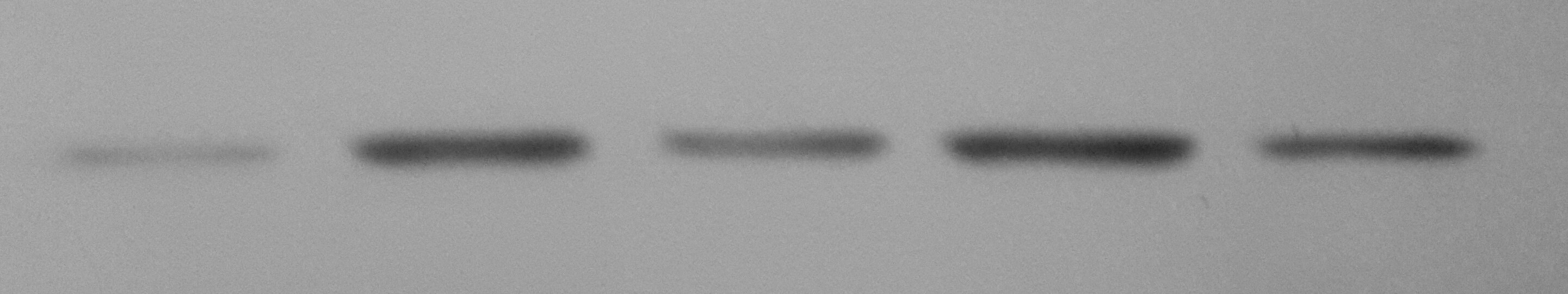
![Western Blot: Beclin 1 AntibodyBSA Free [NB500-249]](https://resources.rndsystems.com/images/products/Beclin-1-Antibody---BSA-Free-Western-Blot-NB500-249-img0018.jpg "Western Blot: Beclin 1 AntibodyBSA Free [NB500-249]")
Key Product Details
Validated by
Species Reactivity
Validated:
Cited:
Applications
Validated:
Cited:
Label
Antibody Source
Format
Product Specifications
Immunogen
Localization
Clonality
Host
Isotype
Scientific Data Images for Beclin 1 Antibody - BSA Free
Western Blot: Beclin 1 AntibodyBSA Free [NB500-249]
Beclin-1-Antibody---BSA-Free-Western-Blot-NB500-249-img0018.jpg![Immunocytochemistry/ Immunofluorescence: Beclin 1 Antibody - BSA Free [NB500-249]](https://resources.rndsystems.com/images/products/Beclin-1-Antibody---BSA-Free-Immunocytochemistry-Immunofluorescence-NB500-249-img0013.jpg "Immunocytochemistry/ Immunofluorescence: Beclin 1 Antibody - BSA Free [NB500-249]")
Immunocytochemistry/ Immunofluorescence: Beclin 1 Antibody - BSA Free [NB500-249]
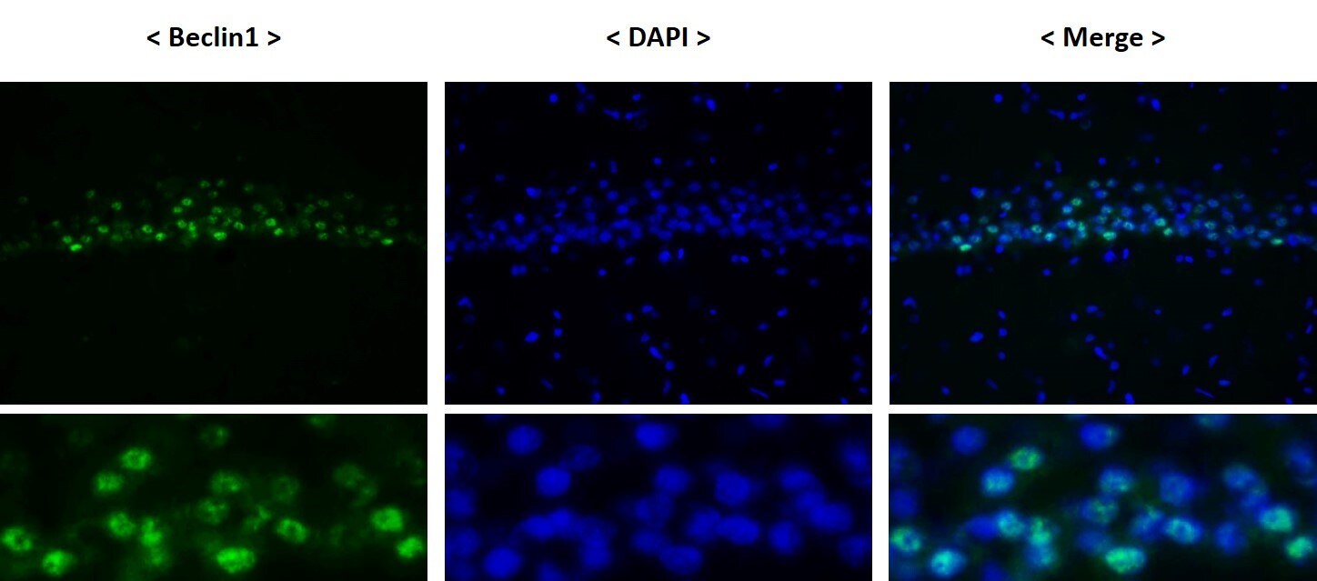
Immunocytochemistry/Immunofluorescence: Beclin 1 Antibody - BSA Free [NB500-249] - Beclin 1/ATG6 Antibody [NB500-249] - Beclin 1 antibody was tested in HeLa cells with Dylight 488 (green). Nuclei and alpha-tubulin were counterstained with DAPI (blue) and Dylight 550 (red).![Immunohistochemistry: Beclin 1 Antibody - BSA Free [NB500-249]](https://resources.rndsystems.com/images/products/Beclin-1-Antibody---BSA-Free-Immunohistochemistry-NB500-249-img0020.jpg "Immunohistochemistry: Beclin 1 Antibody - BSA Free [NB500-249]")
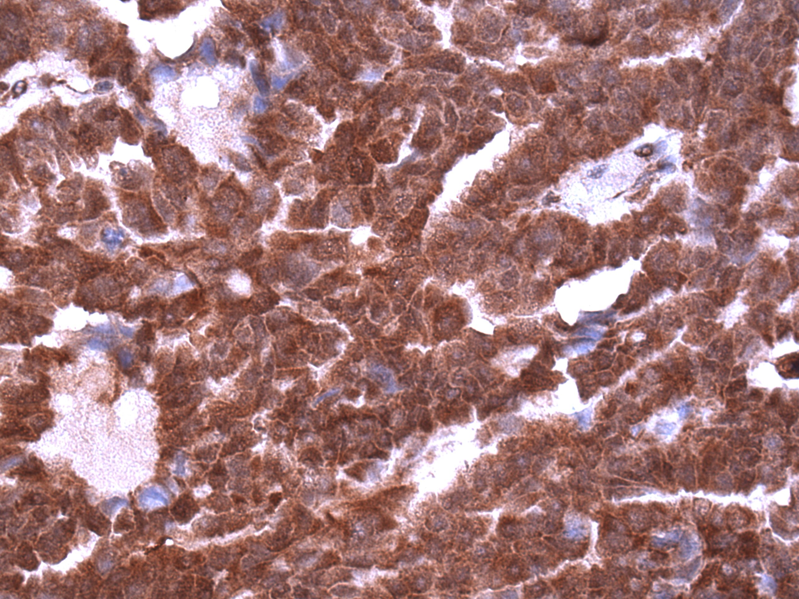
Immunohistochemistry: Beclin 1 Antibody - BSA Free [NB500-249]
Immunohistochemistry: Beclin 1 Antibody - BSA Free [NB500-249] - Detection of Beclin 1 (red) in Pheochromocytes of the Adrenal Medulla 40x.![Flow Cytometry: Beclin 1 Antibody - BSA Free [NB500-249]](https://resources.rndsystems.com/images/products/Beclin-1-Antibody---BSA-Free-Flow-Cytometry-NB500-249-img0024.jpg "Flow Cytometry: Beclin 1 Antibody - BSA Free [NB500-249]")
Flow Cytometry: Beclin 1 Antibody - BSA Free [NB500-249]
Flow Cytometry: Beclin 1 Antibody - BSA Free [NB500-249] - An intracellular stain was performed on U-87MG cells with Beclin 1 Antibody NB500-249 (blue) and a matched isotype control NBP2-24891 (orange). Cells were fixed with 4% PFA and then permeabilized with 0.1% saponin. Cells were incubated in an antibody dilution of 2.5 ug/mL for 30 minutes at room temperature, followed by Rabbit IgG (H+L) Cross-Adsorbed Secondary Antibody, Dylight 550 (SA5-10033, Thermo Fisher).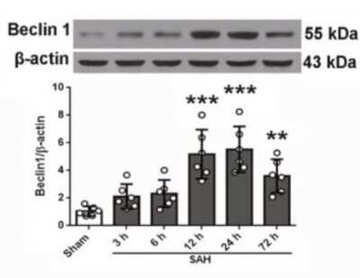
![Western Blot: Beclin 1 AntibodyBSA Free [NB500-249]](https://resources.rndsystems.com/images/products/Beclin-1-Antibody---BSA-Free-Western-Blot-NB500-249-img0011.jpg "Western Blot: Beclin 1 AntibodyBSA Free [NB500-249]")
Western Blot: Beclin 1 AntibodyBSA Free [NB500-249]
Western Blot: Beclin 1 Antibody - BSA Free [NB500-249] - Analysis of Belclin1. Lane 1: human brain. Lane 2: mouse brain.![Immunohistochemistry-Paraffin: Beclin 1 Antibody - BSA Free [NB500-249]](https://resources.rndsystems.com/images/products/Beclin-1-Antibody---BSA-Free-Immunohistochemistry-Paraffin-NB500-249-img0014.jpg "Immunohistochemistry-Paraffin: Beclin 1 Antibody - BSA Free [NB500-249]")
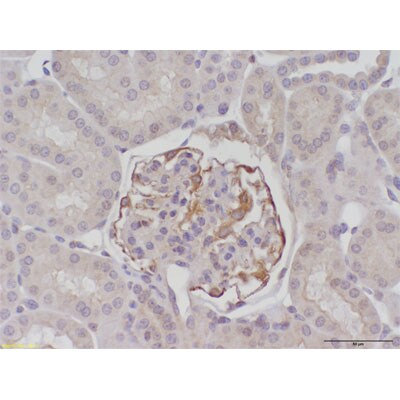
Immunohistochemistry-Paraffin: Beclin 1 Antibody - BSA Free [NB500-249]
Immunohistochemistry-Paraffin: Beclin 1 Antibody - BSA Free [NB500-249] - Beclin 1/ATG6 Antibody [NB500-249] - Analysis of Beclin1 in mouse kidney. Image courtsey of product review submitted by Kelly Hudkins.![Immunohistochemistry-Frozen: Beclin 1 Antibody - BSA Free [NB500-249]](https://resources.rndsystems.com/images/products/Beclin-1-Antibody---BSA-Free-Immunohistochemistry-Frozen-NB500-249-img0015.jpg "Immunohistochemistry-Frozen: Beclin 1 Antibody - BSA Free [NB500-249]")
Immunohistochemistry-Frozen: Beclin 1 Antibody - BSA Free [NB500-249]
Immunohistochemistry-Frozen: Beclin 1 Antibody - BSA Free [NB500-249] - Merged immunostaining of frozen section of Rat brain tissue. Image from verified customer review.![Flow Cytometry: Beclin 1 Antibody - BSA Free [NB500-249]](https://resources.rndsystems.com/images/products/Beclin-1-Antibody---BSA-Free-Flow-Cytometry-NB500-249-img0016.jpg "Flow Cytometry: Beclin 1 Antibody - BSA Free [NB500-249]")
Flow Cytometry: Beclin 1 Antibody - BSA Free [NB500-249]
Flow Cytometry: Beclin 1 Antibody - BSA Free [NB500-249] - An intracellular stain was performed on HeLa cells with NB500-249AF700 (blue) and a matched isotype control (orange). Cells were fixed with 4% PFA and then permeabilized with 0.1% saponin. Cells were incubated in an antibody dilution of 5 ug/mL for 30 minutes at room temperature. Both antibodies were conjugated to Alexa Fluor 700.![Flow Cytometry: Beclin 1 Antibody - BSA Free [NB500-249]](https://resources.rndsystems.com/images/products/Beclin-1-Antibody---BSA-Free-Flow-Cytometry-NB500-249-img0021.jpg "Flow Cytometry: Beclin 1 Antibody - BSA Free [NB500-249]")
Flow Cytometry: Beclin 1 Antibody - BSA Free [NB500-249]
Flow Cytometry: Beclin 1 Antibody - BSA Free [NB500-249] - An intracellular stain was performed on HepG2 cells with Beclin 1 Antibody NB500-249 (blue) and a matched isotype control NBP2-24891 (orange). Cells were fixed with 4% PFA and then permeabilized with 0.1% saponin. Cells were incubated in an antibody dilution of 1.0 ug/mL for 30 minutes at room temperature, followed by Rabbit IgG (H+L) Cross-Adsorbed Secondary Antibody, Dylight 550 (SA5-10033, Thermo Fisher).![Flow Cytometry: Beclin 1 Antibody - BSA Free [NB500-249]](https://resources.rndsystems.com/images/products/Beclin-1-Antibody---BSA-Free-Flow-Cytometry-NB500-249-img0022.jpg "Flow Cytometry: Beclin 1 Antibody - BSA Free [NB500-249]")
Flow Cytometry: Beclin 1 Antibody - BSA Free [NB500-249]
Flow Cytometry: Beclin 1 Antibody - BSA Free [NB500-249] - An intracellular stain was performed on THP-1 cells with Beclin 1 Antibody NB500-249 (blue) and a matched isotype control NBP2-24891 (orange). Cells were fixed with 4% PFA and then permeabilized with 0.1% saponin. Cells were incubated in an antibody dilution of 1.0 ug/mL for 30 minutes at room temperature, followed by Rabbit IgG (H+L) Cross-Adsorbed Secondary Antibody, Dylight 550 (SA5-10033, Thermo Fisher).![Flow Cytometry: Beclin 1 Antibody - BSA Free [NB500-249]](https://resources.rndsystems.com/images/products/Beclin-1-Antibody---BSA-Free-Flow-Cytometry-NB500-249-img0023.jpg "Flow Cytometry: Beclin 1 Antibody - BSA Free [NB500-249]")
Flow Cytometry: Beclin 1 Antibody - BSA Free [NB500-249]
Flow Cytometry: Beclin 1 Antibody - BSA Free [NB500-249] - An intracellular stain was performed on Neuro2a cells with Beclin 1 Antibody NB500-249 (blue) and a matched isotype control NBP2-24891 (orange). Cells were fixed with 4% PFA and then permeabilized with 0.1% saponin. Cells were incubated in an antibody dilution of 1.0 ug/mL for 30 minutes at room temperature, followed by Rabbit IgG (H+L) Cross-Adsorbed Secondary Antibody, Dylight 550 (SA5-10033, Thermo Fisher).![Simple Western: Beclin 1 AntibodyBSA Free [NB500-249]](https://resources.rndsystems.com/images/products/Beclin-1-Antibody---BSA-Free-Simple-Western-NB500-249-img0012.jpg "Simple Western: Beclin 1 AntibodyBSA Free [NB500-249]")
Simple Western: Beclin 1 AntibodyBSA Free [NB500-249]
Simple Western: Beclin 1 Antibody - BSA Free [NB500-249] - Image shows a specific band for Beclin1 in 1.0 mg/mL of HeLa lysate. This experiment was performed under reducing conditions using the 12-230 kDa separation system.![Knockdown Validated: Beclin 1 Antibody - BSA Free [NB500-249]](https://resources.rndsystems.com/images/products/Beclin-1-Antibody---BSA-Free-Knockdown-Validated-NB500-249-img0019.jpg "Western Blot: Beclin 1 Antibody - BSA Free [NB500-249]")

Western Blot: Beclin 1 Antibody - BSA Free [NB500-249] -
Differences in neuronal autophagy & dendrite varicosity following HIV-1 Tat protein & morphine treatment. (A) Representative images of neurons transfected with a fluorescent reporter plasmid to monitor autophagic flux at 8 h following the indicated treatments. GFP (green) & GFP + mRFP (yellow) fluorescence are observed prior to the fusion of autophagosomes with lysosomes whereas only mRFP (red) fluorescence is present in post-fusion autolysosomes. DIC, differential interference contrast microscopy image. DAPI (blue) staining indicates cell nuclei. (B) Quantification of autolysosomes (red puncta) from (A). F(3,13) = 8.756, p = 0.0019; ∗p < 0.05 when compared to all other groups. (C) Western blotting analysis of the indicated autophagy associated protein levels at 24 h following the indicated treatments. GAPDH was used as a loading control. Blots are representative of three independent experiments. (D) Quantification of dendrite beading from (A). F(3,77) = 6.429, p = 0.0006; ∗p < 0.05 when compared to control cells. Error bars show the SEM. Image collected & cropped by CiteAb from the following publication (http://journal.frontiersin.org/Article/10.3389/fmicb.2015.00653/abstract), licensed under a CC-BY license. Not internally tested by Novus Biologicals.
Western Blot: Beclin 1 Antibody - BSA Free [NB500-249] -
Metabolic profiling & biochemical assay. (A) Relative abundance of the substrates in the glycolytic pathway & TCA cycle in ORP compared to OCL by targeted metabolic profiling. When compared with OCL heart, ORP hearts have significantly lower glucose-6-phosphate & fructose-6-phosphate (both are glycolytic metabolites), & significantly higher a-ketoglutarate, fumarate, malate, & citrate (all are TCA cycle metabolites). *P < 0.05 compared with OCL. See Table S6 for numerical data. (B) A schematic diagram summarizing the changes in metabolism by rapamycin in old heart. (C) Western blots of autophagic markers show no significant change of LC3 II/I, p62, or beclin-1 in cardiac aging. However, OCR has significantly lower p62 than that in OCL. #P < 0.05 compared with OCL. (D) Both CR & RP significantly reduce the age-dependent increase in protein carbonyls (nmol mL−1). #P < 0.05 compared with OCL. (E). Both CR & RP significantly reduce the age-dependent increase in protein ubiquitination.*P < 0.05 compared with YCL & #P < 0.05 compared with OCL. n = 3–8. G6P: glucose 6-phosphate; G1P: glucose 1-phosphate; F6P: fructose 6-phosphate; F1P: fructose 1-phosphate; F16BP: fructose 1,6-bisphosphate; F26BP: fructose 2,6-biphosphate; G3P: glyceraldehyde 3-phosphate; DHAP:dihydroxyacetone phosphate; 2(3)-PGA: 2- or 3-phosphoglycerate; & PEP: phosphoenolpyruvate. Isomers of same molecular weight, that is, G6P versus G1P, F6P versus F1P, & F16BP versus F26BP, were not distinguishable by the LC-MS/MS-based metabolic profiling method. Image collected & cropped by CiteAb from the following publication (https://pubmed.ncbi.nlm.nih.gov/24612461), licensed under a CC-BY license. Not internally tested by Novus Biologicals.
Immunocytochemistry/ Immunofluorescence: Beclin 1 Antibody - BSA Free [NB500-249] -
Autophagy associated protein immunoreactivity in HIV-infected brain tissue. (A) Representative images from five randomly selected fields of cells each examined in duplicate frontal lobe white matter sections for the indicated subject groups. The indicated proteins were labeled red & microglia with the cell-type-specific marker Iba1 (green). Blue staining indicates cell nuclei. Arrow heads indicate examples of higher Iba1 immunoreactivity whereas arrows indicate more focal (punctal) vs. diffuse (filamentous) patterns of autophagy associated protein expression. Scale bar = 10 μm. (B) Quantification of relative Iba1 immunoreactivity from (A). F(3,20) = 6.450, p = 0.0031; ∗p < 0.05 when compared to all other subject groups. Error bars show the SEM for the average values of 2–6 regions from each subject group across the six autophagy associated proteins examined. (C) Quantification of the indicated autophagy associated protein relative immunoreactivity from (A). Beclin 1: F(3,12) = 11.29, p = 0.0008; LC3B: F(3,12) = 1.994, p = 0.1687; APG7/ATG7: F(3,12) = 84.20, p = < 0.0001; ATG5: F(3,12) = 6.218, p = 0.0086; p62/SQSTM1: F(3,12) = 87.04, p = < 0.0001; LAMP1: F(3,12) = 8.317, p = 0.0029. ∗p < 0.05 when compared to HIV-negative; #p < 0.05 when compared to HIV-positive; & Ωp < 0.05 when compared to HIV-positive/NCI subjects. Error bars show the SEM for four regions from each subject group. Image collected & cropped by CiteAb from the following publication (http://journal.frontiersin.org/Article/10.3389/fmicb.2015.00653/abstract), licensed under a CC-BY license. Not internally tested by Novus Biologicals.
Western Blot: Beclin 1 Antibody - BSA Free [NB500-249] -
Western Blot: Beclin 1 Antibody - BSA Free [NB500-249] - Excessive activation of P62-mediated autophagic degradation by the phosphorylated ER alpha -ERK cascades may lead to the autophagic cell death induced by gemcitabine in ER positive MCF-7 cells. B. MCF-7 cells treated by gemcitabine, gemcitabine+ PD98059 (30 μmol/L, added before gemcitabine treatment for 1 h) or DMSO for 48 h. Then total cell lysates subjected to immunoblot analysis with indicated antibodies. The data represented a typical experiment conducted 3times with similar results. Image collected & cropped by CiteAb from the following publication (https://www.oncotarget.com/lookup/doi/10.18632/oncotarget.10363), licensed under a CC-BY license. Not internally tested by Novus Biologicals.
Western Blot: Beclin 1 Antibody - BSA Free [NB500-249] -
Western Blot: Beclin 1 Antibody - BSA Free [NB500-249] - Fucoxanthin failed to suppress oxidative stress & activate autophagy in Nrf2−/− mice following TBI.Fucoxanthin treatment had no effect on change the level of MDA & the activity of GPx (A) & the expression of Beclin-1, LC3 & p62 (B) in Nrf2−/− mice compared to the vehicle-treated group. n = 6 per group. @p > 0.05 versus TBI + vehicle group. beta -actin was used as a loading control. Image collected & cropped by CiteAb from the following publication (https://pubmed.ncbi.nlm.nih.gov/28429775), licensed under a CC-BY license. Not internally tested by Novus Biologicals.
Western Blot: Beclin 1 Antibody - BSA Free [NB500-249] -
Western Blot: Beclin 1 Antibody - BSA Free [NB500-249] - AMPK knockout interrupts & decreases apoptosis in cochlea. (C) WB results show changes in autophagy-related proteins in cochleae of aging mice. There is a remarkable decline of mTOR signaling (Tg-B1 vs. AMPK+/−/Tg-B1, p<0.0001; Tg-B1 vs. WT, p=0.0001) & more Beclin-1 (Tg-B1 vs. AMPK+/−/Tg-B1, p<0.0001, one-way ANOVA followed by Bonferroni post-test) expressed in cochleae of Tg-B1 mice. (D) The histograms of WB analyses show knockouts of AMPK relieve the ROS-induced autophagic stress in Tg-B1 mice. Analysis performed by using Image J software & one-way ANOVA followed by Bonferroni post-test. * P<0.05, ** P<0.01, ***P<0.001, **** P<0.0001; n=3 per group. Image collected & cropped by CiteAb from the following publication (https://pubmed.ncbi.nlm.nih.gov/32240104), licensed under a CC-BY license. Not internally tested by Novus Biologicals.
Western Blot: Beclin 1 Antibody - BSA Free [NB500-249] -
Western Blot: Beclin 1 Antibody - BSA Free [NB500-249] - Baclofen reversed the changes of protein markers characteristic for autophagy in hippocampal CA1 area under chronic cerebral hypoperfusion.(a–c) Five weeks after induction of hypoperfusion, p-mTOR was significantly decreased, & LC3-II, Beclin 1, atg5 & atg7 were significantly increased, & baclofen could reverse the changes of these proteins expression. Treatment with baclofen at 12.5 mg/kg & 25 mg/kg in sham-operated rats did not change the expression of LC3-II, mTOR, p-mTOR, Beclin 1, atg5 & atg7 compared with sham-operated rats (n = 4 in each group). Blots shown have been cropped to fit space requirements & run under the same experimental conditions. *P < 0.05 & **P < 0.01 vs sham-operated rats; ##P < 0.01 vs 2VO rats. Image collected & cropped by CiteAb from the following publication (https://www.nature.com/articles/srep14474), licensed under a CC-BY license. Not internally tested by Novus Biologicals.
Western Blot: Beclin 1 Antibody - BSA Free [NB500-249] -
Western Blot: Beclin 1 Antibody - BSA Free [NB500-249] - Fucoxanthin protected primary cultured neurons from TBI.(A) Primary cortical neurons were subjected to scratch injury & then treated with 5, 10 or 20 μM fucoxanthin or DMSO for 1 day. The LDH release assay was used to evaluate cell viability. The percentage of survival cells significantly decreased after TBI compared to the control group. Fucoxanthin treatment significantly increased survival cells after TBI. (B) Fucoxanthin repressed the production of ROS in primary cultured cells after TBI. Cells were subjected to scratch injury & subsequently treated with 100 μM edaravone or 5, 10 or 20 μM fucoxanthin or DMSO for 1 day. Then cells were incubated with DCFH-DA & subjected to fluorescence spectrophotometer analysis. The intracellular ROS was significantly increased after TBI compared to the sham group, & administration of edaravone or fucoxanthin significantly repressed ROS production as compared to the TBI + DMSO group. (C) Fucoxanthin inhibited apoptosis & activated autophagy in primary cultured neurons. Primary cortical neurons were subjected to scratch injury & then treated with 5, 10 or 20 μM fucoxanthin or DMSO for 1 day, the expression of cleaved caspase-3, Beclin-1, LC3 & p62 was measured by western blot. Fucoxanthin significantly decreased the expression of cleaved caspase-3 & p62 while increased the expression of Beclin-1 & LC3-II. Data are presented as mean ± SEM, n = 6 per group; *p < 0.05, **p < 0.01, ***p < 0.001 versus control group; #p < 0.05, ##p < 0.01, ###p < 0.001 versus TBI + DMSO group; @p > 0.05 versus TBI + DMSO group. beta -actin was used as a loading control. Image collected & cropped by CiteAb from the following publication (https://pubmed.ncbi.nlm.nih.gov/28429775), licensed under a CC-BY license. Not internally tested by Novus Biologicals.
Western Blot: Beclin 1 Antibody - BSA Free [NB500-249] -
Western Blot: Beclin 1 Antibody - BSA Free [NB500-249] - Inhibition of lysosomes leads to G1 arrest by inactivating cyclin E/CDK2 complex. (A) CQ treatment (10, 50, or 100 μM, 24 h) leads to cell cycle G1 arrest. Quantitation of different cell cycle stages in TM3 cells in the presence or absence of CQ. (B–D) EdU incorporation & mitotic index are reduced in CQ-treated TM3 cells. (B) Immunostaining of EdU (red) & DAPI (blue) in scramble control (CTL) or CQ treated TM3 cells. Quantitation of EdU incorporation (C) or mitotic index (D) in scramble control (CTL) or CQ-treated TM3 cells. These results are mean +/− SD from three independent experiments; more than 1000 cells were counted in each individual group. (E–G) CQ inhibited cyclin E1 expression & CDK2 activation. (E,F) Whole cell extracts of CQ-treated TM3 cells at the concentration of 10, 50, or 100 μM are analyzed by immunoblot with antibodies against Beclin1, cyclin D, cyclin A, cyclin E1, CDK2, phosphorylated CDK2 at Thr160 (pCDK2) & alpha -tubulin. (G) Quantitation of relative intensity of cyclin E & pCDK2 in (F). *P < 0.05; **P < 0.01; ***P < 0.001. Image collected & cropped by CiteAb from the following publication (https://www.nature.com/articles/s41598-017-00393-4), licensed under a CC-BY license. Not internally tested by Novus Biologicals.
Western Blot: Beclin 1 Antibody - BSA Free [NB500-249] -
Western Blot: Beclin 1 Antibody - BSA Free [NB500-249] - 8-Cl-Ado-induces autophagic cell killing. (A) Western blot analysis of beclin1 & ATG7 levels in MCF-7 cells transfected with either a pool of control siRNA (siCONT), siRNA targeting the expression of the beclin1 gene (siBECN1), or targeting the expression of the ATG7 gene (siATG7). Immunoblot analysis of LC3B lipidation & PARP cleavage were assessed as markers of autophagosome formation & apoptosis, respectively. GAPDH was used as loading control. Flow cytometric analysis of cells transfected with siCONT, solid bars, siBECN1, hatched bars, or siATG7, checkered bars, treated with 10 μM 8-Cl-Ado & stained with (B) annexin V & PI, as well as (C) acridine orange. Effect of autophagy on 8-Cl-Ado-inhibiton of clonogenic survival. Cells transfected with (D) siCONT, ○, or siBECN1, ●, & with (E) siCONT, ○, or siATG7, ●, were treated with the indicated doses of 8-Cl-Ado for 3 days, washed with PBS, & cultured in fresh medium for 10 days. Colonies of >50 cells were counted under a dissecting microscope. Image collected & cropped by CiteAb from the following publication (https://jhoonline.biomedcentral.com/articles/10.1186/1756-8722-7-23), licensed under a CC-BY license. Not internally tested by Novus Biologicals.
Western Blot: Beclin 1 Antibody - BSA Free [NB500-249] -
Western Blot: Beclin 1 Antibody - BSA Free [NB500-249] - Fucoxanthin activated autophagy after TBI.(A) Representative images of immunofluorescence for LC3 surrounding the injured cortex. LC3 punctate dots were observed in the cytoplasm by immunofluorescent staining of LC3 (red). Neuron cells & nuclei are labeled with NeuN (green) & DAPI (blue), respectively. Magnification: 40 x. Scale bar: 50 mm. (B) Mice brain tissues were collected 1 day after TBI in different groups, & the expression of LC3, Beclin-1 & p62 was measured by western blot. Fucoxanthin treatment significantly increased the level of LC3-II & Beclin-1 while decreasing the level of p62 after TBI. (C) 3-MA (400 nM) was injected i.c.v. 30 min before TBI. Mice were then subjected to TBI & treatment of fucoxanthin 30 min after TBI. Pretreatment with 3-MA significantly attenuated fucoxanthin-induced activation of autophagy & suppression of apoptosis & oxidative stress in the ipsilateral cortex. Data are presented as mean ± SEM, n = 6 per group; **p < 0.01, ***p < 0.001 versus sham group; #p < 0.05, ##p < 0.01 versus TBI + vehicle group; &&p < 0.01, &&&p < 0.001 versus TBI + fucoxanthin group. beta -actin was used as a loading control. Image collected & cropped by CiteAb from the following publication (https://pubmed.ncbi.nlm.nih.gov/28429775), licensed under a CC-BY license. Not internally tested by Novus Biologicals.
Western Blot: Beclin 1 Antibody - BSA Free [NB500-249] -
Western Blot: Beclin 1 Antibody - BSA Free [NB500-249] - GABARAP interacts with AC3 via LIRs of AC3 in a ciliary expression‐dependent manner. A‐E) WB (A) & densitometric quantification of the expression of GABARAP (B), ATG5 (C), VPS34 (D), & Beclin1(E) in the hypothalami of AC3+/+, AC3−/−, & hAC3 mice (n = 3 mice per group). Actin served as the loading control. F) Representative IF co‐staining with AC3 & GABARAP antibodies in the VMHs of WT mice. F′) A higher magnification of the boxed region. Scale bars: F) 20 µm; F′) 5 µm. G) Representative images showing the expression levels of GABARAP & AC3 in the VMHs of VMH pIFT88‐AC3 KD mice & the controls. Scale bars: 20 µm. H) Schematic representation of GABARAP binding LIRs at aa488‐aa493 & aa958‐aa963 of AC3. I) Co‐IP analysis of GABARAP & AC3 (WT), AC3 (LIR1 Mut), or AC3 (LIR2 Mut). LgG served as the negative control. J) Pull‐down analysis of GABARAP & AC3. K) Co‐IP analysis of GABARAP & AC3 (WT), AC3 (296 Mut), or AC3 (465 Mut). LgG served as the negative control. L) Schematic representation of AC3 regulating GABARAP. Data represent the mean ± SEM; *p < 0.05 & **p < 0.01; one‐way ANOVA & Bonferroni pairwise comparisons. Image collected & cropped by CiteAb from the following publication (https://pubmed.ncbi.nlm.nih.gov/34783461), licensed under a CC-BY license. Not internally tested by Novus Biologicals.
Western Blot: Beclin 1 Antibody - BSA Free [NB500-249] -
Western Blot: Beclin 1 Antibody - BSA Free [NB500-249] - EMPA treatment enhanced autophagy in failing hearts. (A) Transmission electron microscopy (TEM) showed autophagosomes in the hearts. Red arrows point to autophagosomes. (B) EMPA activated autophagy pathway by inhibiting mTOR pathway. (C) RT-PCR results showed that the mRNA expression of Beclin1, Atg7 & LC3 were increased by EMPA treatment after TAC. Results are expressed as mean ± SEM, n = 5–7, *p < 0.05 vs. corresponding sham group, †p < 0.05 vs. corresponding TAC vehicle group. One-way ANOVA & Tukey post hoc test. EMPA, empagliflozin; SEM, standard error of the mean; TAC, transverse aortic constriction; AMPK, AMP-activated protein kinase; mTOR, mammalian target of rapamycin; Ulk, Unc-51 like autophagy activating kinase; Atg7, autophagy related 7; LC3, light chain 3; GAPDH, glyceraldehyde 3-phosphate dehydrogenase. Image collected & cropped by CiteAb from the following publication (https://pubmed.ncbi.nlm.nih.gov/35647080), licensed under a CC-BY license. Not internally tested by Novus Biologicals.
Beclin 1 in HepG2 Human Cell Line.
Beclin 1 was detected in immersion fixed HepG2 human hepatocellular carcinoma cell line using Rabbit anti-Beclin 1 Affinity Purified Polyclonal Antibody conjugated to FITC (Catalog # NB500-249F) (green) at 10 µg/mL overnight at 4C. Cells were counterstained with DAPI (dark blue). Cells were imaged using a 100X objective and digitally deconvolved.Applications for Beclin 1 Antibody - BSA Free
Flow Cytometry
Immunocytochemistry/ Immunofluorescence
Immunohistochemistry
Immunohistochemistry-Frozen
Immunohistochemistry-Paraffin
Immunoprecipitation
Simple Western
Western Blot
Reviewed Applications
Read 9 reviews rated 3.9 using NB500-249 in the following applications:
Flow Cytometry Panel Builder
Bio-Techne Knows Flow Cytometry
Save time and reduce costly mistakes by quickly finding compatible reagents using the Panel Builder Tool.
Advanced Features
- Spectra Viewer - Custom analysis of spectra from multiple fluorochromes
- Spillover Popups - Visualize the spectra of individual fluorochromes
- Antigen Density Selector - Match fluorochrome brightness with antigen density

Formulation, Preparation, and Storage
Purification
Formulation
Format
Preservative
Concentration
Shipping
Stability & Storage
Background: Beclin 1
Long Name
Alternate Names
Gene Symbol
Additional Beclin 1 Products
Product Documents for Beclin 1 Antibody - BSA Free
Certificate of Analysis
To download a Certificate of Analysis, please enter a lot or batch number in the search box below.
Product Specific Notices for Beclin 1 Antibody - BSA Free
This product is for research use only and is not approved for use in humans or in clinical diagnosis. Primary Antibodies are guaranteed for 1 year from date of receipt.
Related Research Areas
Citations for Beclin 1 Antibody - BSA Free
 Powered by Bioz
Customer Reviews for Beclin 1 Antibody - BSA Free (9)
Have you used Beclin 1 Antibody - BSA Free?
Submit a review and receive an Amazon gift card!
$25/€18/£15/$25CAN/¥2500 Yen for a review with an image
$10/€7/£6/$10CAN/¥1110 Yen for a review without an image
Submit a review
Customer Images

-
Application: Western BlotSample Tested: MicrogliaSpecies: RatVerified Customer | Posted 05/20/2021LYSATES OF RAT MICROGLIA (10 MICROG)

-
Application: Immunohistochemistry-FrozenSample Tested: Brain tissueSpecies: RatVerified Customer | Posted 02/21/2018
-
Application: Western BlotSample Tested: Brain tissueSpecies: HumanVerified Customer | Posted 12/11/2017
-
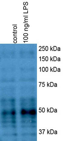
Application: Western BlotSample Tested: Primary mouse hepatocytesSpecies: MouseVerified Customer | Posted 12/08/2017mouse hepatocytes were treated with 100 ng/ml LPS for 4 hoursmouse hepatocytes were treated with 100 ng/ml LPS for 4 hours
-
Application: Western BlotSample Tested: Chondrocyte cell lysateSpecies: RatVerified Customer | Posted 01/18/2017
-
Application: Immunohistochemistry-ParaffinSample Tested: Human ovarian carcinoma -patient derived xenograftSpecies: HumanVerified Customer | Posted 03/05/2014Beclin 1 staining on ovarian cancer PDX
-
Application: Immunohistochemistry-ParaffinSample Tested: Mouse kidneySpecies: MouseVerified Customer | Posted 10/08/2012
-
Application: Western BlotSample Tested: Mouse whole cell lysateSpecies: MouseVerified Customer | Posted 10/06/2012
-
Application: ImmunohistochemistrySample Tested: Human Esophagus tissueSpecies: HumanVerified Customer | Posted 09/28/2010
There are no reviews that match your criteria.
Protocols
View specific protocols for Beclin 1 Antibody - BSA Free (NB500-249):
Culture cells to appropriate density in 35 mm culture dishes or 6-well plates.
1. Remove culture medium and wash the cells briefly in PBS. Add 10% formalin to the dish and fix at room temperature for 10 minutes.
2. Remove the formalin and wash the cells in PBS.
3. Permeablize the cells with 0.1% Triton X100 or other suitable detergent for 10 min.
4. Remove the permeablization buffer and wash three times for 10 minutes each in PBS. Be sure to not let the specimen dry out.
5. To block nonspecific antibody binding, incubate in 10% normal goat serum from 1 hour to overnight at room temperature.
6. Add primary antibody at appropriate dilution and incubate overnight at 4C.
7. Remove primary antibody and replace with PBS. Wash three times for 10 minutes each.
8. Add secondary antibody at appropriate dilution. Incubate for 1 hour at room temperature.
9. Remove secondary antibody and replace with PBS. Wash three times for 10 minutes each.
10. Counter stain DNA with DAPi if required.
Antigen Unmasking:
Bring slides to a boil in 10 mM sodium citrate buffer (pH 6.0) then maintain at a sub-boiling temperature for 10 minutes. Cool slides on bench-top for 30 minutes (keep slides in the sodium citrate buffer at all times).
Staining:
1. Wash sections in deionized water three times for 5 minutes each.
2. Wash sections in PBS for 5 minutes.
3. Block each section with 100-400 ul blocking solution (1% BSA in PBS) for 1 hour at room temperature.
4. Remove blocking solution and add 100-400 ul diluted primary antibody. Incubate overnight at 4 C.
5. Remove antibody solution and wash sections in wash buffer three times for 5 minutes each.
6. Add 100-400 ul HRP polymer conjugated secondary antibody. Incubate 30 minutes at room temperature.
7. Wash sections three times in wash buffer for 5 minutes each.
8. Add 100-400 ul DAB substrate to each section and monitor staining closely.
9. As soon as the sections develop, immerse slides in deionized water.
10. Counterstain sections in hematoxylin.
11. Wash sections in deionized water two times for 5 minutes each.
12. Dehydrate sections.
13. Mount coverslips.
Immunoprecipitation Protocol:
1. Cells in 2x 75cm flasks (60% confluency) are scraped with 0.5ml of Tris lysis Buffer (50mM Tris, 150mM NaCl, 1mM EDTA, 100ug/ml PMSF, 1% triton).
2. Lyse 1h at 4C, with gentle agitation.
3. Centrifuge to clear the lysates.
4. 0.1 ml of lysate is kept aside for Western Blot experiments.
5. IP : Add 5ul of polyclonal beclin antibody (NB 500-249) to 0.4ml of lysate (1:80 dilution).
6. Incubate overnight at 4C, with gentle agitation.
7. Next day, add 60ul of protein A sepharose beads to the lysate.
8. Incubate for one hour at 4C.
9. Wash beads 3X with Tris lysis buffer.
10. Beads are re-suspended with 15ul of Laemmli buffer and boiled.
11. A SDS-PAGE gel is run and the proteins are transferred to a membrane.
12. The efficiency of IP is determined by using a monoclonal anti-beclin antibody.
1. Perform SDS-PAGE on samples to be analyzed, loading 10-25 ug of total protein per lane.
2. Transfer proteins to PVDF membrane according to the instructions provided by the manufacturer of the membrane and transfer apparatus.
3. Stain the membrane with Ponceau S (or similar product) to assess transfer success, and mark molecular weight standards where appropriate.
4. Rinse the blot TBS -0.05% Tween 20 (TBST).
5. Block the membrane in 5% Non-fat milk in TBST (blocking buffer) for at least 1 hour.
6. Wash the membrane in TBST three times for 10 minutes each.
7. Dilute primary antibody in blocking buffer and incubate overnight at 4C with gentle rocking.
8. Wash the membrane in TBST three times for 10 minutes each.
9. Incubate the membrane in diluted HRP conjugated secondary antibody in blocking buffer (as per manufacturer's instructions) for 1 hour at room temperature.
10. Wash the blot in TBST three times for 10 minutes each (this step can be repeated as required to reduce background).
11. Apply the detection reagent of choice in accordance with the manufacturer's instructions.
Find general support by application which include: protocols, troubleshooting, illustrated assays, videos and webinars.
- 7-Amino Actinomycin D (7-AAD) Cell Viability Flow Cytometry Protocol
- Antigen Retrieval Protocol (PIER)
- Antigen Retrieval for Frozen Sections Protocol
- Appropriate Fixation of IHC/ICC Samples
- Cellular Response to Hypoxia Protocols
- Chromogenic IHC Staining of Formalin-Fixed Paraffin-Embedded (FFPE) Tissue Protocol
- Chromogenic Immunohistochemistry Staining of Frozen Tissue
- ClariTSA™ Fluorophore Kits
- Detection & Visualization of Antibody Binding
- Extracellular Membrane Flow Cytometry Protocol
- Flow Cytometry Protocol for Cell Surface Markers
- Flow Cytometry Protocol for Staining Membrane Associated Proteins
- Flow Cytometry Staining Protocols
- Flow Cytometry Troubleshooting Guide
- Fluorescent IHC Staining of Frozen Tissue Protocol
- Graphic Protocol for Heat-induced Epitope Retrieval
- Graphic Protocol for the Preparation and Fluorescent IHC Staining of Frozen Tissue Sections
- Graphic Protocol for the Preparation and Fluorescent IHC Staining of Paraffin-embedded Tissue Sections
- Graphic Protocol for the Preparation of Gelatin-coated Slides for Histological Tissue Sections
- ICC Cell Smear Protocol for Suspension Cells
- ICC Immunocytochemistry Protocol Videos
- ICC for Adherent Cells
- IHC Sample Preparation (Frozen sections vs Paraffin)
- Immunocytochemistry (ICC) Protocol
- Immunocytochemistry Troubleshooting
- Immunofluorescence of Organoids Embedded in Cultrex Basement Membrane Extract
- Immunofluorescent IHC Staining of Formalin-Fixed Paraffin-Embedded (FFPE) Tissue Protocol
- Immunohistochemistry (IHC) and Immunocytochemistry (ICC) Protocols
- Immunohistochemistry Frozen Troubleshooting
- Immunohistochemistry Paraffin Troubleshooting
- Immunoprecipitation Protocol
- Intracellular Flow Cytometry Protocol Using Alcohol (Methanol)
- Intracellular Flow Cytometry Protocol Using Detergents
- Intracellular Nuclear Staining Flow Cytometry Protocol Using Detergents
- Intracellular Staining Flow Cytometry Protocol Using Alcohol Permeabilization
- Intracellular Staining Flow Cytometry Protocol Using Detergents to Permeabilize Cells
- Preparing Samples for IHC/ICC Experiments
- Preventing Non-Specific Staining (Non-Specific Binding)
- Primary Antibody Selection & Optimization
- Propidium Iodide Cell Viability Flow Cytometry Protocol
- Protocol for Heat-Induced Epitope Retrieval (HIER)
- Protocol for Liperfluo
- Protocol for Making a 4% Formaldehyde Solution in PBS
- Protocol for VisUCyte™ HRP Polymer Detection Reagent
- Protocol for the Characterization of Human Th22 Cells
- Protocol for the Characterization of Human Th9 Cells
- Protocol for the Fluorescent ICC Staining of Cell Smears - Graphic
- Protocol for the Fluorescent ICC Staining of Cultured Cells on Coverslips - Graphic
- Protocol for the Preparation & Fixation of Cells on Coverslips
- Protocol for the Preparation and Chromogenic IHC Staining of Frozen Tissue Sections
- Protocol for the Preparation and Chromogenic IHC Staining of Frozen Tissue Sections - Graphic
- Protocol for the Preparation and Chromogenic IHC Staining of Paraffin-embedded Tissue Sections
- Protocol for the Preparation and Chromogenic IHC Staining of Paraffin-embedded Tissue Sections - Graphic
- Protocol for the Preparation and Fluorescent ICC Staining of Cells on Coverslips
- Protocol for the Preparation and Fluorescent ICC Staining of Non-adherent Cells
- Protocol for the Preparation and Fluorescent ICC Staining of Stem Cells on Coverslips
- Protocol for the Preparation and Fluorescent IHC Staining of Frozen Tissue Sections
- Protocol for the Preparation and Fluorescent IHC Staining of Paraffin-embedded Tissue Sections
- Protocol for the Preparation of Gelatin-coated Slides for Histological Tissue Sections
- Protocol for the Preparation of a Cell Smear for Non-adherent Cell ICC - Graphic
- Protocol: Annexin V and PI Staining by Flow Cytometry
- Protocol: Annexin V and PI Staining for Apoptosis by Flow Cytometry
- R&D Systems Quality Control Western Blot Protocol
- TUNEL and Active Caspase-3 Detection by IHC/ICC Protocol
- The Importance of IHC/ICC Controls
- Troubleshooting Guide: Fluorokine Flow Cytometry Kits
- Troubleshooting Guide: Immunohistochemistry
- Troubleshooting Guide: Western Blot Figures
- Western Blot Conditions
- Western Blot Protocol
- Western Blot Protocol for Cell Lysates
- Western Blot Troubleshooting
- Western Blot Troubleshooting Guide
- View all Protocols, Troubleshooting, Illustrated assays and Webinars
FAQs for Beclin 1 Antibody - BSA Free
-
Q: Can a Lightning-Link HRP labeling kit be used to label the beclin 1 antibody NB500-249, I've used it to label mouse isotypes but wasn't sure if it will work on a rabbit IgG antibody like this beclin antibody?
A: I can't find any specific mention of this beclin 1 antibody being labeled with a lightning link kit; however lightning link kits are extreamly versatile in terms of labeling ability and should be able to label this beclin 1 antibody without any trouble.
-
Q: I am interested in Novus' Beclin 1 peptide NB500-249PEP. Would you please send me the sequence information, and any data you may have for this peptide?
A: In regards to your inquiry, this Beclin 1 peptide is useful as a blocking peptide to our primary antibody NB500-249. This peptide contains amino acids that fall within the range of 1-100. Unfortunately the exact sequence is deemed proprietary by the lab.
-
Q: I'm doning an autophagy study using CHO-K1 cells, do you know if this beclin 1 antibody will detect beclin in that cell line?
A: Based on tissue expression data from protein atlas beclin 1 is not highly expressed in ovarian tissue; however in ovarian cancer tissues beclin 1 expression is much greater. We don't have any beclin 1 antibodies that have been validated for use in hamster so you may want to take advantage of our innovator's reward program.
-
Q: I'm looking for a Beclin 1 antibody, which do you recommend for use in western blot?
A: Our best selling beclin 1 antibody NB500-249, has been cited in 41 publications where it was used in western blot. Additionally this beclin 1 antibody is typically available for immediate shipment.
-
Q: I'm using the ATDC5 murine carcinoma cell line, has beclin 1 antibody been documented as working in this cell line?
A: From a cursory look through our publication list I don't see that this beclin 1 antibody has been published as being used in that cell line. We show this beclin 1 antibody was published 19 times as being used in mouse, if I can get your contact info I will take a more in depth look to see if the beclin 1 antibody has been used in that cell line.
-
Q: I've had good results with your beclin antibody in IHC but now I need a beclin one antibody to use in ELISA, will this beclin antibody also work as a capture antibody in ELISA?
A: This particular beclin 1 antibody has not yet been validated for use in ELISA, we do however have 2 beclin 1 antibodies that are validated in ELISA, NBP2-22176 and NBP1-76648. Alternatively you could take advantage of our innovator's reward program which would allow you to test this beclin 1 antibody in ELISA with out financial risk. Just purchase the beclin antibody and test in ELISA, share your results (negative or positive) and we will issue a voucher for the purchase price of the antibody.
-
Q: Is your belcin 1 antibody NB500-249 available as an atto 488 direct conjugate?
A: Unfortunately we do not offer Atto conjugates but we do offer this beclin 1 antibody as an AlexaFlour 488 direct conjugate and as a DyLight 488 conjugated antibody.
-
Q: The beclin 1 antibody NB500-249 has good data but I need a beclin atibody that will work with bovine tissue but I don't see bovine listed as reactive with the beclin 1 antibody. Is bovine not listed because you know this beclin one antibody will not recognize?
A: Although reactivity with bovine has not been experimentally validated, a comparison of the human and bovine sequences within the immunogen range indicates that cross reactivity is likely. An alignment of the human beclin 1 sequence and bovine beclin 1 sequence shows there is 95% match in the region that was used as the immunogen.
-
Q: Will this beclin 1 antibody detect beclin that is cleaved at the last 50 aa of the c-terminal?
A: Because the immunogen used to make this beclin 1 antibody falls within the first 100 amino acids of the N-terminal it should detect beclin 1 that is cleaved at the c-terminal.
-
Q: Can a Lightning-Link HRP labeling kit be used to label the beclin 1 antibody NB500-249, I've used it to label mouse isotypes but wasn't sure if it will work on a rabbit IgG antibody like this beclin antibody?
A: I can't find any specific mention of this beclin 1 antibody being labeled with a lightning link kit; however lightning link kits are extreamly versatile in terms of labeling ability and should be able to label this beclin 1 antibody without any trouble.
-
Q: I am interested in Novus' Beclin 1 peptide NB500-249PEP. Would you please send me the sequence information, and any data you may have for this peptide?
A: In regards to your inquiry, this Beclin 1 peptide is useful as a blocking peptide to our primary antibody NB500-249. This peptide contains amino acids that fall within the range of 1-100. Unfortunately the exact sequence is deemed proprietary by the lab.
-
Q: I'm doning an autophagy study using CHO-K1 cells, do you know if this beclin 1 antibody will detect beclin in that cell line?
A: Based on tissue expression data from protein atlas beclin 1 is not highly expressed in ovarian tissue; however in ovarian cancer tissues beclin 1 expression is much greater. We don't have any beclin 1 antibodies that have been validated for use in hamster so you may want to take advantage of our innovator's reward program.
-
Q: I'm looking for a Beclin 1 antibody, which do you recommend for use in western blot?
A: Our best selling beclin 1 antibody NB500-249, has been cited in 41 publications where it was used in western blot. Additionally this beclin 1 antibody is typically available for immediate shipment.
-
Q: I'm using the ATDC5 murine carcinoma cell line, has beclin 1 antibody been documented as working in this cell line?
A: From a cursory look through our publication list I don't see that this beclin 1 antibody has been published as being used in that cell line. We show this beclin 1 antibody was published 19 times as being used in mouse, if I can get your contact info I will take a more in depth look to see if the beclin 1 antibody has been used in that cell line.
-
Q: I've had good results with your beclin antibody in IHC but now I need a beclin one antibody to use in ELISA, will this beclin antibody also work as a capture antibody in ELISA?
A: This particular beclin 1 antibody has not yet been validated for use in ELISA, we do however have 2 beclin 1 antibodies that are validated in ELISA, NBP2-22176 and NBP1-76648. Alternatively you could take advantage of our innovator's reward program which would allow you to test this beclin 1 antibody in ELISA with out financial risk. Just purchase the beclin antibody and test in ELISA, share your results (negative or positive) and we will issue a voucher for the purchase price of the antibody.
-
Q: Is your belcin 1 antibody NB500-249 available as an atto 488 direct conjugate?
A: Unfortunately we do not offer Atto conjugates but we do offer this beclin 1 antibody as an AlexaFlour 488 direct conjugate and as a DyLight 488 conjugated antibody.
-
Q: The beclin 1 antibody NB500-249 has good data but I need a beclin atibody that will work with bovine tissue but I don't see bovine listed as reactive with the beclin 1 antibody. Is bovine not listed because you know this beclin one antibody will not recognize?
A: Although reactivity with bovine has not been experimentally validated, a comparison of the human and bovine sequences within the immunogen range indicates that cross reactivity is likely. An alignment of the human beclin 1 sequence and bovine beclin 1 sequence shows there is 95% match in the region that was used as the immunogen.
-
Q: Will this beclin 1 antibody detect beclin that is cleaved at the last 50 aa of the c-terminal?
A: Because the immunogen used to make this beclin 1 antibody falls within the first 100 amino acids of the N-terminal it should detect beclin 1 that is cleaved at the c-terminal.
Associated Pathways
