beta-Actin Antibody - BSA Free
Novus Biologicals | Catalog # NB600-503
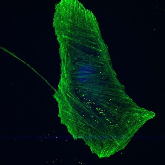
![Immunocytochemistry/ Immunofluorescence: beta-Actin Antibody [NB600-503]](https://resources.rndsystems.com/images/products/beta-Actin-Antibody-Immunocytochemistry-Immunofluorescence-NB600-503-img0012.jpg "Immunocytochemistry/ Immunofluorescence: beta-Actin Antibody [NB600-503]")
Key Product Details
Validated by
Biological Validation
Species Reactivity
Validated:
Human, Mouse, Rat, Porcine, Avian, Bovine, Chinese Hamster, Fish, Primate, Rabbit
Cited:
Human, Mouse, Rat, Porcine, Avian, Bovine, Fish, Hamster - Cricetulus (Chinese Hamster), Primate, Rabbit
Applications
Validated:
Immunohistochemistry, Immunohistochemistry-Paraffin, Western Blot, ELISA, Block/Neutralize, Flow Cytometry, Immunocytochemistry/ Immunofluorescence, Simple Western, Immunoprecipitation, Proximity Ligation Assay
Cited:
Immunohistochemistry-Paraffin, Western Blot, Block/Neutralize, Immunocytochemistry/ Immunofluorescence, Simple Western, Proximity Ligation Assay, IF/IHC, Electron Microscopy
Label
Unconjugated
Antibody Source
Polyclonal Rabbit IgG
Format
BSA Free
Loading...
Product Specifications
Immunogen
This beta-Actin Antibody was made from a synthetic peptide made to an N-terminal region of human Beta Actin. [UniProt P60709]
Reactivity Notes
Rabbit reactivity and Fish reactivity reported in scientific literature (PMID: 23813946 and 25842206 respectively). Expected to cross-react with a wide range of species due to sequence identity. Bovine reactivity reported in scientific literature (PMID:33066332).
Localization
Cytoplasmic
Clonality
Polyclonal
Host
Rabbit
Isotype
IgG
Theoretical MW
42 kDa.
Disclaimer note: The observed molecular weight of the protein may vary from the listed predicted molecular weight due to post translational modifications, post translation cleavages, relative charges, and other experimental factors.
Disclaimer note: The observed molecular weight of the protein may vary from the listed predicted molecular weight due to post translational modifications, post translation cleavages, relative charges, and other experimental factors.
Scientific Data Images for beta-Actin Antibody - BSA Free
Immunocytochemistry/ Immunofluorescence: beta-Actin Antibody [NB600-503]
Immunocytochemistry/Immunofluorescence: beta-Actin Antibody [NB600-503] - Cultured pig trabecular meshwork cells stained with beta-Actin antibody at a dilution of 1:500 followed by an anti-rabbit Alexa Fluor488 at 1:1000 dilution (green). Image from verified customer review.![Western Blot: beta-Actin Antibody [NB600-503]](https://resources.rndsystems.com/images/products/beta-Actin-Antibody-Western-Blot-NB600-503-img0004.jpg "Western Blot: beta-Actin Antibody [NB600-503]")
Western Blot: beta-Actin Antibody [NB600-503]
Western Blot: beta-Actin Antibody [NB600-503] - Rabbit polyclonal at 1/5000.![Simple Western: beta-Actin Antibody [NB600-503]](https://resources.rndsystems.com/images/products/beta-Actin-Antibody-Simple-Western-NB600-503-img0008.jpg "Simple Western: beta-Actin Antibody [NB600-503]")
Simple Western: beta-Actin Antibody [NB600-503]
Simple Western: beta-Actin Antibody [NB600-503] - Simple Western lane view shows a specific band for Beta Actin in 0.1 mg/ml of HeLa lysate. This experiment was performed under reducing conditions using the 12-230 kDa separation system.![Flow Cytometry: beta-Actin Antibody [NB600-503]](https://resources.rndsystems.com/images/products/beta-Actin-Antibody-Flow-Cytometry-NB600-503-img0013.jpg "Flow Cytometry: beta-Actin Antibody [NB600-503]")
Flow Cytometry: beta-Actin Antibody [NB600-503]
Flow Cytometry: beta-Actin Antibody [NB600-503] - An intracellular stain was performed on HeLa cells with beta-actin Antibody NB600-503AF488 (blue) and a matched isotype control (orange). Cells were fixed with 4% PFA and then permeabilized with 0.1% saponin. Cells were incubated in an antibody dilution of 5 ug/mL for 30 minutes at room temperature. Both antibodies were conjugated to Alexa Fluor 488.![Western Blot: beta-Actin Antibody [NB600-503]](https://resources.rndsystems.com/images/products/beta-Actin-Antibody-Western-Blot-NB600-503-img0006.jpg "Western Blot: beta-Actin Antibody [NB600-503]")
Western Blot: beta-Actin Antibody [NB600-503]
Western Blot: beta-Actin Antibody [NB600-503] - Western blot analysis of Jurkat, COS, and CHO cell lysate using beta actin antibody at 1:100.![Immunohistochemistry-Paraffin: beta-Actin Antibody [NB600-503]](https://resources.rndsystems.com/images/products/beta-Actin-Antibody-Immunohistochemistry-Paraffin-NB600-503-img0009.jpg "Immunohistochemistry-Paraffin: beta-Actin Antibody [NB600-503]")
Immunohistochemistry-Paraffin: beta-Actin Antibody [NB600-503]
Immunohistochemistry-Paraffin: beta-Actin Antibody [NB600-503] - Analysis of Beta Actin in mouse epidermis using DAB with hematoxylin counterstain.![Immunocytochemistry/ Immunofluorescence: beta-Actin Antibody [NB600-503]](https://resources.rndsystems.com/images/products/beta-Actin-Antibody-Immunocytochemistry-Immunofluorescence-NB600-503-img0007.jpg "Immunocytochemistry/ Immunofluorescence: beta-Actin Antibody [NB600-503]")
Immunocytochemistry/ Immunofluorescence: beta-Actin Antibody [NB600-503]
Immunocytochemistry/Immunofluorescence: beta-Actin Antibody [NB600-503] - IF Confocal analysis of HeLa cells using Beta Actin antibody (NB600-503, 1:5). An Alexa Fluor 488-conjugated Goat to rabbit IgG was used as secondary antibody (green). DAPI was used to stain the cell nuclei (blue).![Flow Cytometry: beta-Actin Antibody [NB600-503]](https://resources.rndsystems.com/images/products/beta-Actin-Antibody-Flow-Cytometry-NB600-503-img0010.jpg "Flow Cytometry: beta-Actin Antibody [NB600-503]")
Flow Cytometry: beta-Actin Antibody [NB600-503]
Flow Cytometry: beta-Actin Antibody [NB600-503] - Analysis of HeLa cells using mouse Monoclonal beta-Actin antibody (Orange) and Isotype control Antibody (Blue).![Simple Western: beta-Actin Antibody [NB600-503]](https://resources.rndsystems.com/images/products/beta-Actin-Antibody-Simple-Western-NB600-503-img0014.jpg "Simple Western: beta-Actin Antibody [NB600-503]")

Western Blot: beta-Actin Antibody [NB600-503] -
Western Blot: beta-Actin Antibody [NB600-503] - Regulation of inflammatory signaling in hSMC.ND (open bars) & T2D (solid bars) cells extracted after 48 hr treatment with LPS, Pioglitazone (Pio), palmitate or oleate. Results expressed relative to untreated control for each individual set of cells, Ave + SEM. (A) IkBa protein, n = 7–12 & 6–15 for ND & T2D, respectively. (B) Total & phospho-p38, n = 6–14 & 5–8. (C) Total & phospho-p44/42, n = 7–13 & 7–11. (D) Total & phospho-JNK, n = 9–12 & 3–8. * p<0.05 T2D vs ND. †p<0.05 T2D response vs ND response Image collected & cropped by CiteAb from the following publication (https://pubmed.ncbi.nlm.nih.gov/27453994), licensed under a CC0-1.0 license. Not internally tested by Novus Biologicals.
Western Blot: beta-Actin Antibody [NB600-503] -
Western Blot: beta-Actin Antibody [NB600-503] - Impact of T2D on inflammatory signaling in hSMC.Proteins extracted from ND (open bars) & T2D (solid bars) hSMC under control conditions. (A) Representative western blots. (B) Quantification of protein expression normalized to loading control ( beta -actin). Ave + SEM, n = 13–21 for ND, 10–16 for T2D. (C) Protein phosphorylation, expressed as ratio of phosphorylated to total protein. Ave + SEM, n = 13–17 for ND, 9–15 for T2D. *p<0.05 vs ND Image collected & cropped by CiteAb from the following publication (https://pubmed.ncbi.nlm.nih.gov/27453994), licensed under a CC0-1.0 license. Not internally tested by Novus Biologicals.
Western Blot: beta-Actin Antibody [NB600-503] -
Western Blot: beta-Actin Antibody [NB600-503] - Early start GM1 administration did not affect alpha -synuclein expression or transport to the striatum. (A,B) When assessed 1 week following AAV-A53T alpha -synuclein injection, levels of striatal alpha -synuclein were no different in saline (N = 6) vs. GM1-treated animals (N = 6), suggesting no influence of GM1 on A53T alpha -synuclein transduction or transport to the striatum. Representative Wes Western blots are shown after cropping (full length images of blots are presented as Supplementary Fig. 2. (C) Double label immunofluorescence 1 week after AAV-A53T alpha -synuclein injection showed no differences between saline & GM1-treated animals in alpha -synuclein accumulation (green) in TH+ neurons (red) in the SNc. Image collected & cropped by CiteAb from the following publication (https://pubmed.ncbi.nlm.nih.gov/31182727), licensed under a CC-BY license. Not internally tested by Novus Biologicals.
Western Blot: beta-Actin Antibody [NB600-503] -
Western Blot: beta-Actin Antibody [NB600-503] - Anti-hnRNP A1 antibodies alter protein levels as measured by Western blot. SK-N-SH cells were cultured & treated with anti-hnRNP A1 antibodies or control IgG. Following a 48 hour incubation, cells were lysed & protein lysate was run on 10% Tris-glycine gels for Western blot analysis & probed for Beta-actin (A) or GAPDH (B)) (control), hnRNP A1, SPG4, SPG7, & SPG20. Results revealed that there was a marked reduction of SPG 4 & SPG 7 protein levels in anti-hnRNP A1 antibody compared to control isotype IgG treated cells. There was a variable response to hnRNP A1 protein & a modest reduction of SPG 20. Parentheses show relative percent reduction of signal comparing anti-hnRNP A1 antibody to control isotype IgG treatment of cells. (L=lysate, IgG=control isotype IgG, A1=anti-hnRNP A1-M9 antibody treatment of cells). Image collected & cropped by CiteAb from the following publication (https://pubmed.ncbi.nlm.nih.gov/27375925), licensed under a CC-BY license. Not internally tested by Novus Biologicals.Applications for beta-Actin Antibody - BSA Free
Application
Recommended Usage
Flow Cytometry
1:10-1:1000
Immunohistochemistry
1:100
Immunohistochemistry-Paraffin
1:100
Immunoprecipitation
1:10-1:500
Simple Western
1:12.5
Western Blot
1:100-1:2000
Application Notes
See Simple Western Antibody Database for Simple Western validation: tested in HeLa lysate (0.1 mg/ml); antibody dilution of 1:12.5; separated by size; detects a band at 50 kDa
Reviewed Applications
Read 4 reviews rated 5 using NB600-503 in the following applications:
Flow Cytometry Panel Builder
Bio-Techne Knows Flow Cytometry
Save time and reduce costly mistakes by quickly finding compatible reagents using the Panel Builder Tool.
Advanced Features
- Spectra Viewer - Custom analysis of spectra from multiple fluorochromes
- Spillover Popups - Visualize the spectra of individual fluorochromes
- Antigen Density Selector - Match fluorochrome brightness with antigen density

Formulation, Preparation, and Storage
Purification
Immunogen affinity purified
Formulation
Tris-Citrate/Phosphate (pH 7.0 - 8.0)
Format
BSA Free
Preservative
0.09% Sodium Azide
Concentration
1.0 mg/ml
Shipping
The product is shipped with polar packs. Upon receipt, store it immediately at the temperature recommended below.
Stability & Storage
Store at 4C. Do not freeze.
Background: beta-Actin
References
1. Vandekerckhove J, Weber K. 1978. At least six different actins are expressed in a higher mammal: an analysis based on the amino acid sequence of the amino-terminal tryptic peptide. J Mol Biol. 126(4):783-802. PMID: 745245
2. Gimona M, Vandekerckhove J, Goethals M, Herzog M, Lando Z, Small JV. (1994) Beta-actin specific monoclonal antibody. Cell Motil Cytoskeleton. 27(2):108-16. PMID: 8162619
3. Holden VI, Lenio S, Kuick R, Ramakrishnan SK, Shah YM, Bachman MA. (2014) Bacterial siderophores that evade or overwhelm Lipocalin 2 induce HIF-1a and pro-inflammatory cytokine secretion in cultured respiratory epithelial cells Infect Immun. 82(9):3826-36 PMID: 24980968
4. Tsai WL, Yeh PH, Tsai CY, Ting CT, Chiu YH, Tao MH, Li WC, Hung SC. (2016) Efficient Programming of Human Mesenchymal Stem Cell Derived Hepatocytes by Epigenetic Regulations. J. Gastroenterol. Hepatol. 32(1):261-269. PMID: 27218433
Alternate Names
ACTB, betaActin
Gene Symbol
ACTB
UniProt
Additional beta-Actin Products
Product Documents for beta-Actin Antibody - BSA Free
Certificate of Analysis
To download a Certificate of Analysis, please enter a lot or batch number in the search box below.
Product Specific Notices for beta-Actin Antibody - BSA Free
This product is for research use only and is not approved for use in humans or in clinical diagnosis. Primary Antibodies are guaranteed for 1 year from date of receipt.
Related Research Areas
Citations for beta-Actin Antibody - BSA Free
 Powered by Bioz
Customer Reviews for beta-Actin Antibody - BSA Free (4)
5 out of 5
4 Customer Ratings
Have you used beta-Actin Antibody - BSA Free?
Submit a review and receive an Amazon gift card!
$25/€18/£15/$25CAN/¥2500 Yen for a review with an image
$10/€7/£6/$10CAN/¥1110 Yen for a review without an image
Submit a review
Customer Images
-(005-ml)_NB600-503_8201.bmp)
-(005-ml)_NB600-503_8196.jpg)
Showing
1
-
4 of
4 reviews
Showing All
Filter By:
-
Application: ImmunocytochemistrySample Tested: pig trabecular meshwork cellsSpecies: PigVerified Customer | Posted 12/05/2016Detection of Actin in cultured pig trabecular meshwork cells using beta-Actin Antibody (NB600-503) at 1:500 dilution overnight and donkey anti-rabbit Alexa 488 (1:1000) for 1 hour.

-
Application: ImmunocytochemistrySample Tested:Species: HumanVerified Customer | Posted 06/11/2014IF Confocal analysis of HeLa cells using Beta Actin antibody (NB600-503, 1:5).
-
Application: Western BlotSample Tested:Species: HumanVerified Customer | Posted 06/11/2014Western blot analysis of extracts from Jurkat, COS and CHO cells using Actin antibody (NB600-503, 1:200).
-
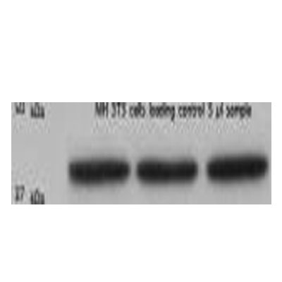
Application: Western BlotSample Tested: NIH 3T3 cell lysate, Sample Amount: 5 ulSpecies: MouseVerified Customer | Posted 11/19/2010
There are no reviews that match your criteria.
Protocols
Find general support by application which include: protocols, troubleshooting, illustrated assays, videos and webinars.
- 7-Amino Actinomycin D (7-AAD) Cell Viability Flow Cytometry Protocol
- Antigen Retrieval Protocol (PIER)
- Antigen Retrieval for Frozen Sections Protocol
- Appropriate Fixation of IHC/ICC Samples
- Cellular Response to Hypoxia Protocols
- Chromogenic IHC Staining of Formalin-Fixed Paraffin-Embedded (FFPE) Tissue Protocol
- Chromogenic Immunohistochemistry Staining of Frozen Tissue
- ClariTSA™ Fluorophore Kits
- Detection & Visualization of Antibody Binding
- ELISA Sample Preparation & Collection Guide
- ELISA Troubleshooting Guide
- Extracellular Membrane Flow Cytometry Protocol
- Flow Cytometry Protocol for Cell Surface Markers
- Flow Cytometry Protocol for Staining Membrane Associated Proteins
- Flow Cytometry Staining Protocols
- Flow Cytometry Troubleshooting Guide
- Fluorescent IHC Staining of Frozen Tissue Protocol
- Graphic Protocol for Heat-induced Epitope Retrieval
- Graphic Protocol for the Preparation and Fluorescent IHC Staining of Frozen Tissue Sections
- Graphic Protocol for the Preparation and Fluorescent IHC Staining of Paraffin-embedded Tissue Sections
- Graphic Protocol for the Preparation of Gelatin-coated Slides for Histological Tissue Sections
- How to Run an R&D Systems DuoSet ELISA
- How to Run an R&D Systems Quantikine ELISA
- How to Run an R&D Systems Quantikine™ QuicKit™ ELISA
- ICC Cell Smear Protocol for Suspension Cells
- ICC Immunocytochemistry Protocol Videos
- ICC for Adherent Cells
- IHC Sample Preparation (Frozen sections vs Paraffin)
- Immunocytochemistry (ICC) Protocol
- Immunocytochemistry Troubleshooting
- Immunofluorescence of Organoids Embedded in Cultrex Basement Membrane Extract
- Immunofluorescent IHC Staining of Formalin-Fixed Paraffin-Embedded (FFPE) Tissue Protocol
- Immunohistochemistry (IHC) and Immunocytochemistry (ICC) Protocols
- Immunohistochemistry Frozen Troubleshooting
- Immunohistochemistry Paraffin Troubleshooting
- Immunoprecipitation Protocol
- Intracellular Flow Cytometry Protocol Using Alcohol (Methanol)
- Intracellular Flow Cytometry Protocol Using Detergents
- Intracellular Nuclear Staining Flow Cytometry Protocol Using Detergents
- Intracellular Staining Flow Cytometry Protocol Using Alcohol Permeabilization
- Intracellular Staining Flow Cytometry Protocol Using Detergents to Permeabilize Cells
- Preparing Samples for IHC/ICC Experiments
- Preventing Non-Specific Staining (Non-Specific Binding)
- Primary Antibody Selection & Optimization
- Propidium Iodide Cell Viability Flow Cytometry Protocol
- Protocol for Heat-Induced Epitope Retrieval (HIER)
- Protocol for Liperfluo
- Protocol for Making a 4% Formaldehyde Solution in PBS
- Protocol for VisUCyte™ HRP Polymer Detection Reagent
- Protocol for the Characterization of Human Th22 Cells
- Protocol for the Characterization of Human Th9 Cells
- Protocol for the Fluorescent ICC Staining of Cell Smears - Graphic
- Protocol for the Fluorescent ICC Staining of Cultured Cells on Coverslips - Graphic
- Protocol for the Preparation & Fixation of Cells on Coverslips
- Protocol for the Preparation and Chromogenic IHC Staining of Frozen Tissue Sections
- Protocol for the Preparation and Chromogenic IHC Staining of Frozen Tissue Sections - Graphic
- Protocol for the Preparation and Chromogenic IHC Staining of Paraffin-embedded Tissue Sections
- Protocol for the Preparation and Chromogenic IHC Staining of Paraffin-embedded Tissue Sections - Graphic
- Protocol for the Preparation and Fluorescent ICC Staining of Cells on Coverslips
- Protocol for the Preparation and Fluorescent ICC Staining of Non-adherent Cells
- Protocol for the Preparation and Fluorescent ICC Staining of Stem Cells on Coverslips
- Protocol for the Preparation and Fluorescent IHC Staining of Frozen Tissue Sections
- Protocol for the Preparation and Fluorescent IHC Staining of Paraffin-embedded Tissue Sections
- Protocol for the Preparation of Gelatin-coated Slides for Histological Tissue Sections
- Protocol for the Preparation of a Cell Smear for Non-adherent Cell ICC - Graphic
- Protocol: Annexin V and PI Staining by Flow Cytometry
- Protocol: Annexin V and PI Staining for Apoptosis by Flow Cytometry
- Quantikine HS ELISA Kit Assay Principle, Alkaline Phosphatase
- Quantikine HS ELISA Kit Principle, Streptavidin-HRP Polymer
- R&D Systems Quality Control Western Blot Protocol
- Sandwich ELISA (Colorimetric) – Biotin/Streptavidin Detection Protocol
- Sandwich ELISA (Colorimetric) – Direct Detection Protocol
- TUNEL and Active Caspase-3 Detection by IHC/ICC Protocol
- The Importance of IHC/ICC Controls
- Troubleshooting Guide: ELISA
- Troubleshooting Guide: Fluorokine Flow Cytometry Kits
- Troubleshooting Guide: Immunohistochemistry
- Troubleshooting Guide: Western Blot Figures
- Western Blot Conditions
- Western Blot Protocol
- Western Blot Protocol for Cell Lysates
- Western Blot Troubleshooting
- Western Blot Troubleshooting Guide
- View all Protocols, Troubleshooting, Illustrated assays and Webinars
FAQs for beta-Actin Antibody - BSA Free
Showing
1
-
5 of
6 FAQs
Showing All
-
Q: Are the beta-Actin antibodies validated in Simple Western?
A: Yes, we offer a few beta actin antibodies that have been tested in Simple Western: NBP1-47423, NB600-501, NB600-532, NB600-503, and NB100-56874.
-
Q: Do Beta-Actin antibodies come in lyophilized format?
A: ACTB antibodies like MAB8969 AND MAB8929 come in lyophilized form.
-
Q: I wanted to know which of the two housekeeping genes B-actin or GAPDH can be used for best results. I intend to do an experiment to determine expression of IL-6 and TNF-alpha in liver and kidney tissues.
A: For homogenized tissue, beta-actin and GAPDH are both fine.
-
Q: What is the immunogen sequence of this Beta-Actin antibody?
A: A sequence within the N-terminal region of Human beta Actin. The exact sequence is proprietary.
-
Q: What is the theoretical molecular weight for Beta-Actin antibodies?
A: The TMW for beta Actin antibodies is approximately 42 kDa.
-
Q: Why is beta actin used as a loading control in blotting ?
A: Beta actin is a highly-expressed protein found in all cells, and is considered a house keeping protein whose expression is necessary for any cell's proper functioning. For this reason it is used as a loading control to confirm that the same amount of total protein is loaded in each lane of an SDS-PAGE gel so that appropriate comparisons can be made between the actual proteins of interest in different samples.
-
Q: Are the beta-Actin antibodies validated in Simple Western?
A: Yes, we offer a few beta actin antibodies that have been tested in Simple Western: NBP1-47423, NB600-501, NB600-532, NB600-503, and NB100-56874.
-
Q: Do Beta-Actin antibodies come in lyophilized format?
A: ACTB antibodies like MAB8969 AND MAB8929 come in lyophilized form.
-
Q: I wanted to know which of the two housekeeping genes B-actin or GAPDH can be used for best results. I intend to do an experiment to determine expression of IL-6 and TNF-alpha in liver and kidney tissues.
A: For homogenized tissue, beta-actin and GAPDH are both fine.
-
Q: What is the immunogen sequence of this Beta-Actin antibody?
A: A sequence within the N-terminal region of Human beta Actin. The exact sequence is proprietary.
-
Q: What is the theoretical molecular weight for Beta-Actin antibodies?
A: The TMW for beta Actin antibodies is approximately 42 kDa.
-
Q: Why is beta actin used as a loading control in blotting ?
A: Beta actin is a highly-expressed protein found in all cells, and is considered a house keeping protein whose expression is necessary for any cell's proper functioning. For this reason it is used as a loading control to confirm that the same amount of total protein is loaded in each lane of an SDS-PAGE gel so that appropriate comparisons can be made between the actual proteins of interest in different samples.
Loading...
Associated Pathways


